
Philippines - Port Barton, Palawan: island-hopping stop 2
Part of the regional summaries: Philippines - Palawan and Philippines
More info
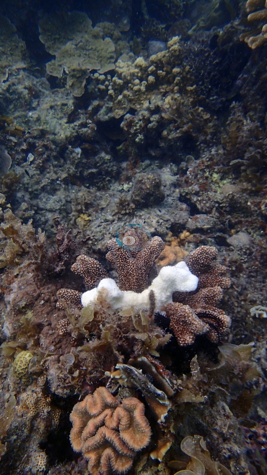
Western Philippines (NOAA CRW virtual station) · Palawan/North Borneo ecoregion mean=4.0°C-heating weeksThe second island-hopping stop was patchier than the first and visibly competing with algae for space, with one small colony showing localized, non-reef-wide tissue bleaching. A photo-derived index puts cover around 43%.
Two other CRD dive sites within 5 km shown in orange — click a marker to open its page.
Site description
Setting
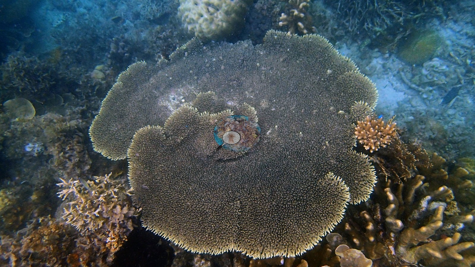
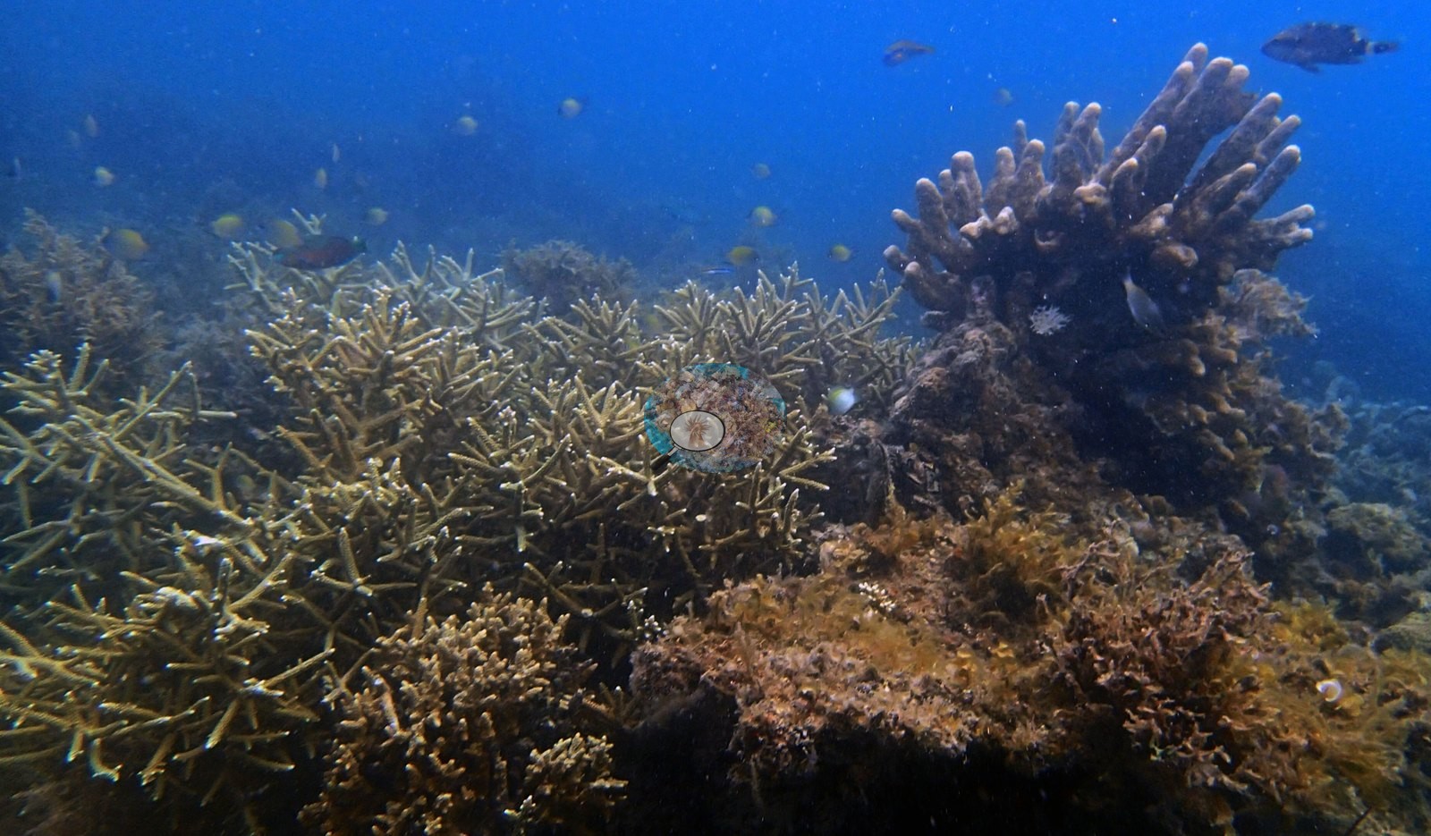
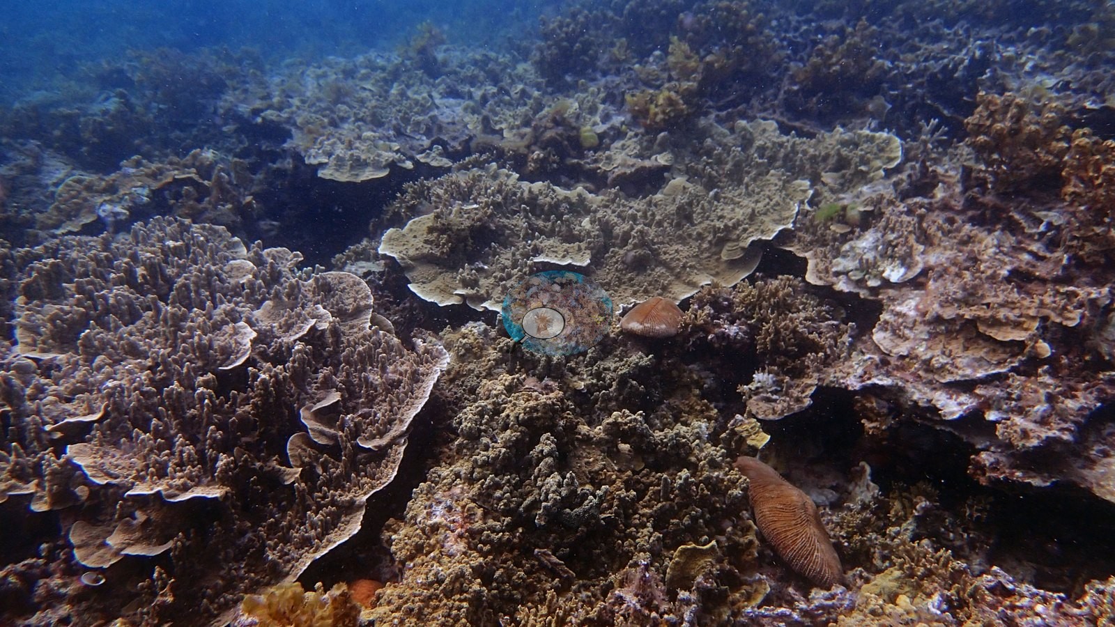
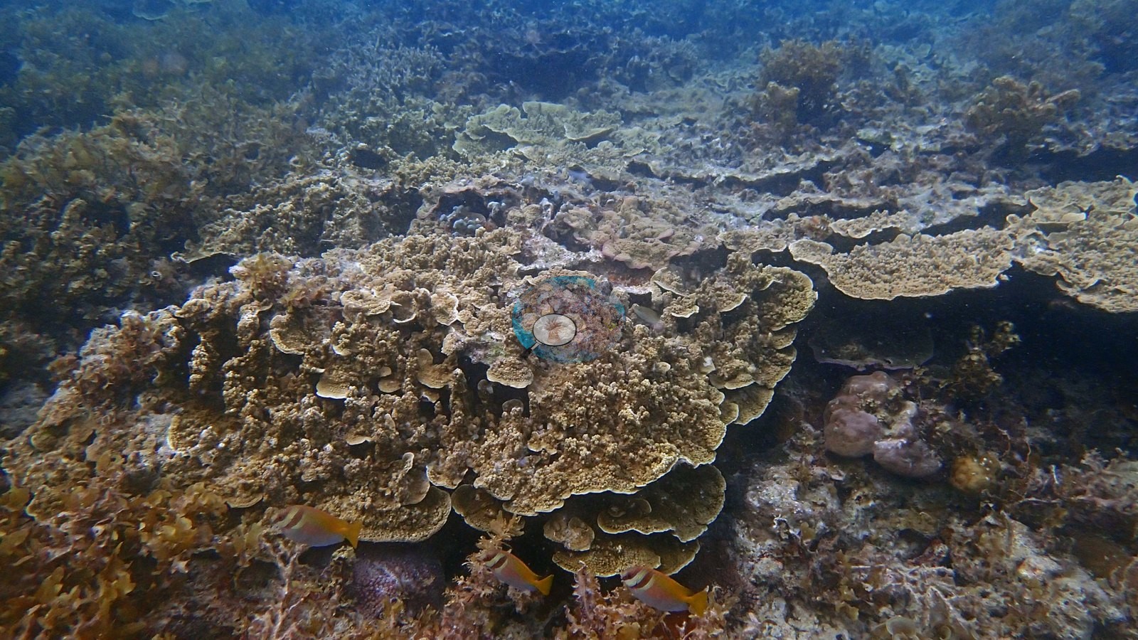
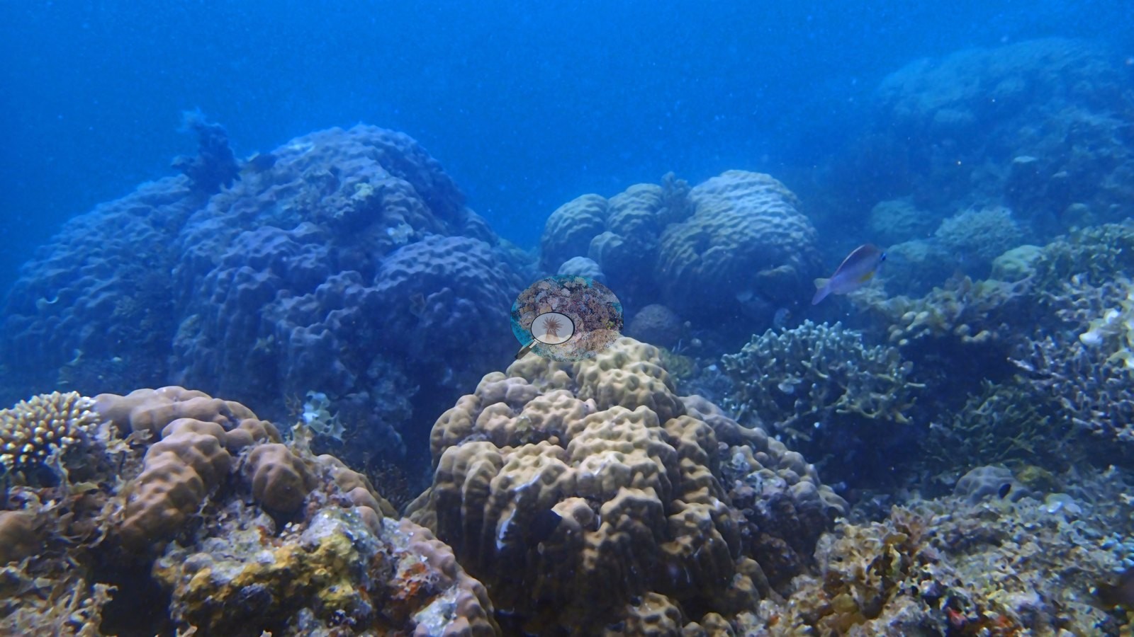
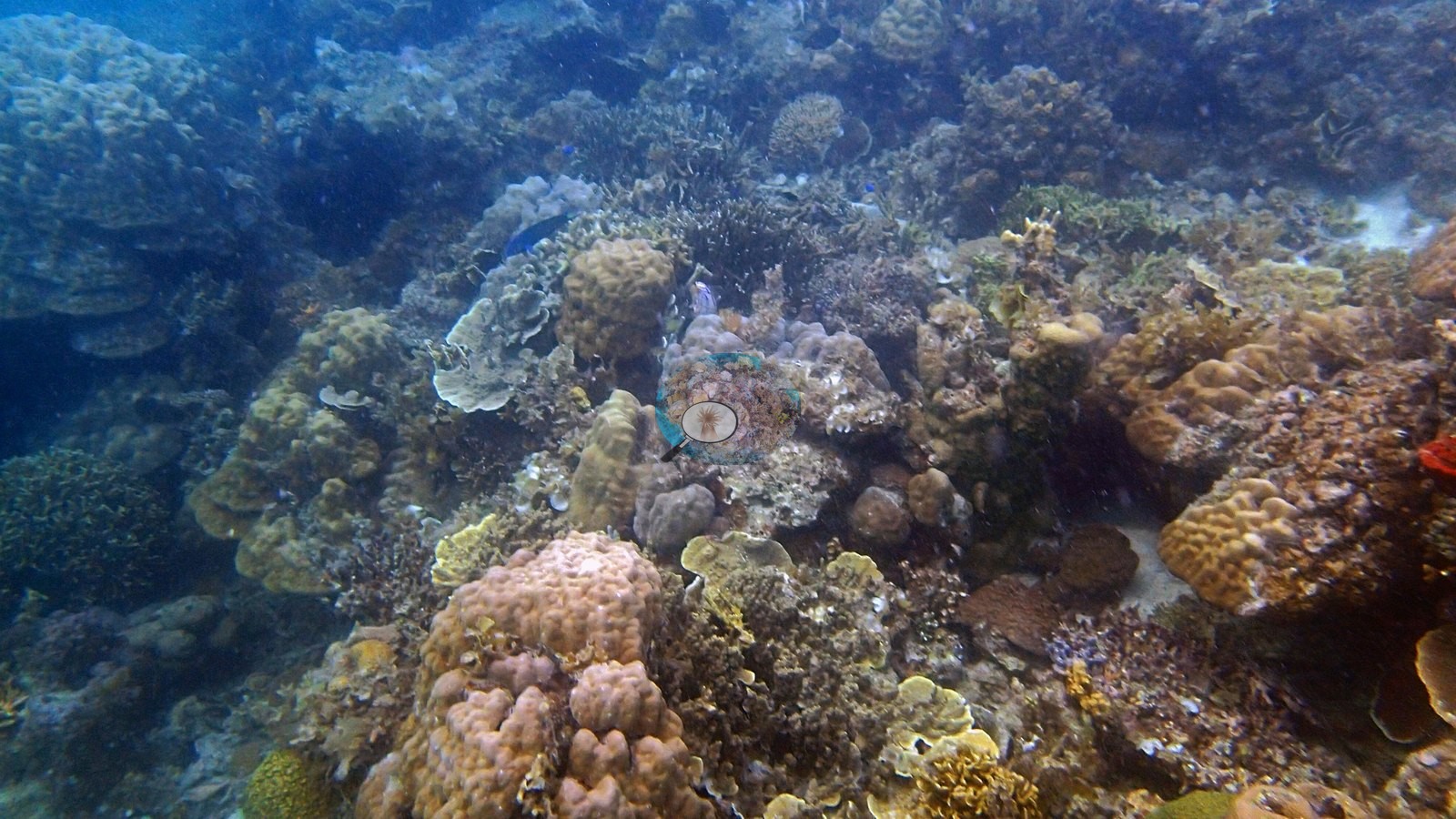
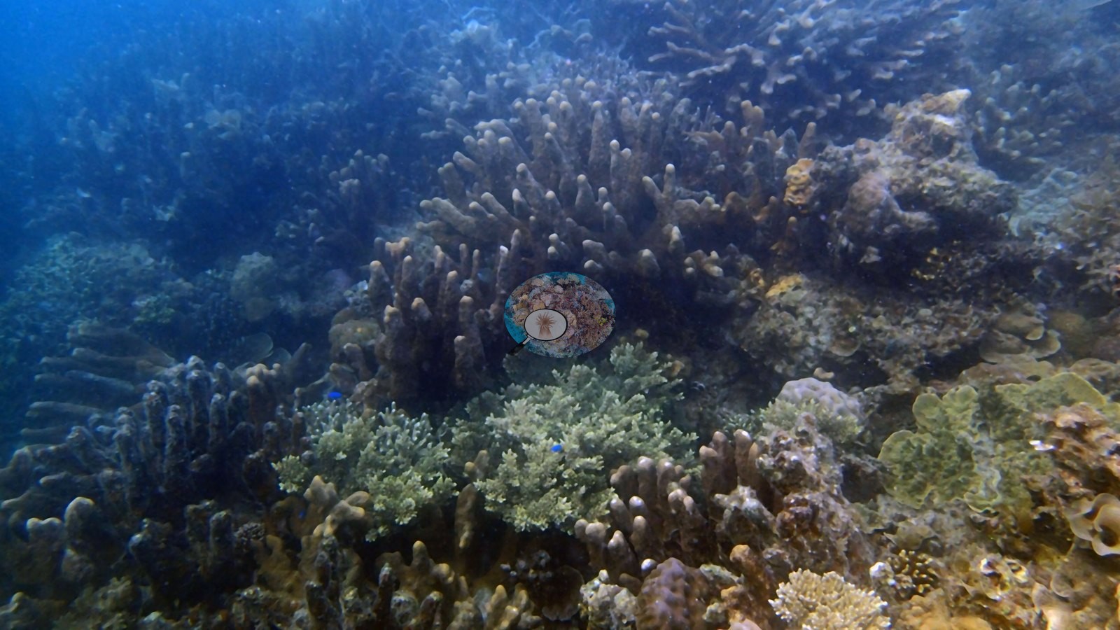
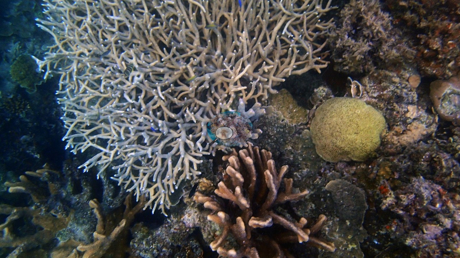
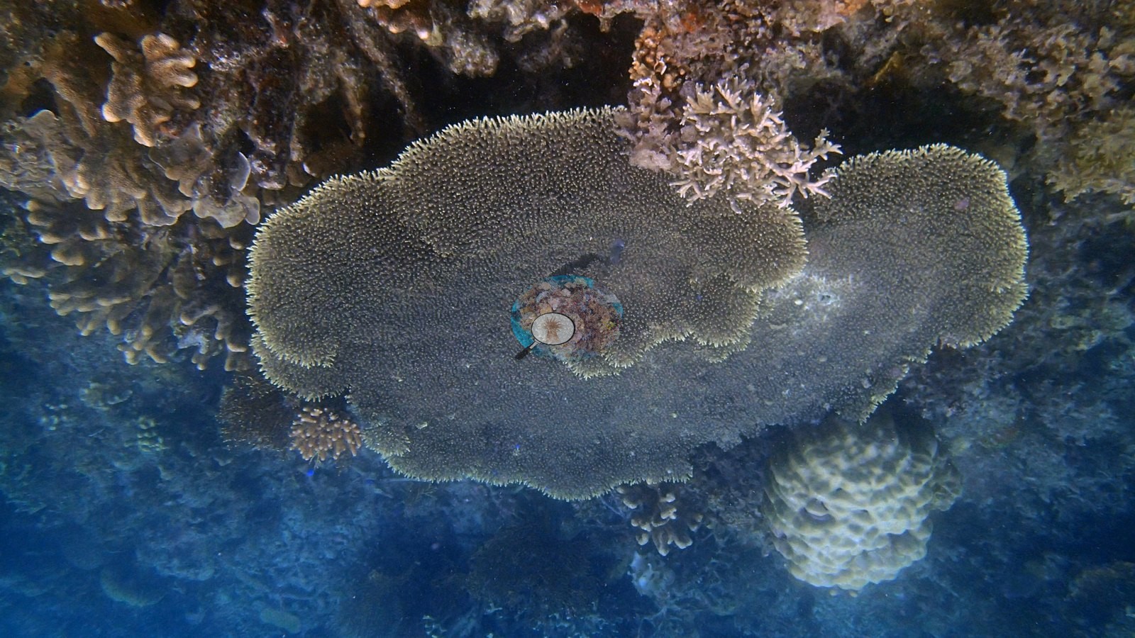
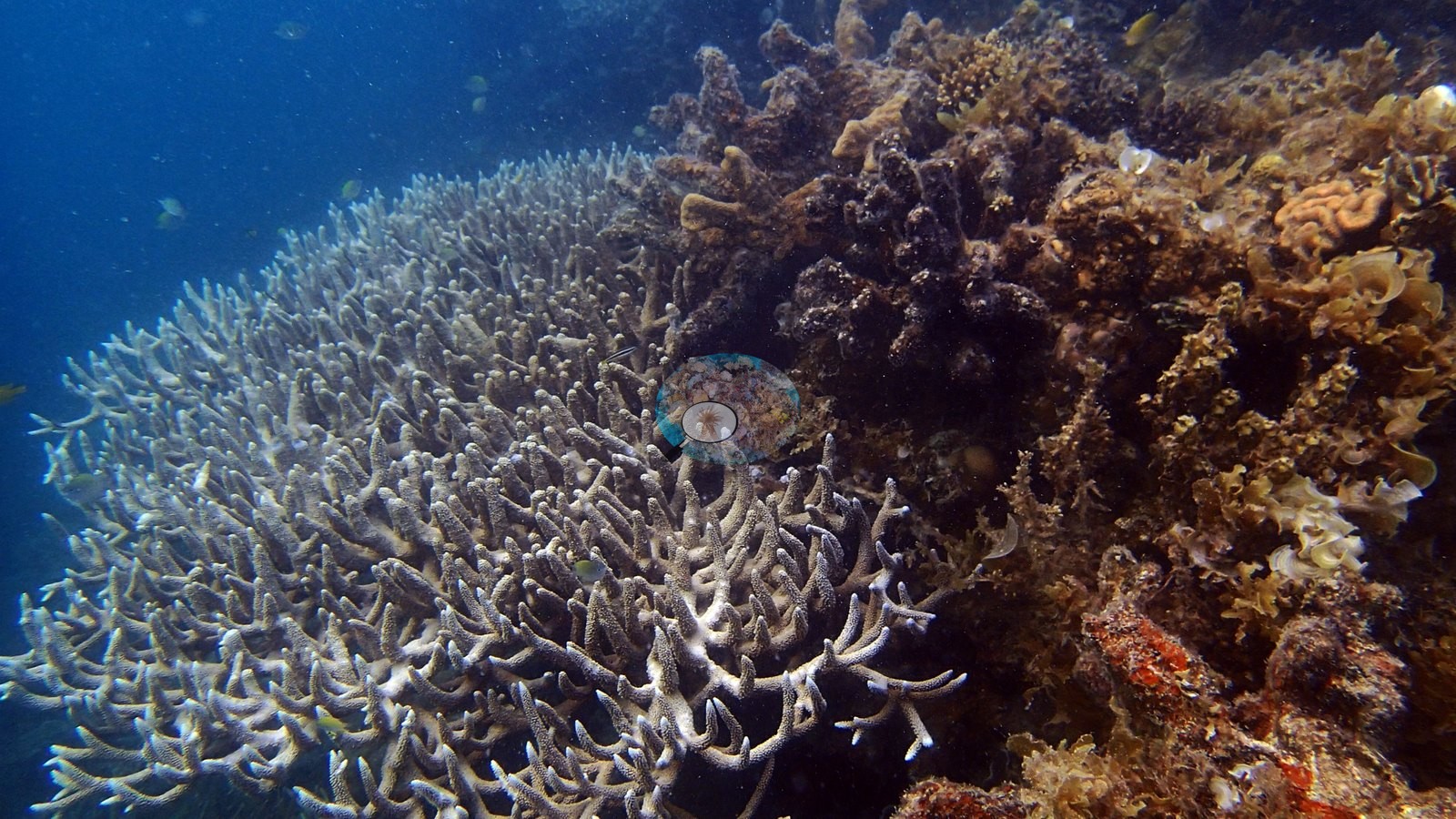
Port Barton, Palawan — the second island-hopping snorkel stop of 2017-01-21, further out along the outer sandbars than stop 1. Across the 20 frames the habitat is a shallow, high-relief reef flat and upper slope of hard framework and rubble, shot in natural light against a blue mid-water background; visibility is good but slightly green-hazy in the wider shots.
General benthic features
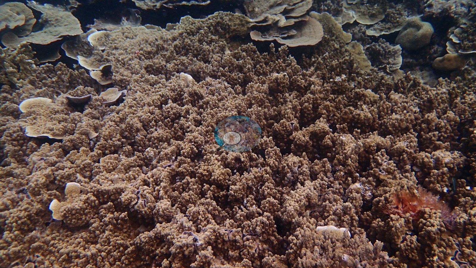
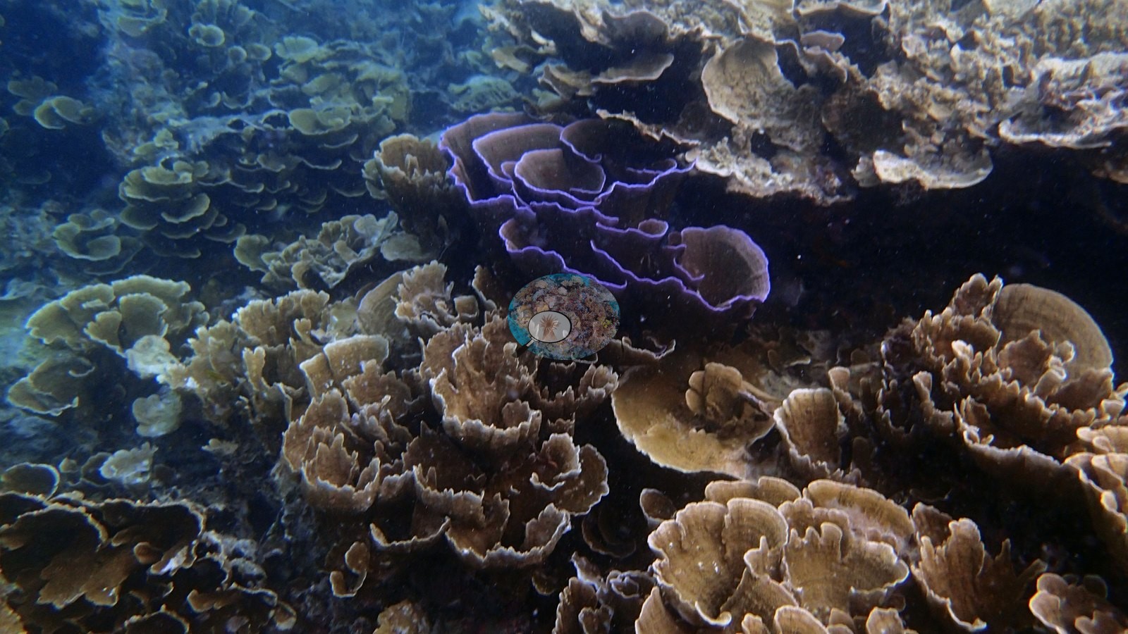
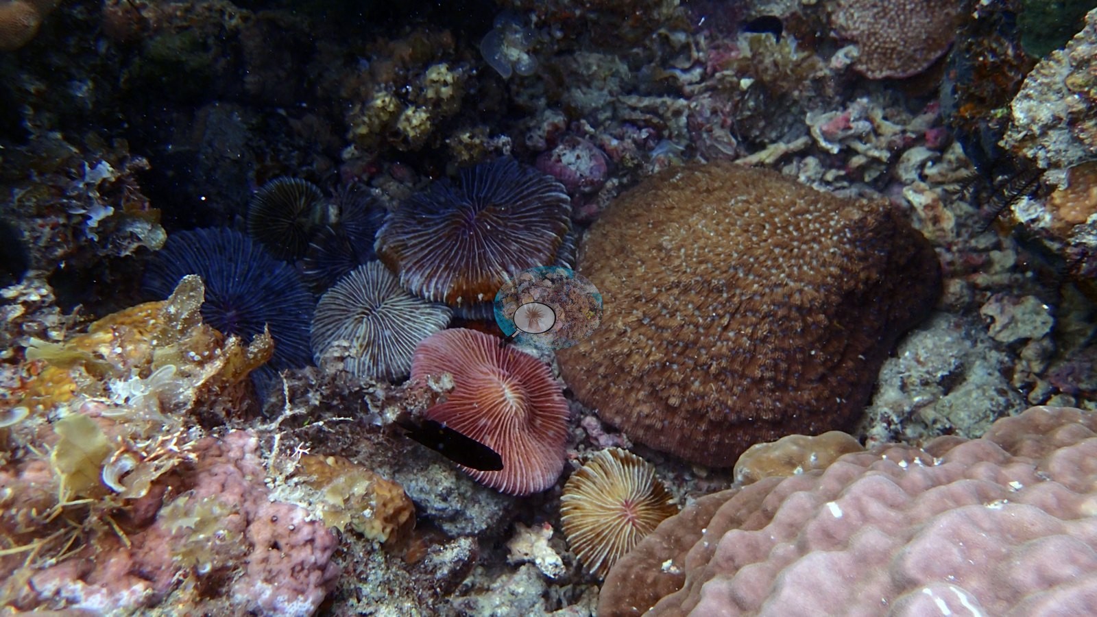
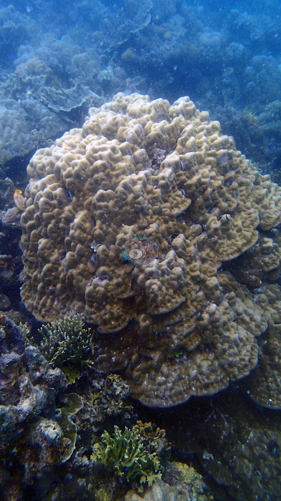
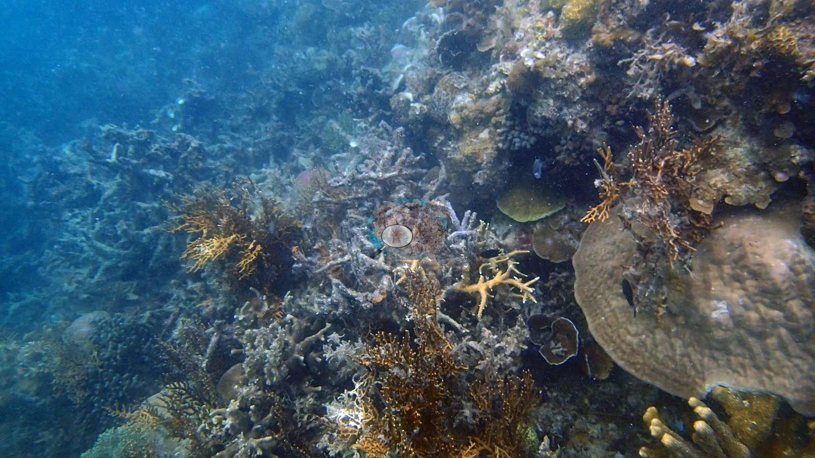
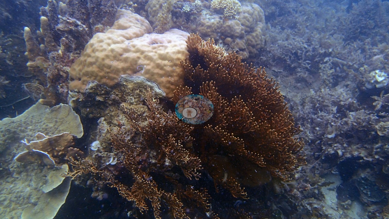
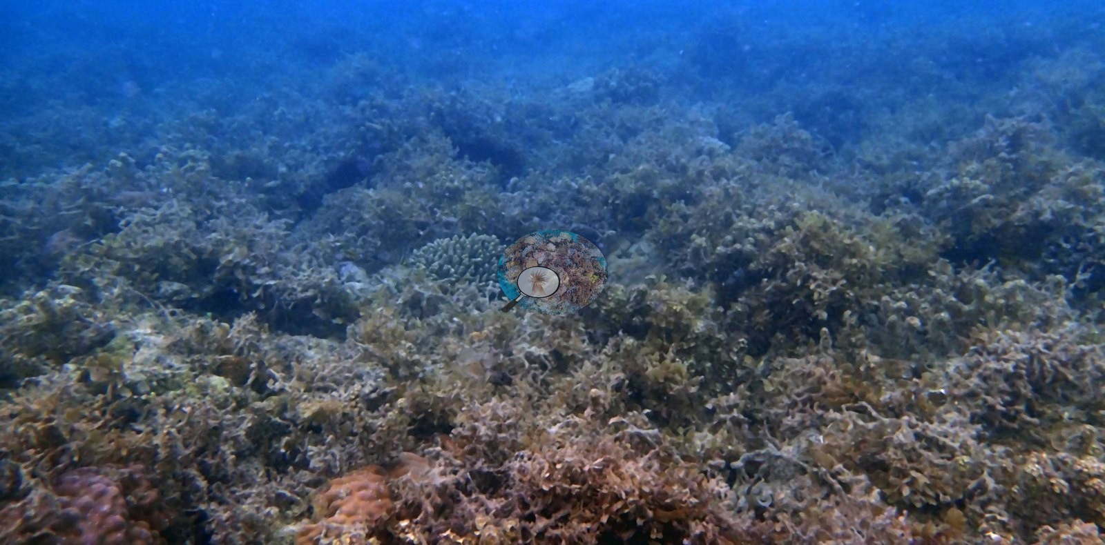
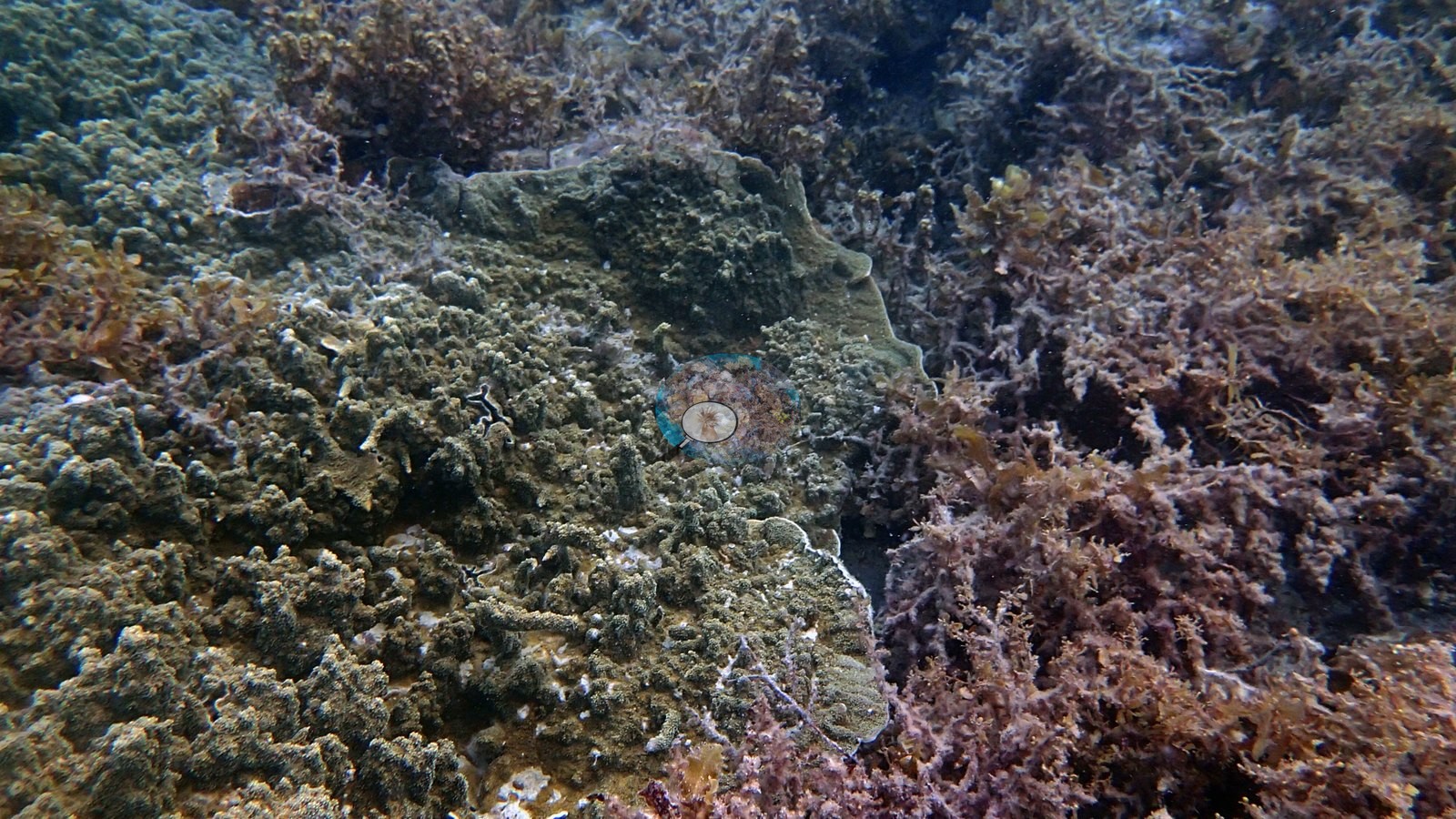
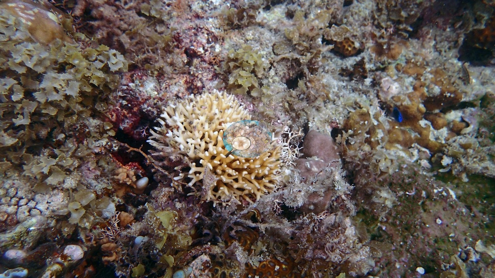
Coral architecture is genuinely varied. Two frames are dominated by broad tabulate Acropora tables a meter or more across, one photographed from above with upright axial corallites around a pale, partly abraded center and one shot from below against open blue. Arborescent staghorn Acropora forms extensive thickets in several frames, some with pale purple-tinted branch tips and small damselfishes and anthias hovering over them. A third register is plating and foliose: whorled, tiered plates compatible with Montipora and Echinopora stacked in shingle-like banks, one striking colony with a vivid violet growing margin, and a large cabbage-form colony of overlapping cupped plates. Massive corals are common — blue-gray Porites bommies, smaller helmet-shaped colonies, and columnar/digitate stands of finger-like Porites in one dense frame. One frame is a fungiid garden: half a dozen free-living mushroom corals in gray, blue-gray, cream, and pink lying among pink crustose coralline algae beside a massive faviid and a pink massive Porites. A bushy brown gorgonian with white expanded polyps occupies one frame, and green calcareous algae and turf occur between colonies throughout. Anderson titled four frames himself, and they carry the site's main message. 'Endangered Seriatopora' shows a single small cream fine-branched Seriatopora colony isolated in a field of rubble and fleshy algae. 'Algal smothered reef' is a near-continuous brown macroalgal canopy over the pavement with one small knobby coral surviving in the middle of it. 'Coral vs. algae' is a close view of the competitive interface — encrusting coral tissue and green-filmed pavement abutting a dense red-brown fleshy macroalgal stand. 'Second snorkel spot' shows a brown verrucose branching colony of Pocillopora form with two branches stark white: partial, colony-level bleaching, not a reef-wide event, and the only bleaching seen in the set. That is the honest summary of the site: real structural coral — tables, staghorn thickets, plate banks, massives, fungiids — interleaved with substantial pavement and rubble that fleshy macroalgae has taken over.
Coral cover
Coral cover: 43% — a single-method photographic index. Quoted verbatim from the pin: 'CoralSCOP computer vision only (no per-stop DINOv3 exists) -- 43.4% over 20 frames.' That figure is computer vision, not a diver survey and not a transect: CoralSCOP sums retained mask fractions with overlaps possible and segments coral-shaped objects rather than living tissue, so standing dead skeleton scores as coral. Against in situ transects it reads high overall (median about 1.46x). Treat it as provisional. Note also that these 20 frames are the same 20 the CV pass ran on, and they are a photographer's selection biased toward the best coral — several frames in the same set show pavement given over to macroalgae, so a swim-through transect here would very likely land lower.
Stressors
No crown-of-thorns, no disease lesions, no physical breakage from anchoring and nothing man-made appear in any frame.
Comparisons with other data
No prior survey data from this site. If you would like to share yours, please email us.
Coral cover: method notes
Data & links
All images from Coral Reef Diagnostics. Data are from Coral Reef Diagnostics.
© Coral Reef Diagnostics.