
Indonesia - Halmahera: Sali Bay-Batu Mangga
Part of the regional summaries: Indonesia - Halmahera and Indonesia - Maluku
More info
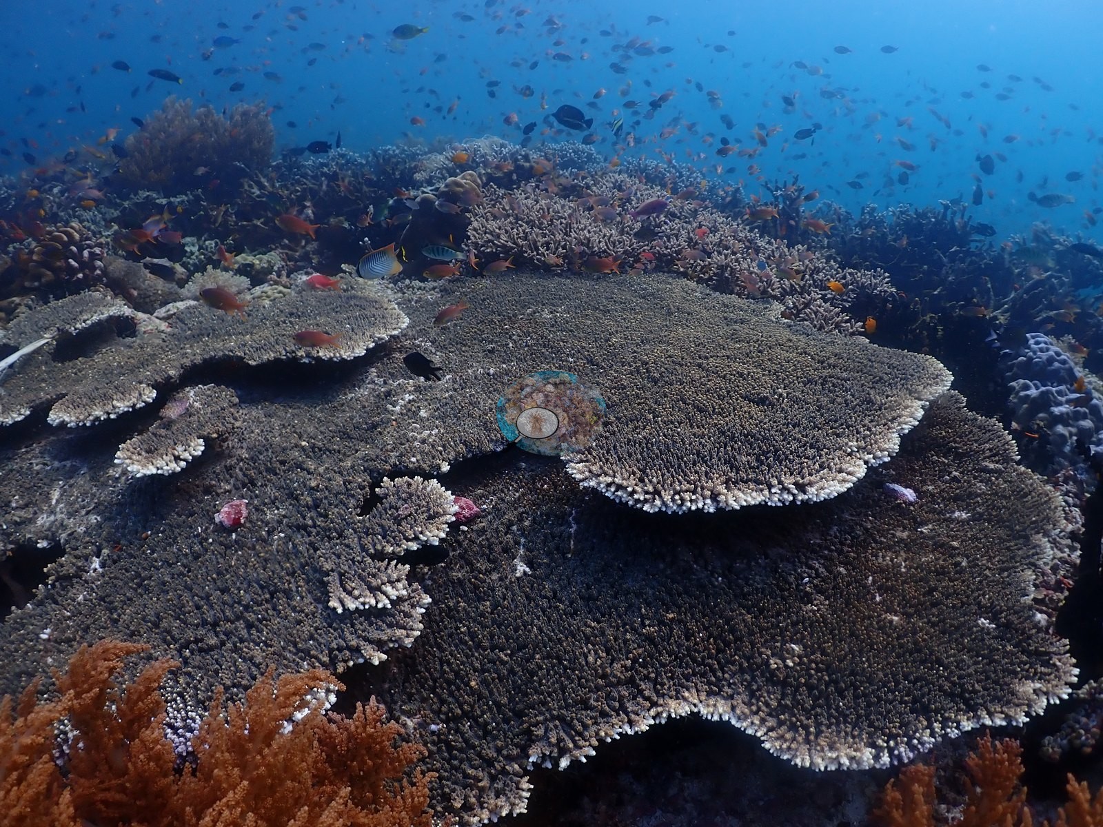
North Maluku (NOAA CRW virtual station)A strong-current reef-hook drift through a dim, current-swept passage opening onto genuinely patchy, colorful reef - a lone red brain coral on white sand, pink tube sponges, and extensive pale staghorn and corymbose Acropora thickets carrying the highest single-frame readings. No bleaching, disease, or crown-of-thorns starfish (COTS) observed. A blind Claude computer-vision point count puts coral cover around 29%.
Hanyutan berarus kuat dengan kait terumbu melewati lorong remang yang tersapu arus, membuka ke terumbu yang benar-benar berbercak dan berwarna — satu karang otak merah soliter di atas pasir putih, spons tabung merah muda, dan rumpun Acropora bercabang serta korimbosa yang pucat dan luas, yang mencatat nilai per-bingkai tertinggi. Tidak teramati pemutihan, penyakit, maupun bintang laut mahkota duri. Penghitungan titik buta visi komputer Claude memperkirakan tutupan karang sekitar 29%.
Terjemahan ringkasan ini oleh AI; versi bahasa Inggris yang berlaku.
Eight other CRD dive sites within 5 km shown in orange — click a marker to open its page.
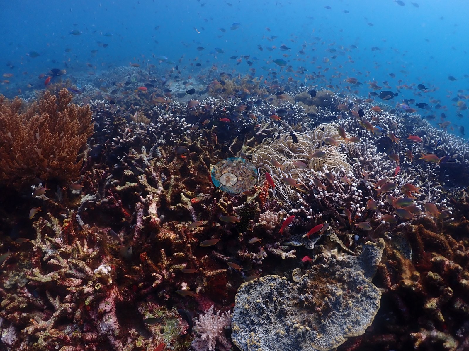
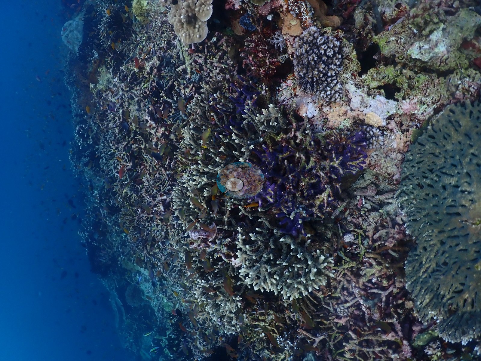
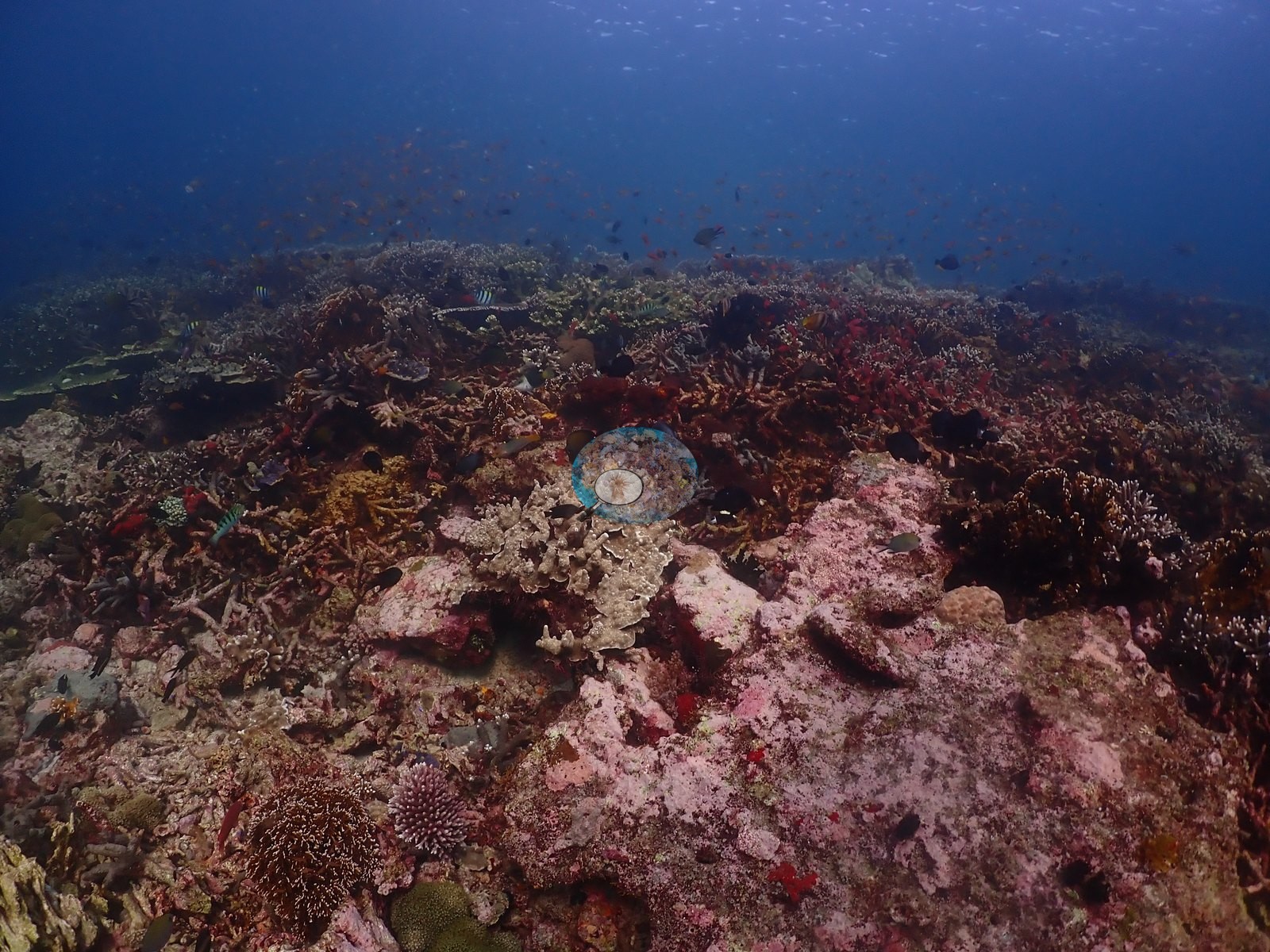
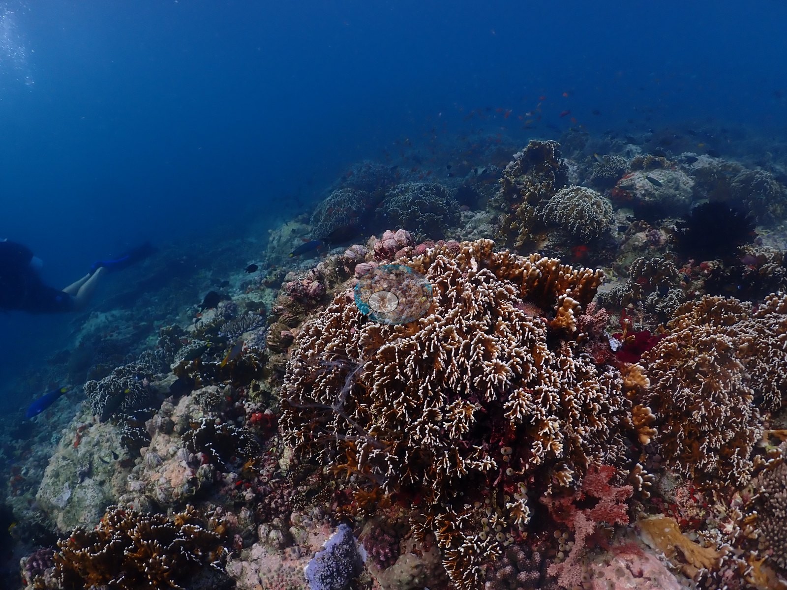
22 of these 52 photographs are shown beside the text that discusses them under "Site description" below.
Site description
Setting
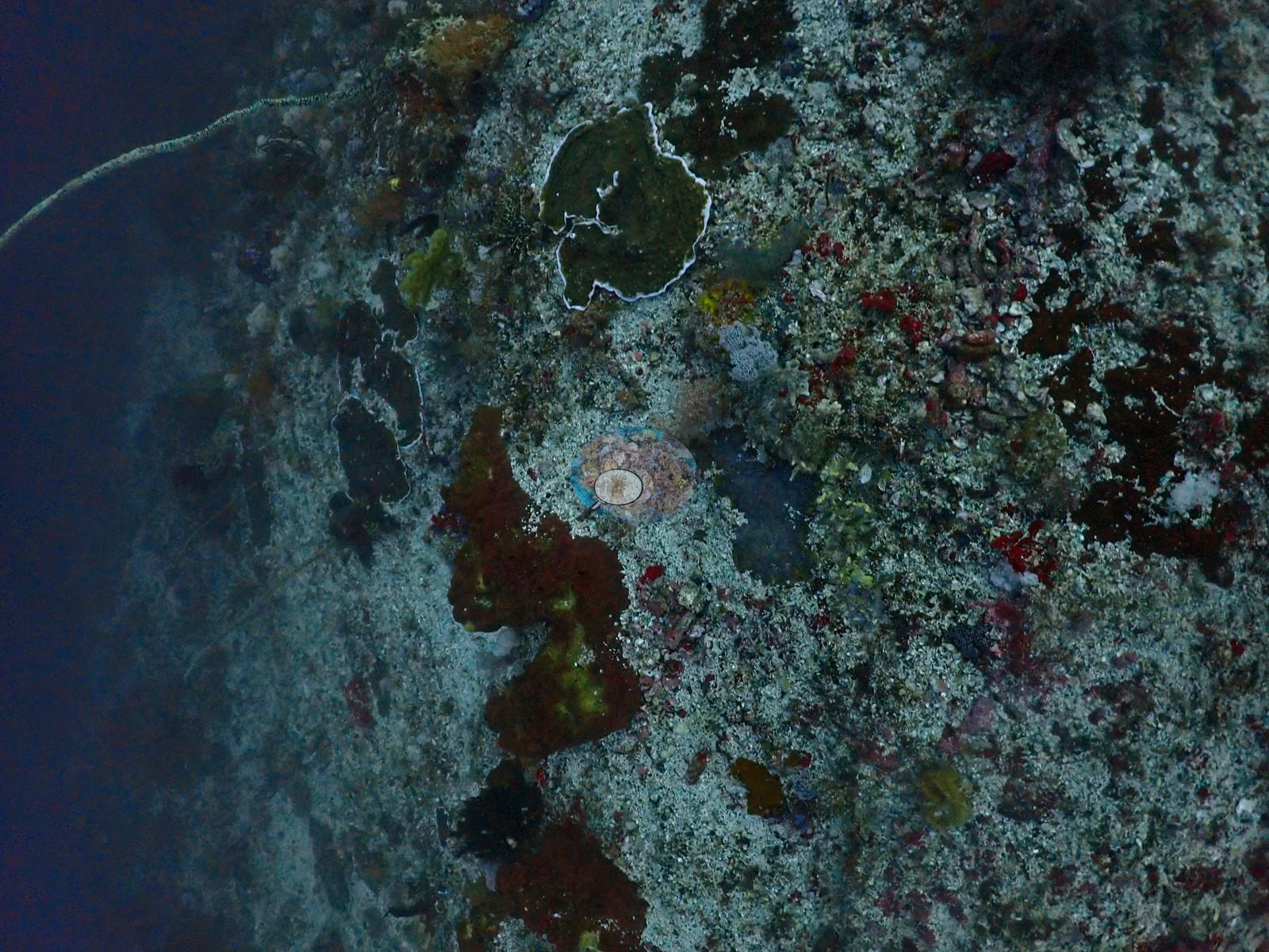
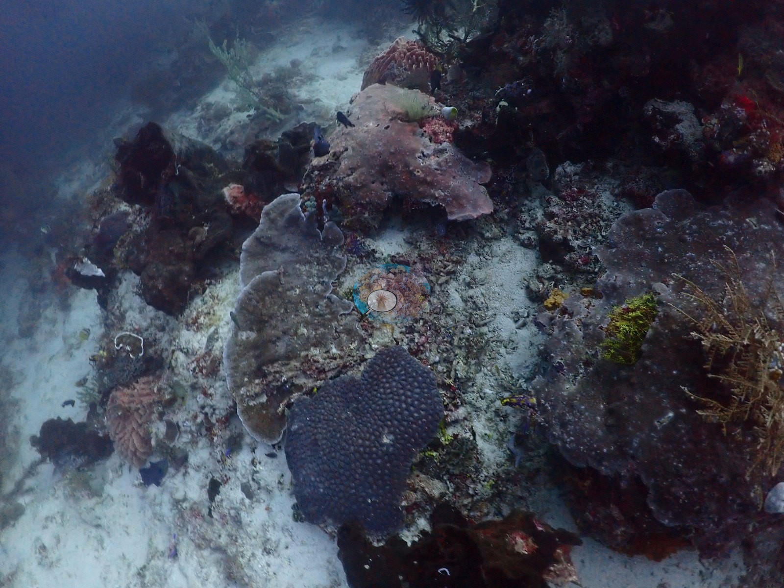
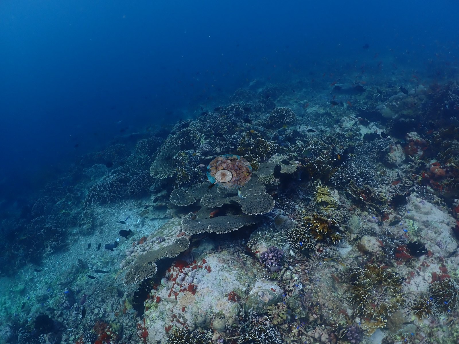
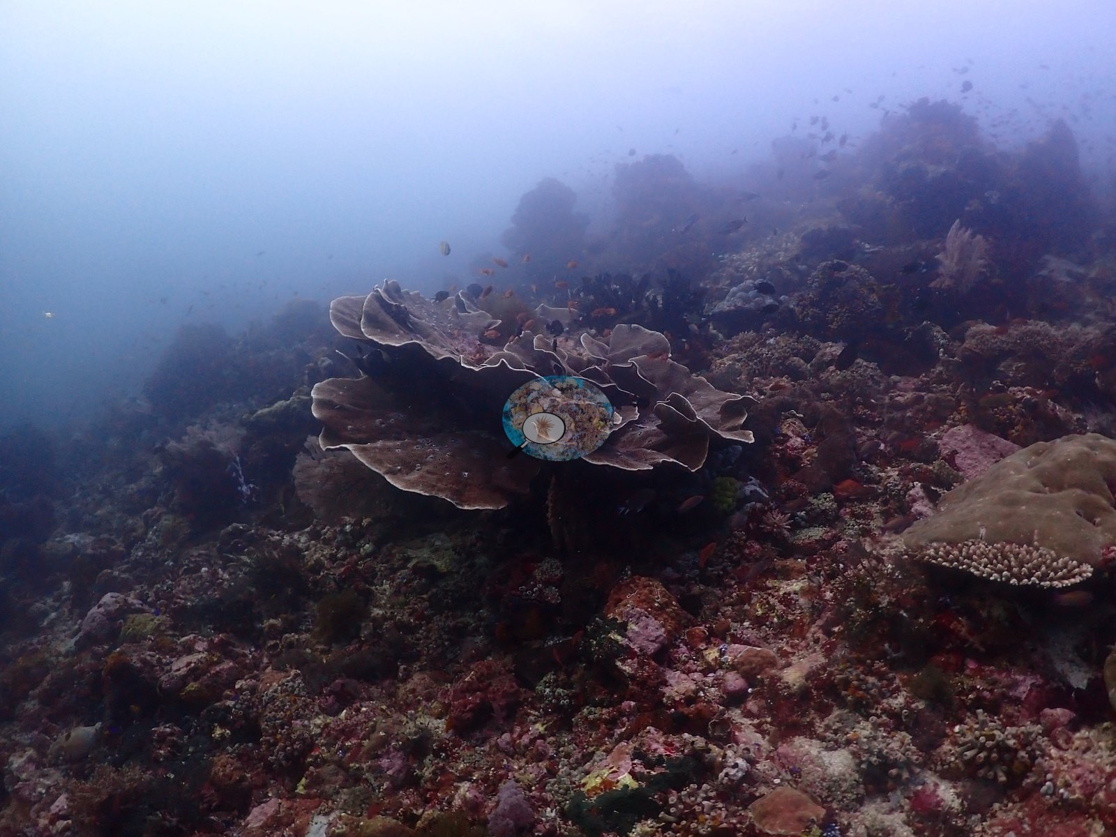
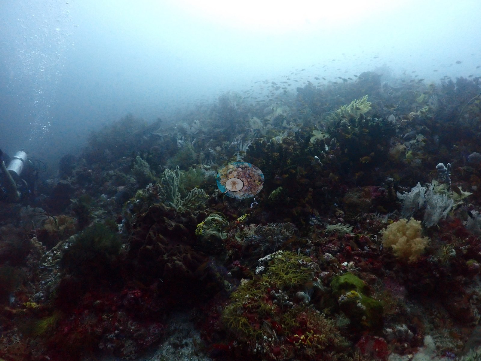
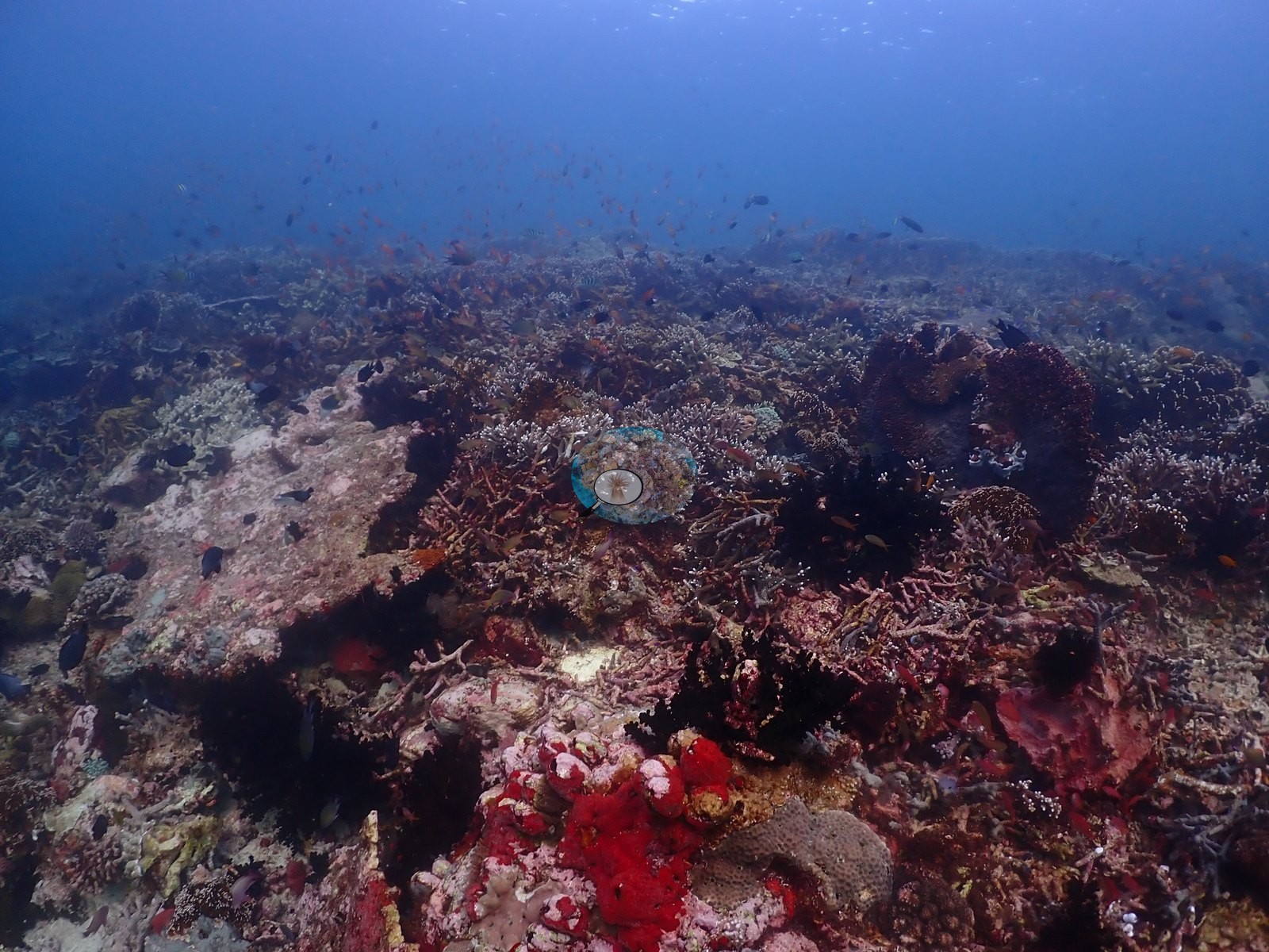
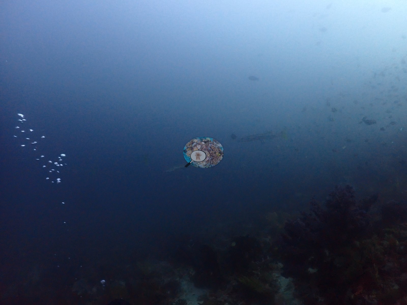
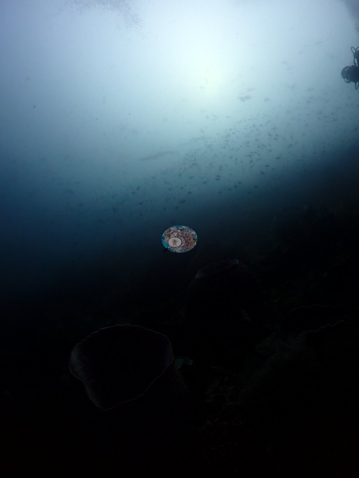
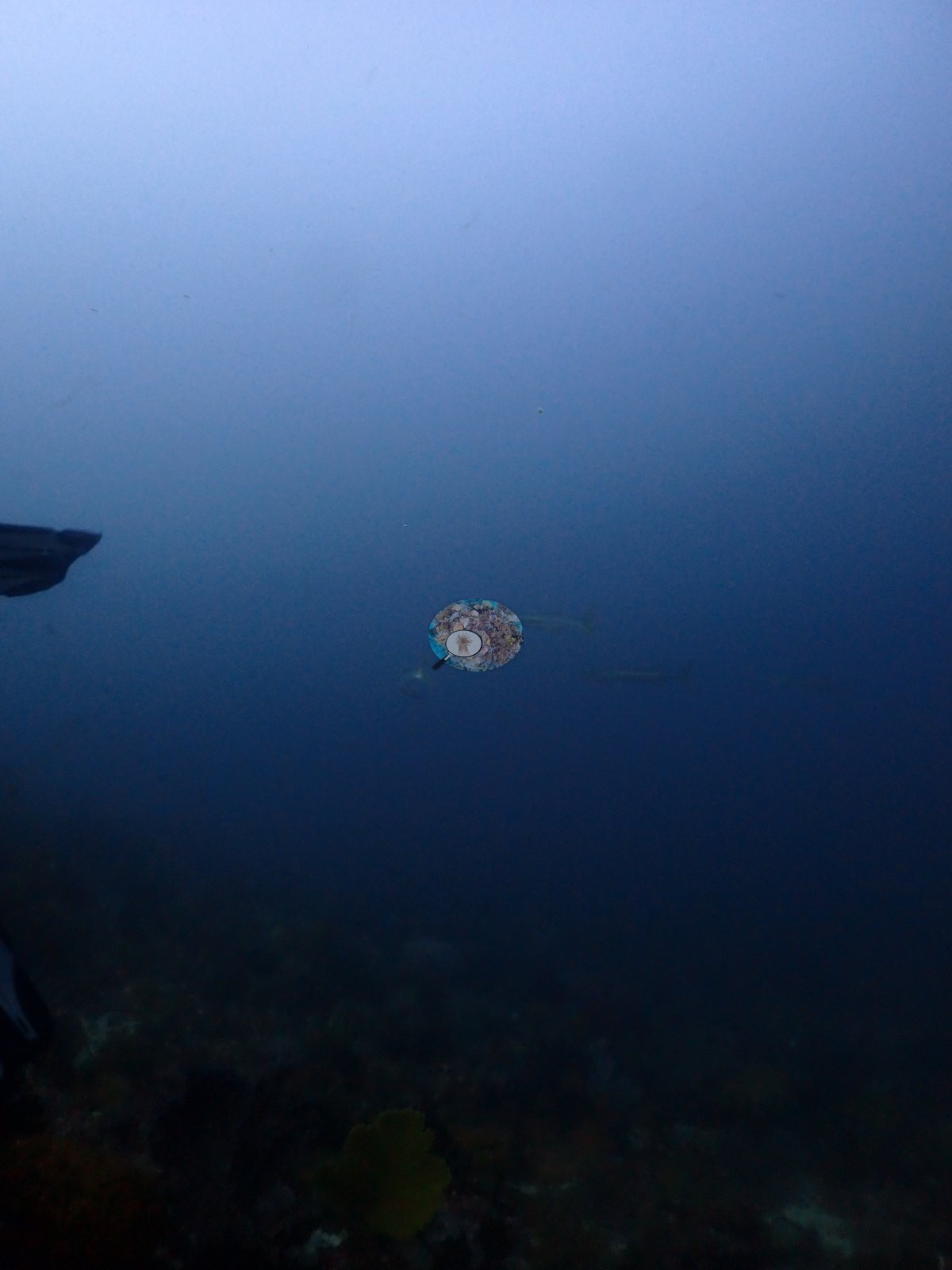
Sali Bay-Batu Mangga (Halmahera dive 9) was a strong-current reef-hook drift, and the imagery shows it: a stretch of near-black, near-zero-cover frames reads as a deep, dim, current-swept passage rather than a data gap (e.g., Image 37, Image 40, Image 48).
General benthic features
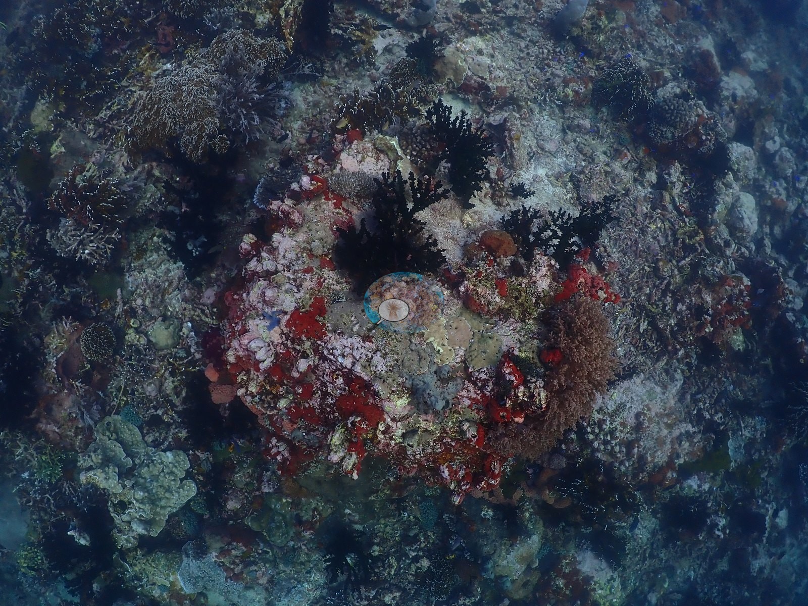
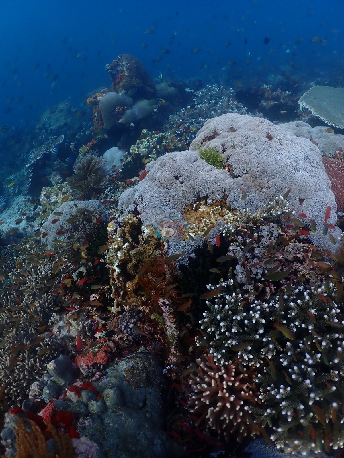
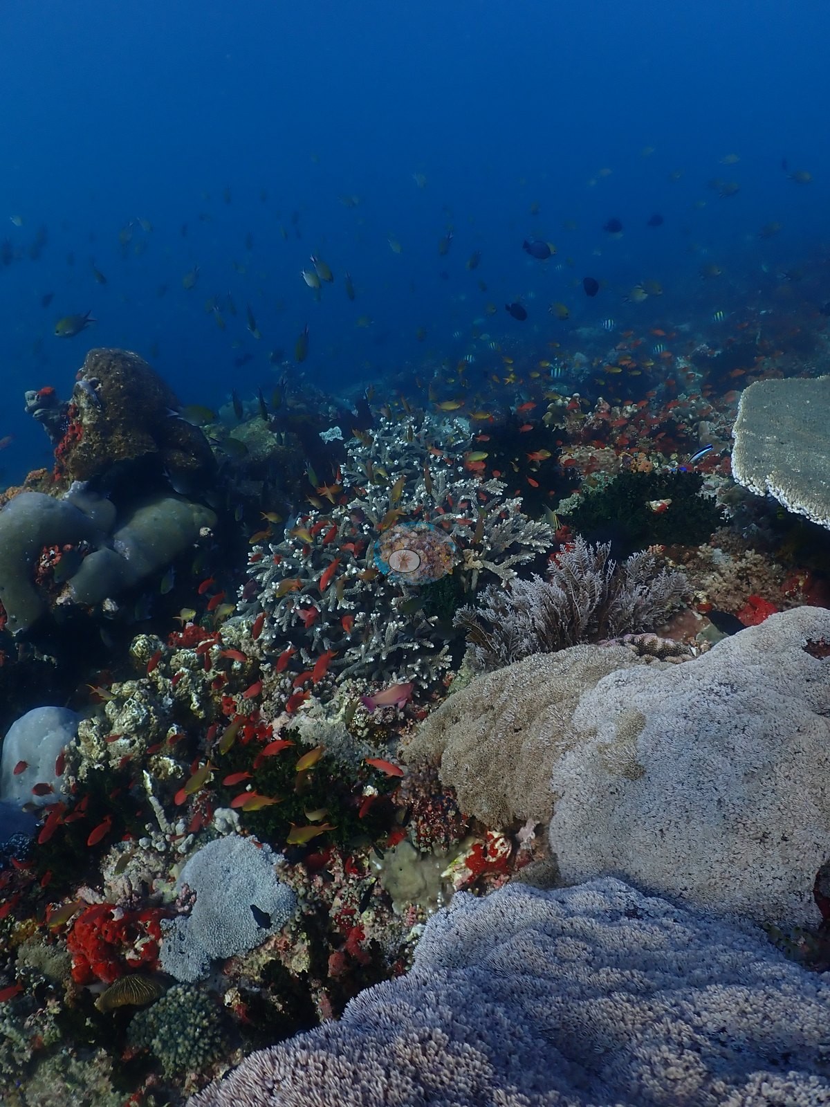
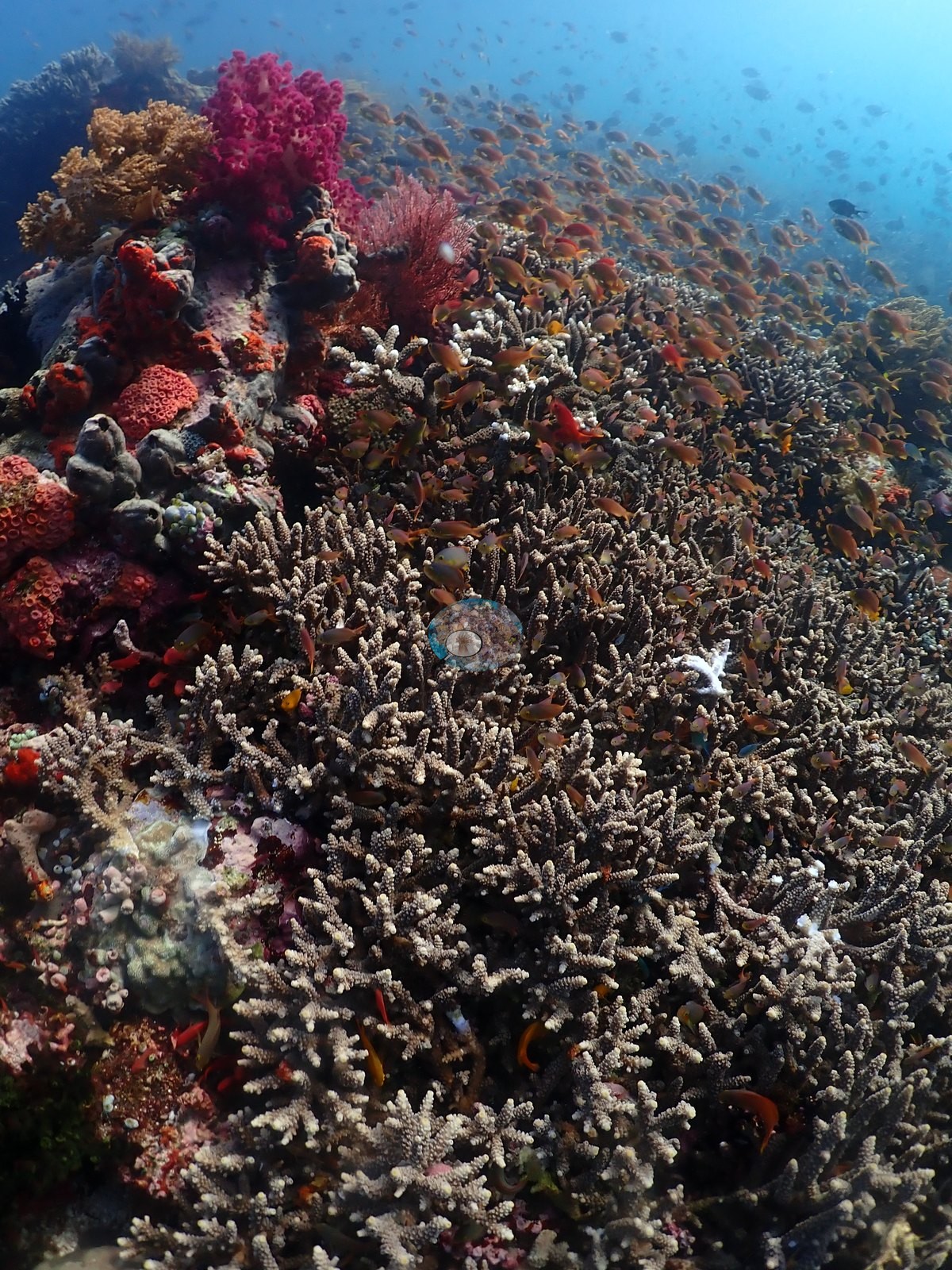
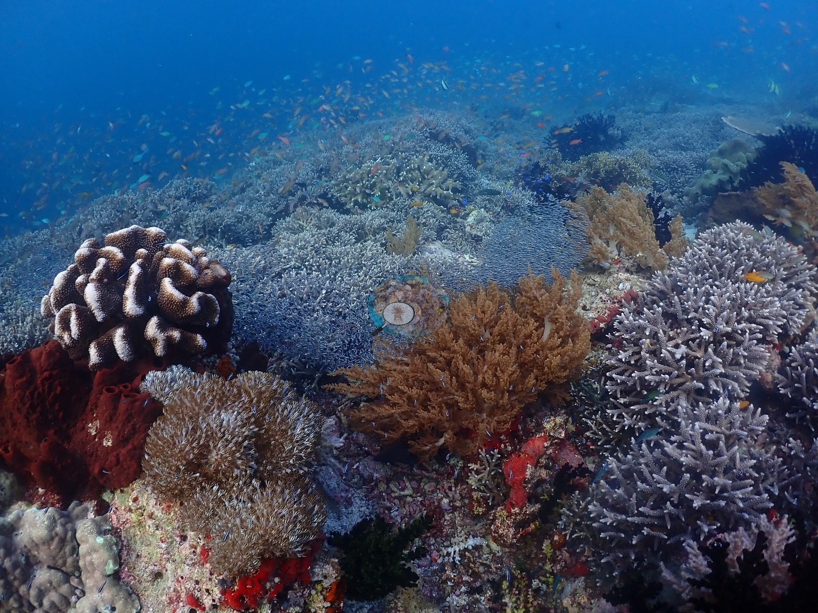
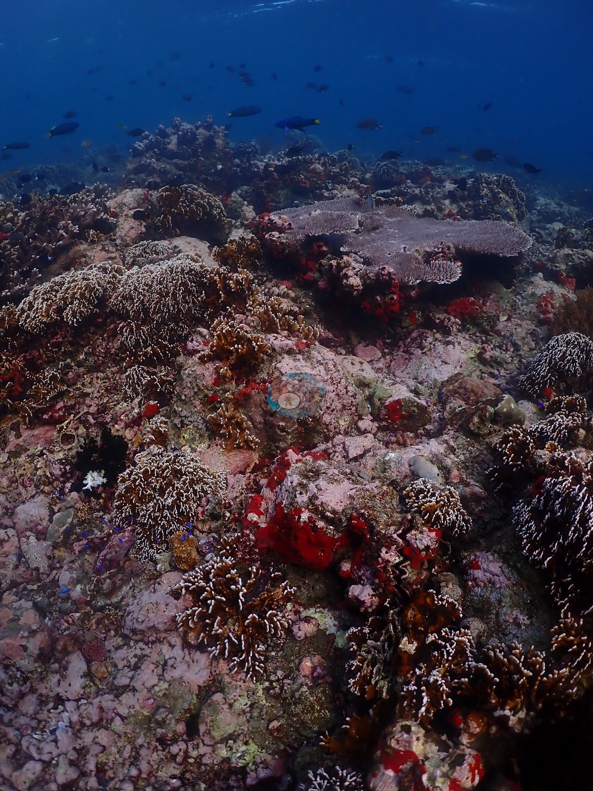
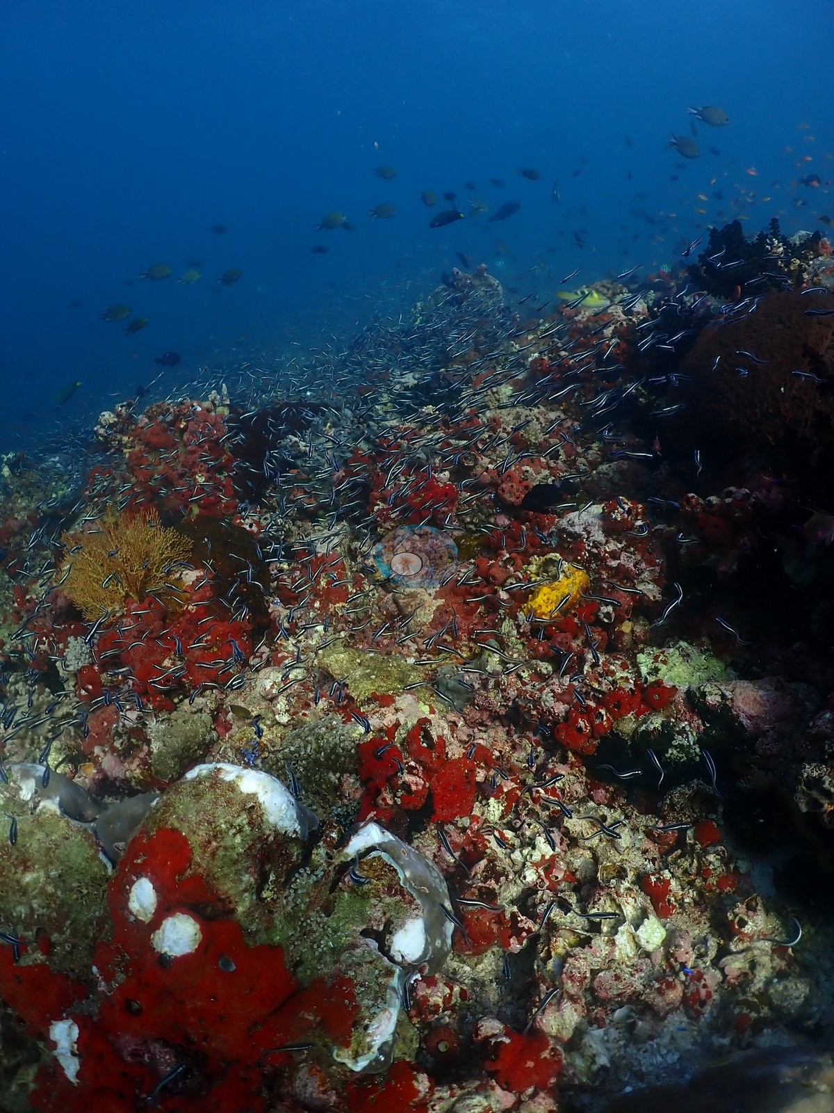
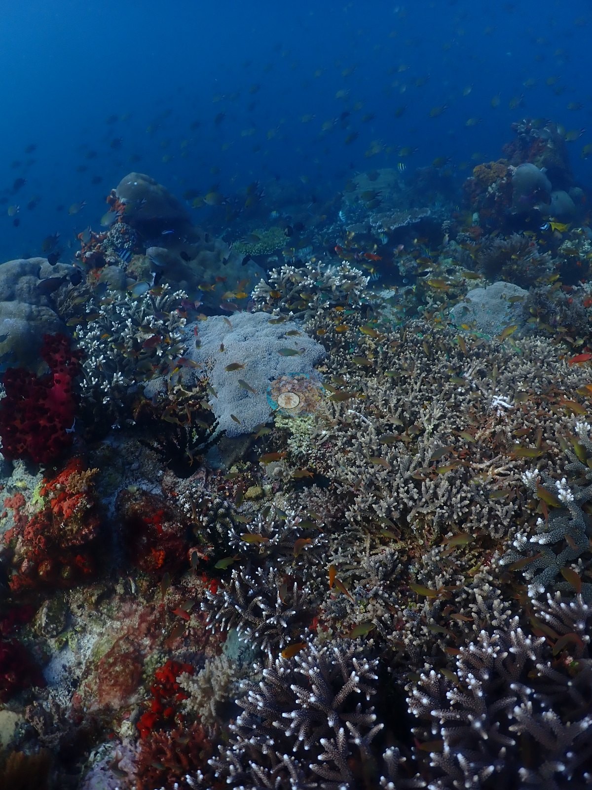
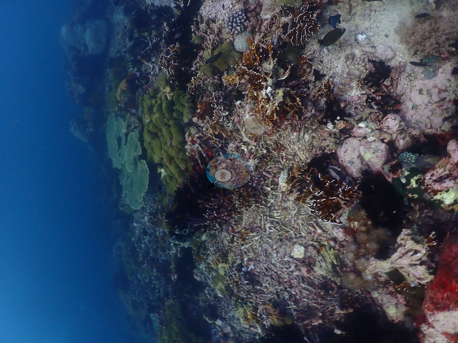
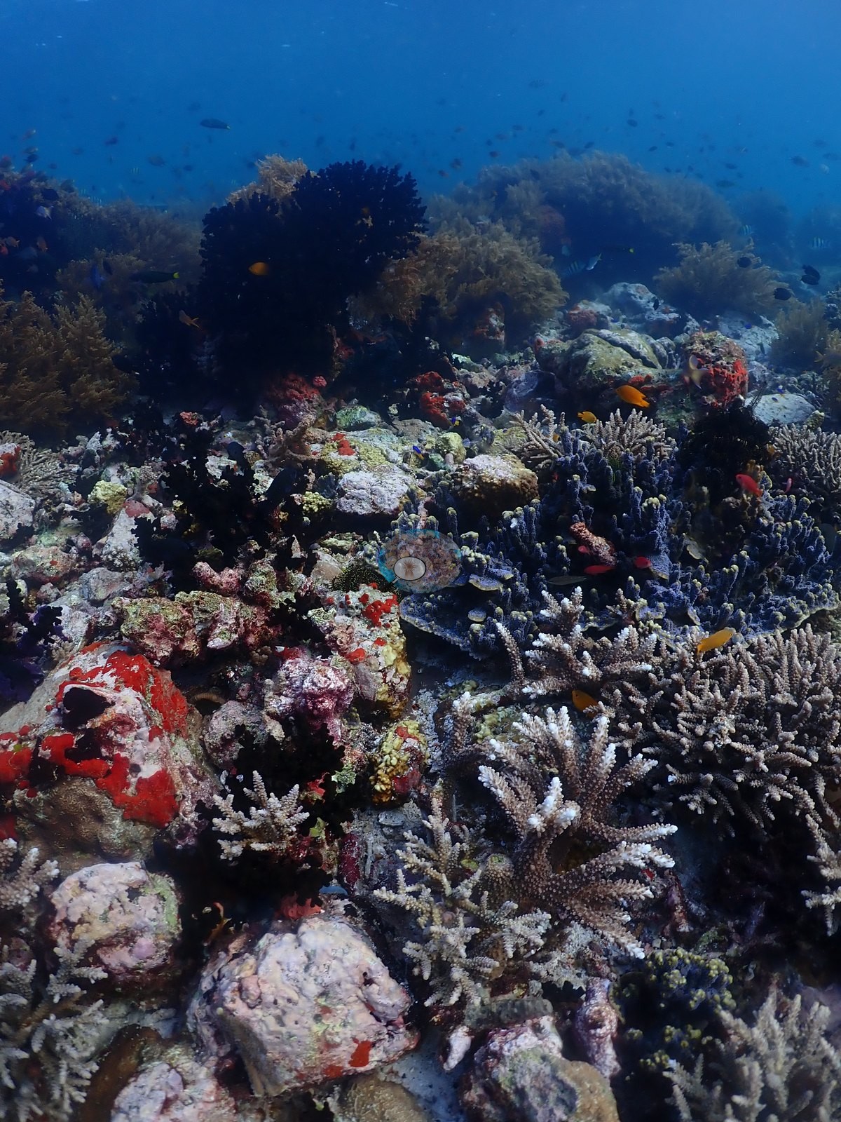
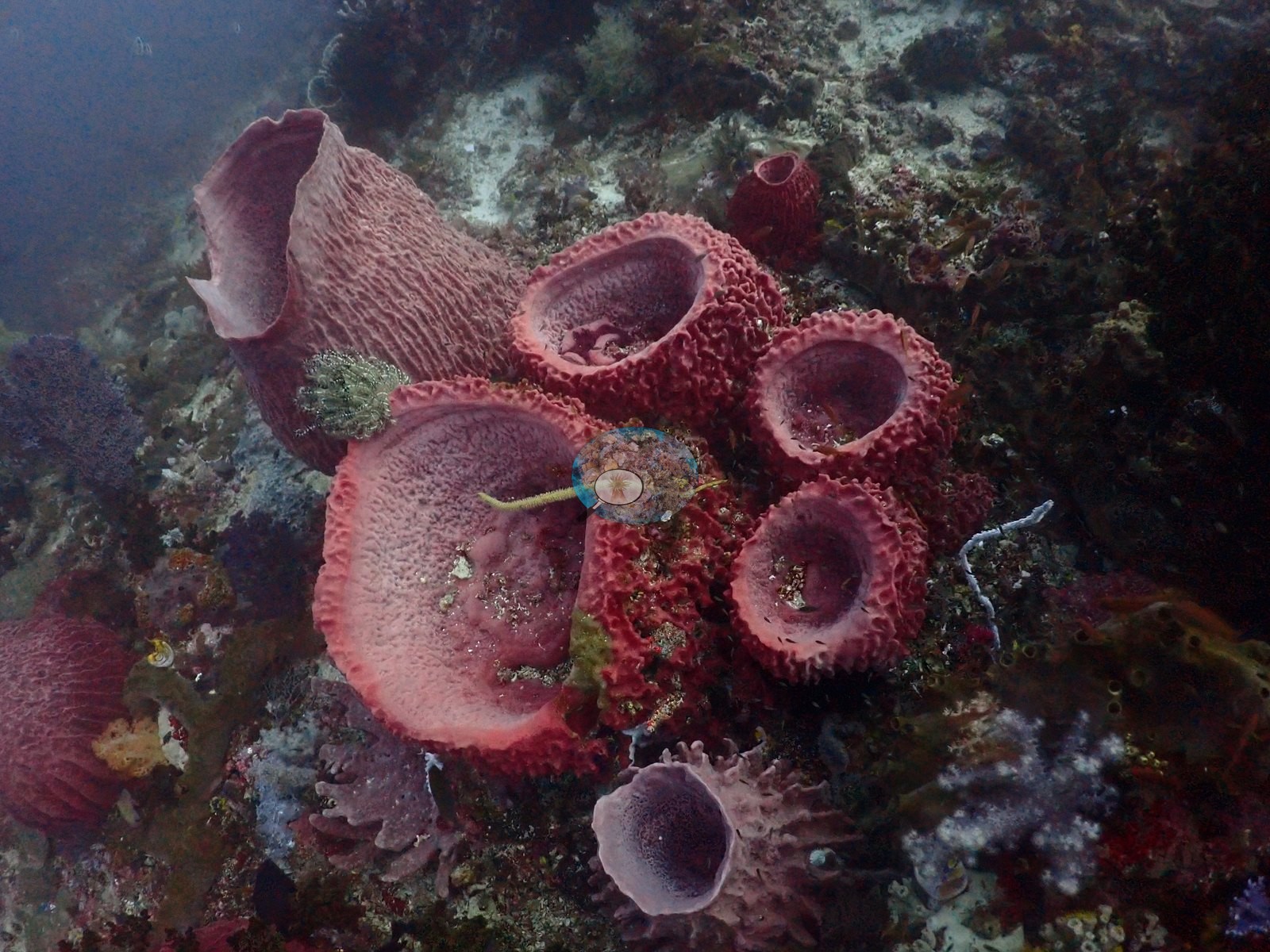
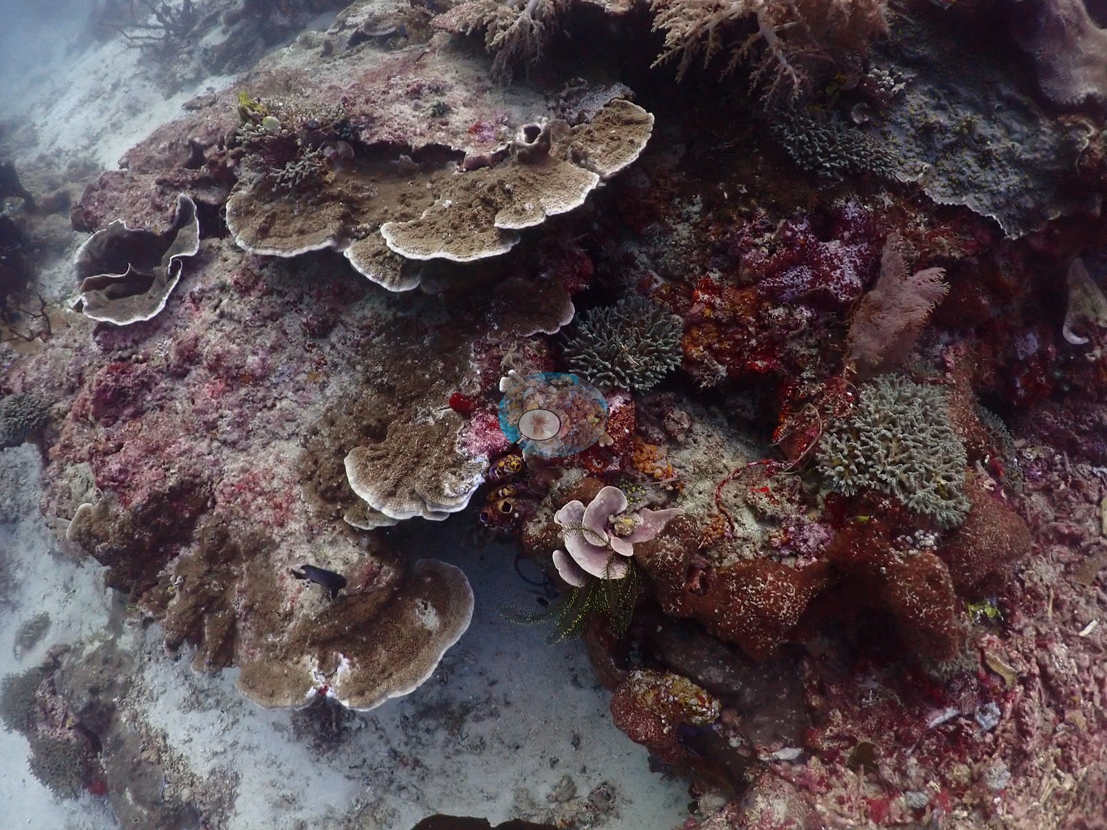
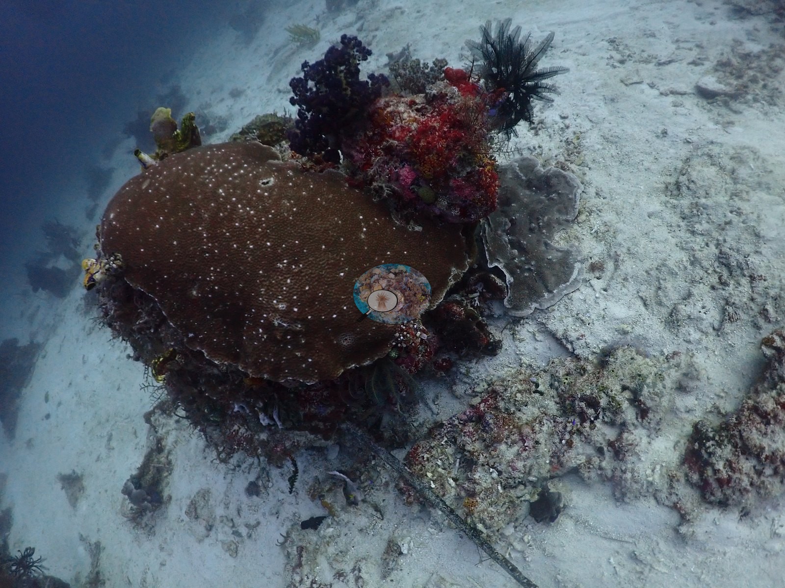
Around it, cover is genuinely patchy and colorful. Standout set-pieces include a vivid red brain coral sitting alone on white sand, clusters of pink tube/barrel sponges, and a striking purple-red-yellow soft coral/sponge assemblage on the wall followed later by a magenta gorgonian-and-soft-coral bouquet and orange soft coral fields (e.g., Image 16, Image 21, Image 31, Image 53).
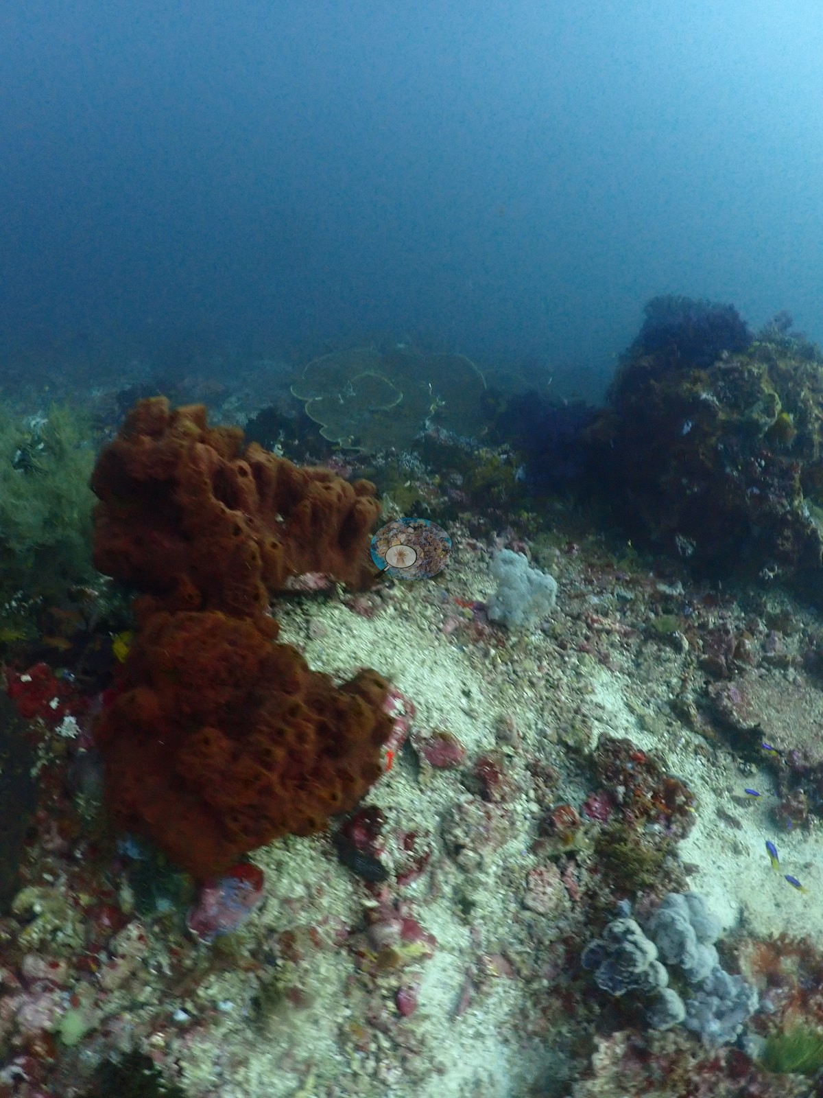
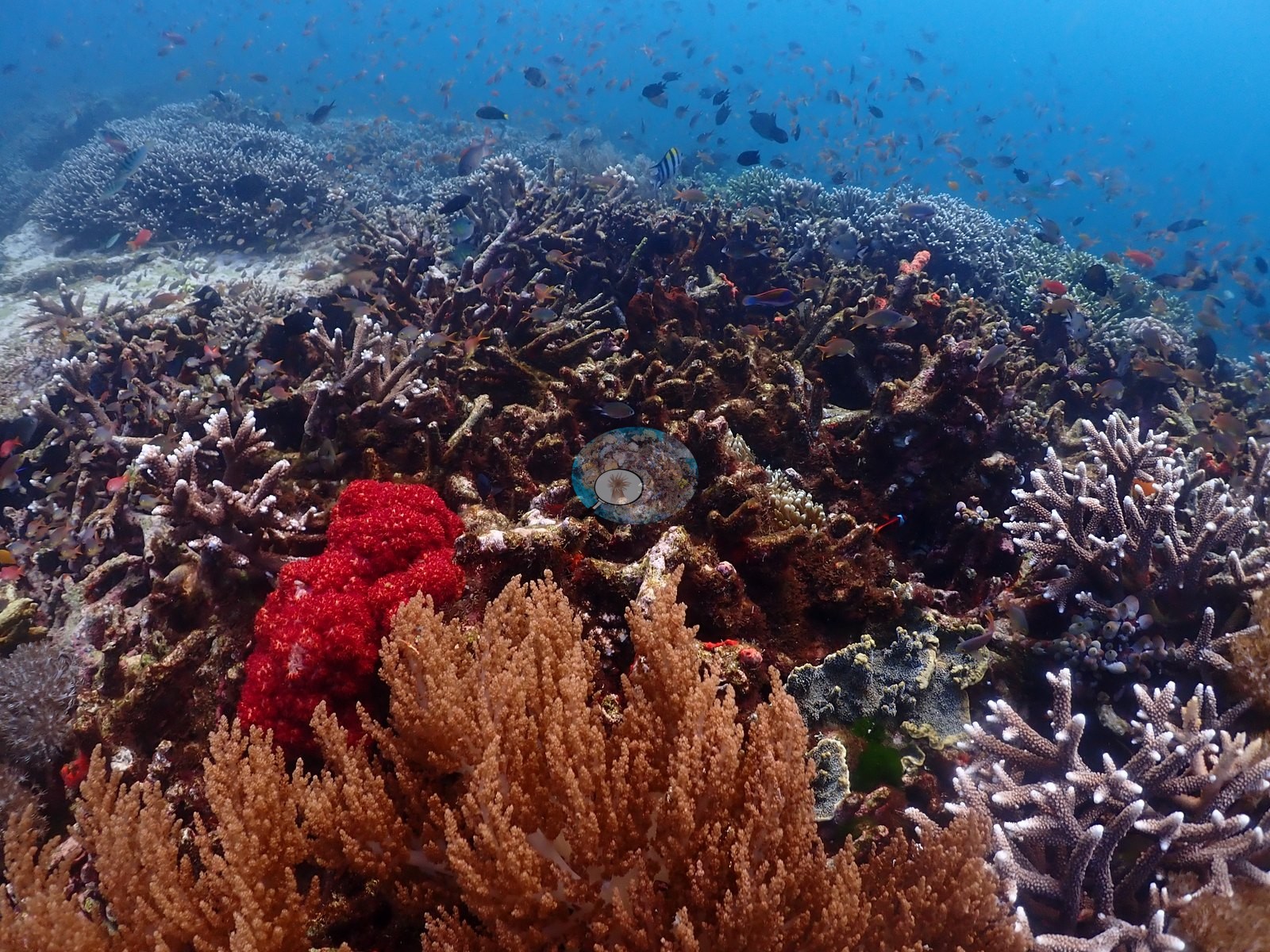
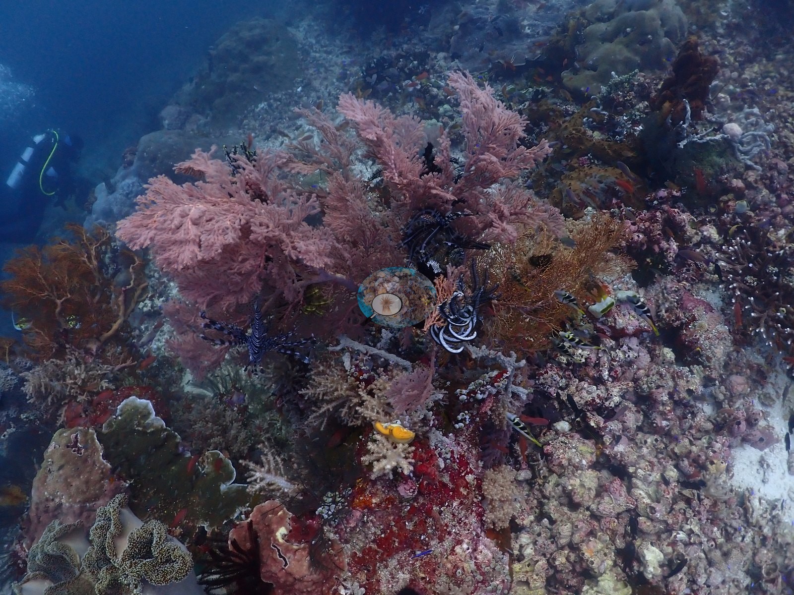
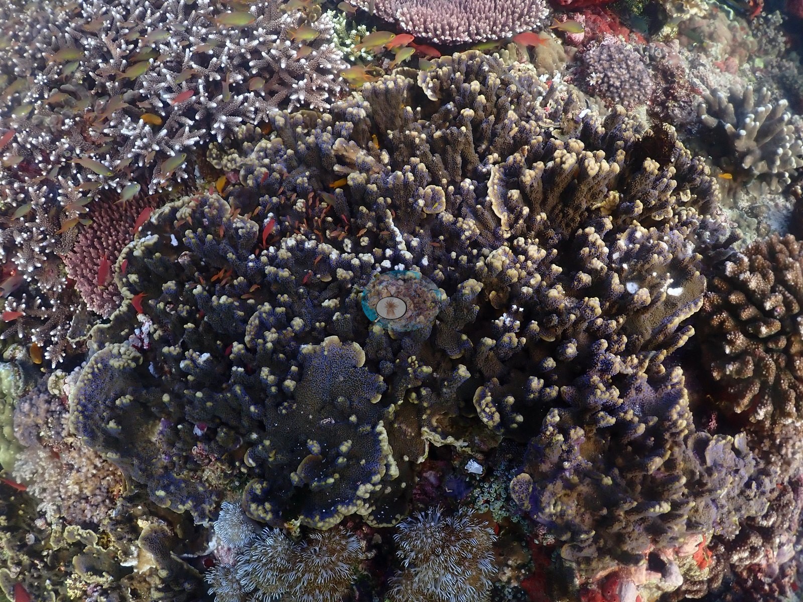
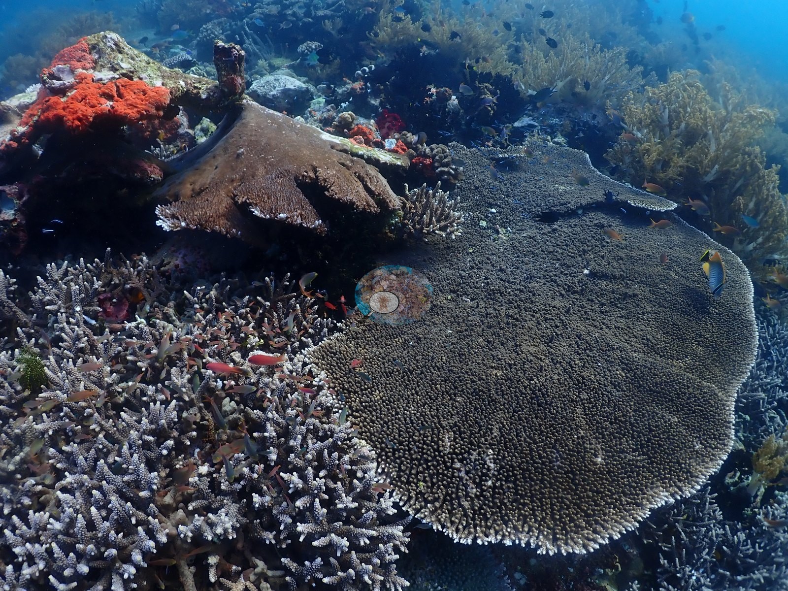
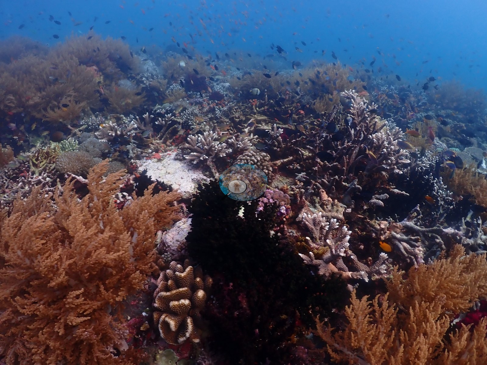
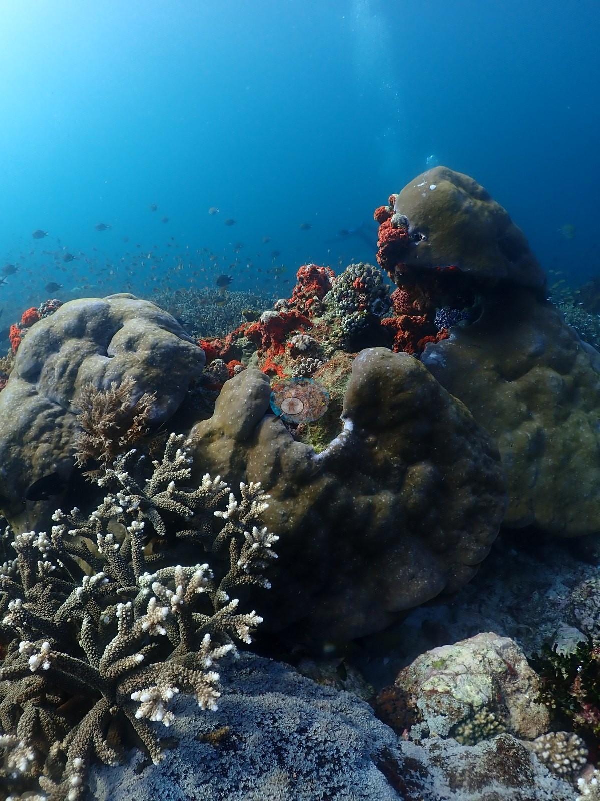
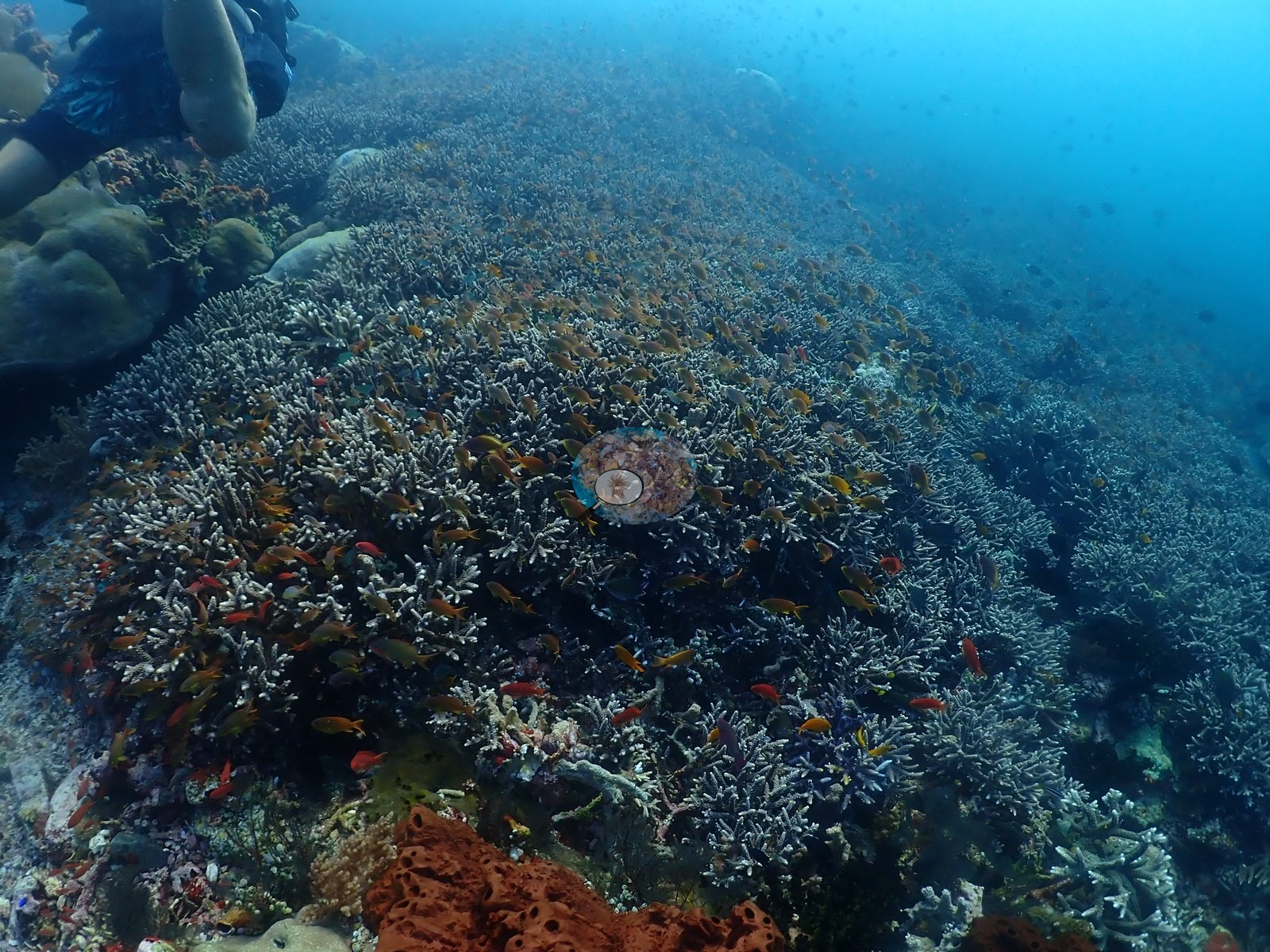
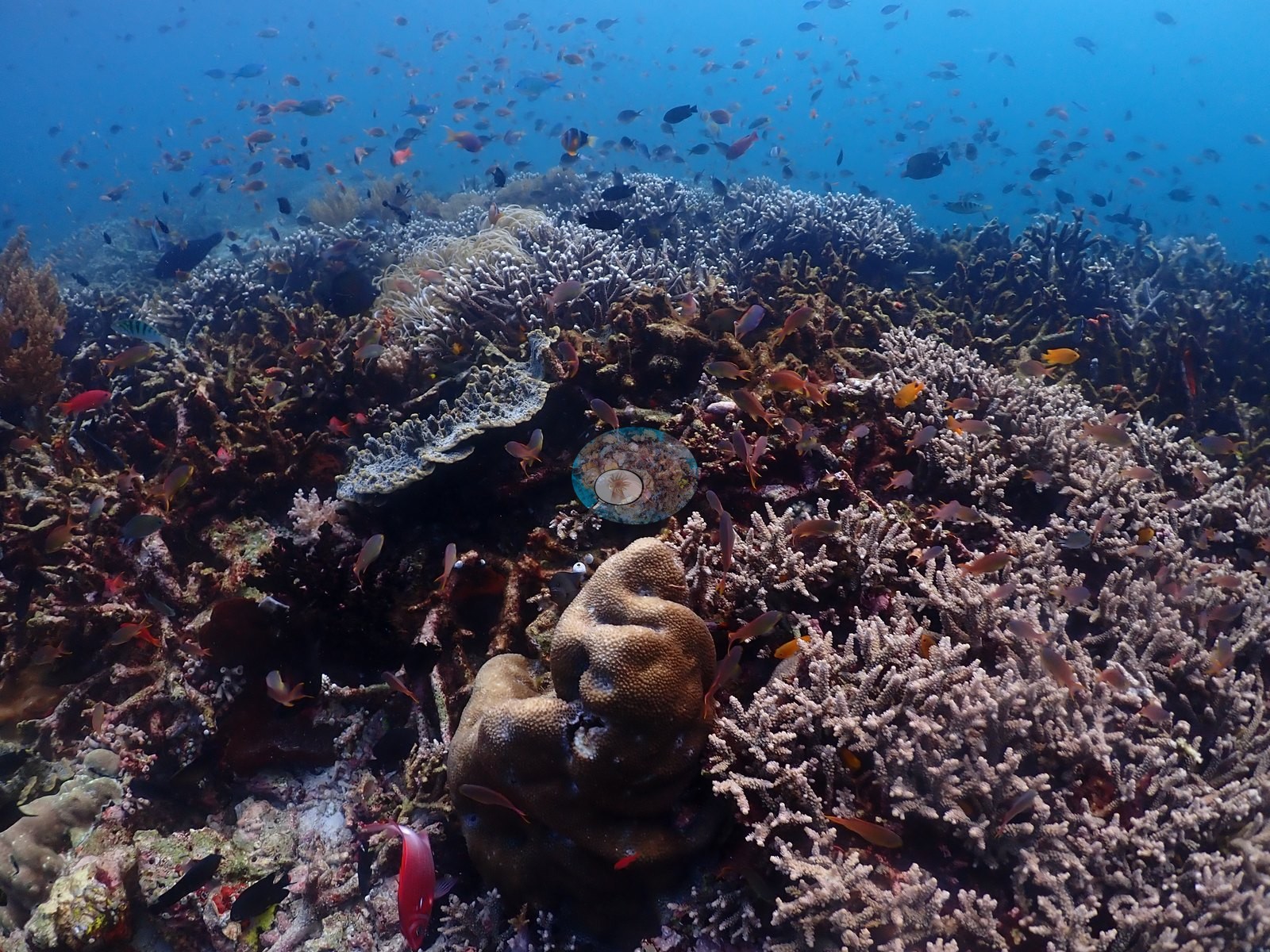
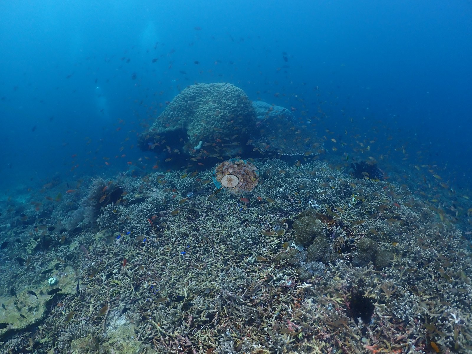
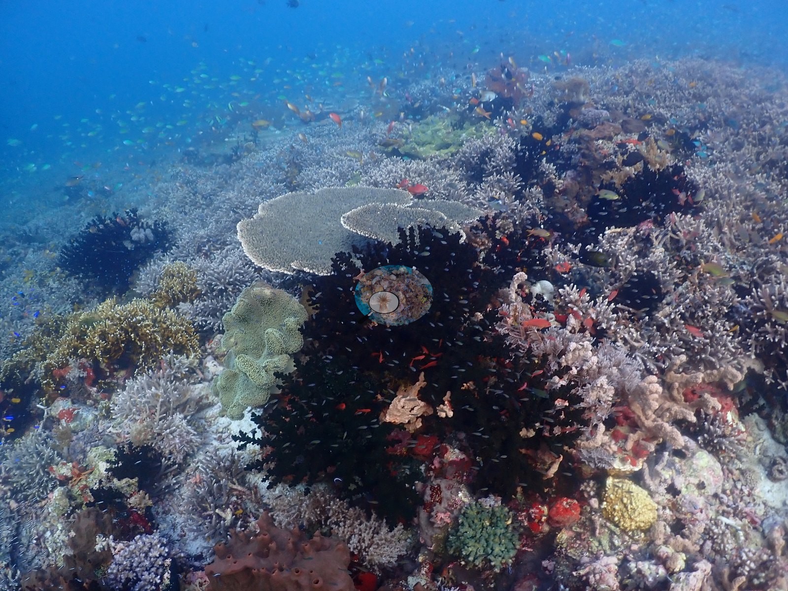
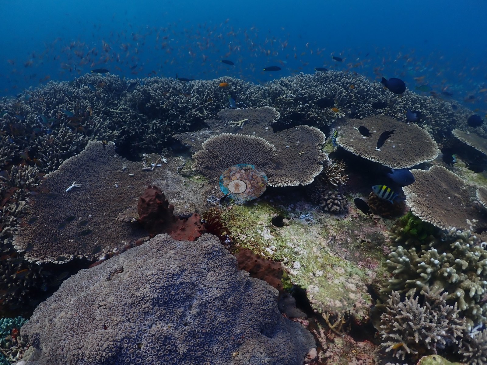
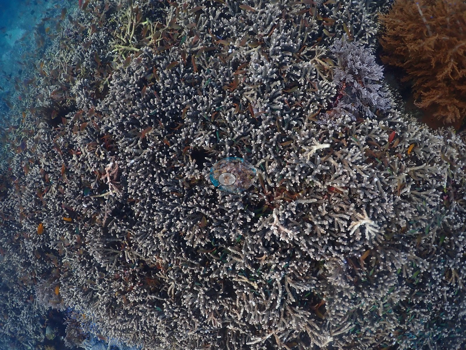
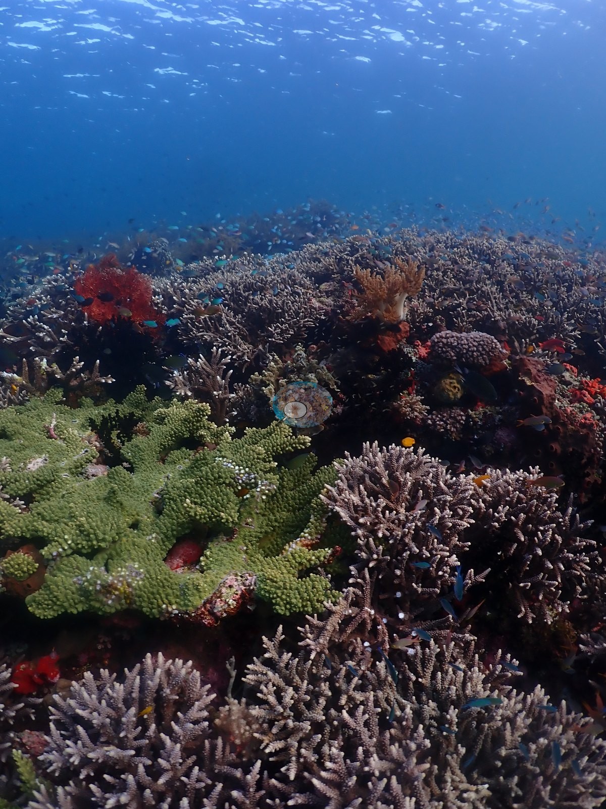
Extensive pale corymbose and staghorn Acropora thickets, mostly cream to pale gray-brown with normal white growth tips, dominate the mid-dive stretch and account for several of the highest single-frame readings (e.g., Image 3, Image 6, Image 9, Image 11, Image 20, Image 24, Image 27, Image 33, Image 42).
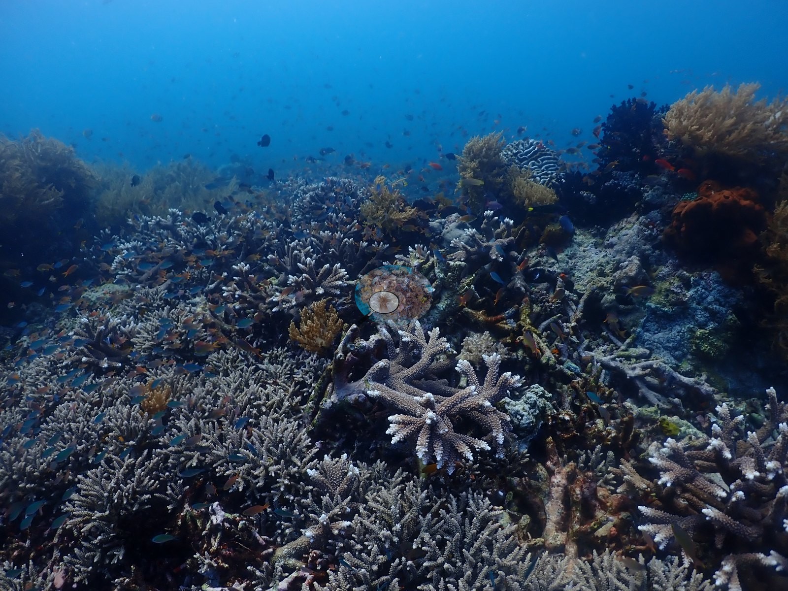
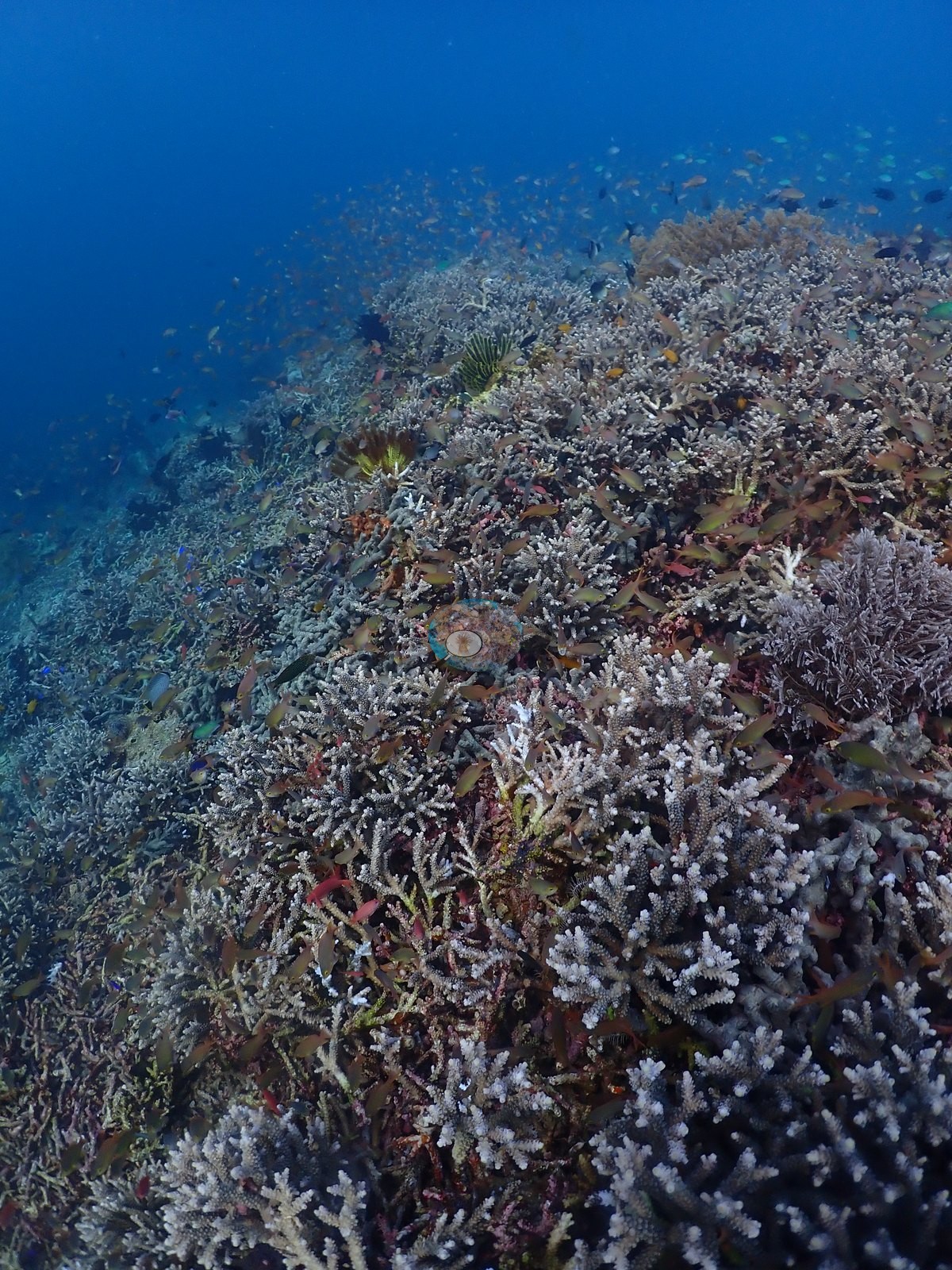
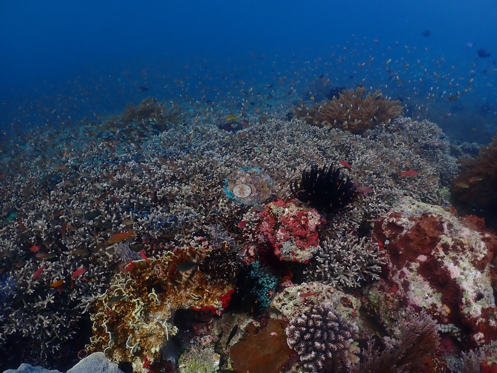
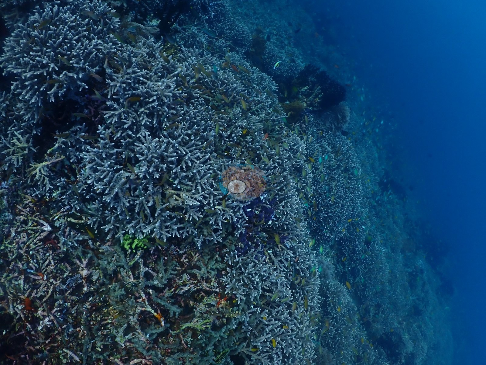
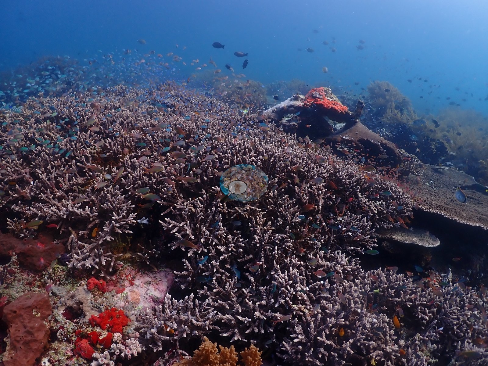
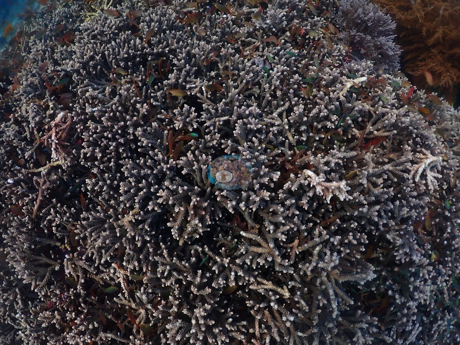
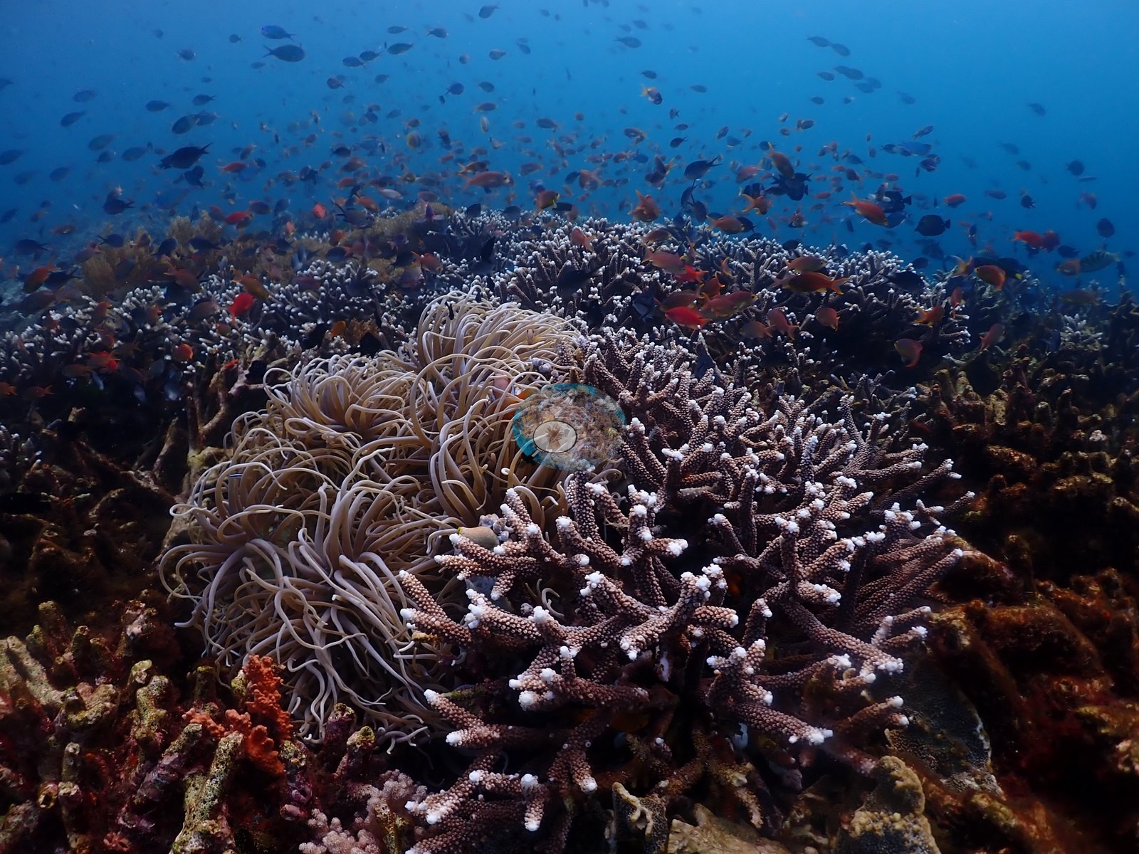
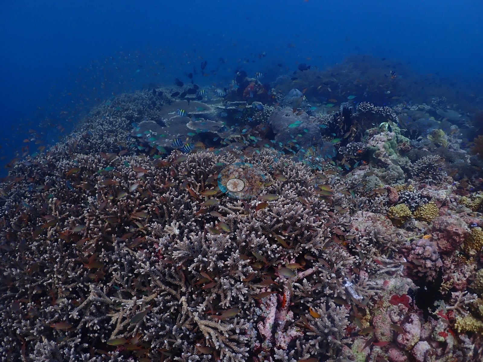
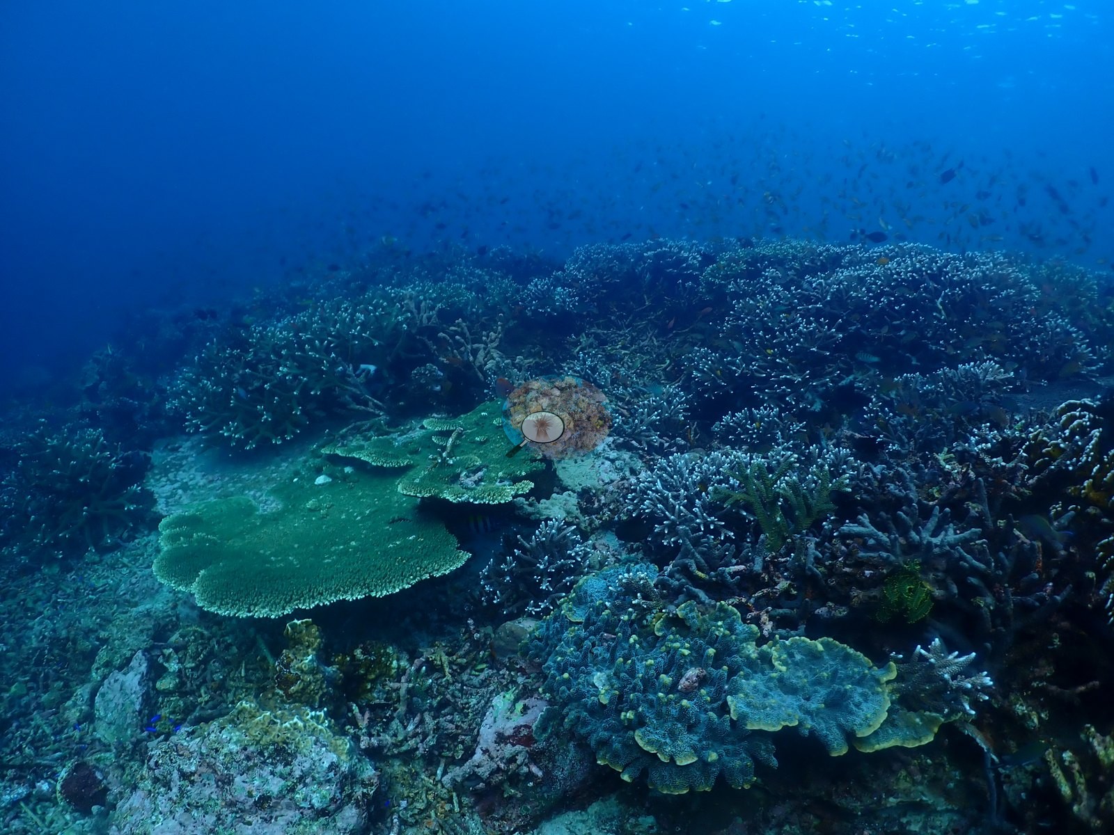
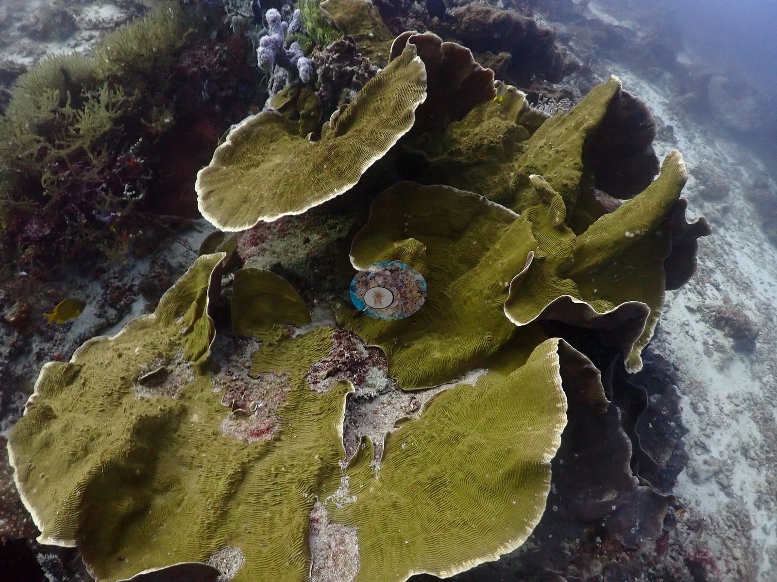
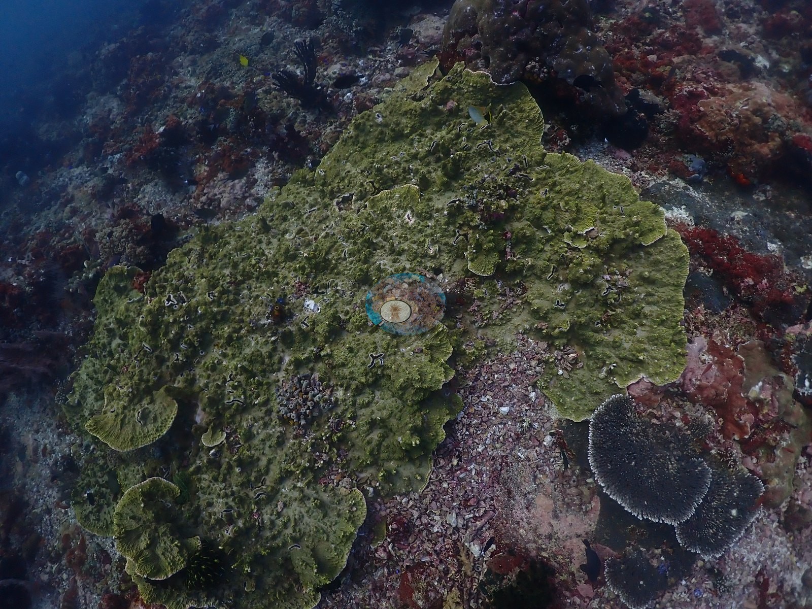
Large ruffled foliose plate corals recur, and a big smooth green-brown massive colony with a hard, glossy surface appears mid-dive (e.g., Image 7, Image 12, Image 23, Image 26, Image 36, Image 45).
Coral cover
Coral cover: 29% — a blind Claude computer-vision point count of 125 of Anderson's unedited photographs (20 points per photo; water, fish, and obstructions excluded). Other computer-vision methods are listed in the method table.
Stressors
Condition: no bleaching, disease, crown-of-thorns, or predation was observed in any of the 127 frames reviewed; the pale coloration on the staghorn fields is a normal growth-tip morph, not tissue loss.
Fish and other fauna
The log-noted turtle is clearly resolved, resting on the reef top in open water. Fish life is heavy throughout the second half — dense damsel and anthias clouds hang over the branching fields. The log-noted sharks were not confidently resolved in the surviving imagery, likely because the current-swept, low-visibility passage cost some of the pelagic-encounter frames.
Comparisons with other data
No prior survey data from this site. If you would like to share yours, please email us.
Other notes
Anderson's field note: “strong current dive (used reef hook), sharks, turtle, adventure”
CV model comparison
More info
DINOv3 and CoralSCOP were run on the same photos and are listed for comparison. Coralscapes v2 and v1 were also run but are not shown: Anderson does not consider Coralscapes reliable for these photos. No diver transect exists for this trip, so agreement between the rows is not accuracy, and as of September 2026 no computer-vision method estimates coral cover accurately on new images; treat every row as indicative.| Method | Hard-coral cover (%) |
|---|---|
| In-house DINOv3 | 29.1 |
| CoralSCOP unedited | 29.0 |
| Claude | 29.2 |
Data & links
All images from Coral Reef Diagnostics. Data are from Coral Reef Diagnostics.
© Coral Reef Diagnostics.