
Part of the regional summaries: Tonga - Niuatoputapu and Tonga
More info
Northern Tonga (NOAA CRW virtual station)A barrier reef on Niuatoputapu's north coast cut by narrow shallow channels barely wider than a diver, which appears to have suffered extensive damage from the tsunami, leaving much rubble now cemented together by coralline algae. Recovery was underway through thin plating Montipora, but many Pocillopora were diseased and large patches of cyanobacteria were present. Surveyed on the Living Oceans Foundation's Global Reef Expedition.
Ko ha hakau fakaā (barrier reef) ʻi he matātahi tokelau ʻo Niuatoputapu, ʻoku vaheʻi ʻe ha ngaahi kiʻi hala vai mamaha ʻoku meimei fālahi pē ʻo hangē ha tokotaha uku, ʻoku hā ngali naʻe maumau lahi ʻi he peau kula (tsunami), ʻo tuku ai ha maka momoa (rubble) lahi kuo pipiki fakataha he taimi ni ʻe he limu feo (coralline algae). Naʻe lolotonga hoko ʻa e fakaakeake ʻi he Montipora fakapaleti manifi (thin plating), ka naʻe puke ʻa e Pocillopora lahi pea naʻe ʻi ai ʻa e ngaahi konga lalahi ʻo e cyanobacteria.
AI translation of this summary; the English is authoritative. (low confidence)
CTD-cast midpoint of 27.6–27.7°C
One other CRD dive site with its own page within 5 km shown in orange — click a marker to open its page.
Site description
Setting
Barrier reef, north coast, west of channel. Large spurs extend from 3-4 m depth to 20 m, ending in sand. In the shallows there are very narrow channels, up to 3-4 m deep. These become wider and lower relief in deeper water.
General benthic features
Image 1 is the site frame and the only wide view.
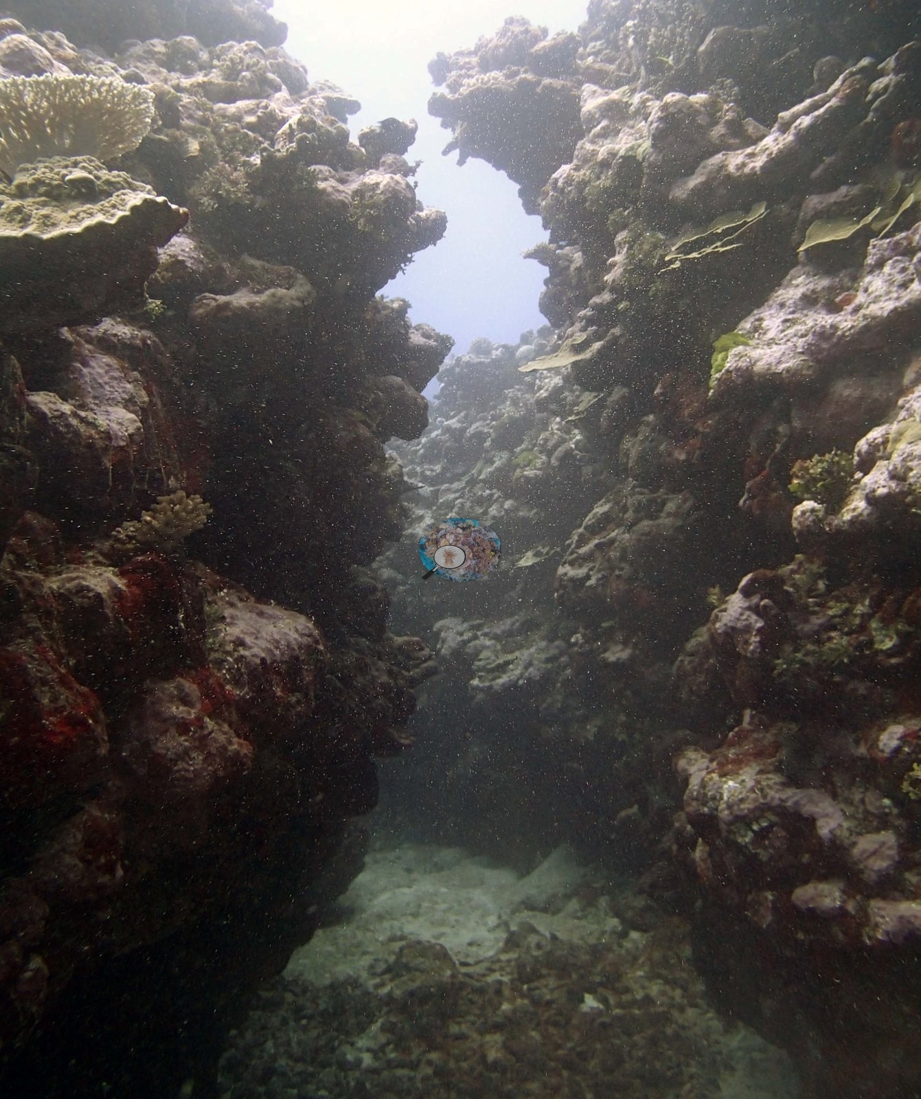
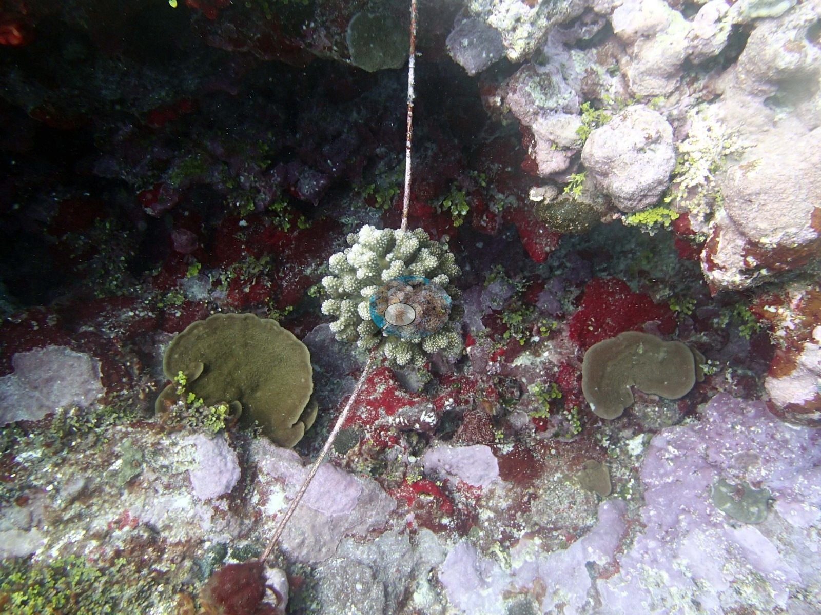
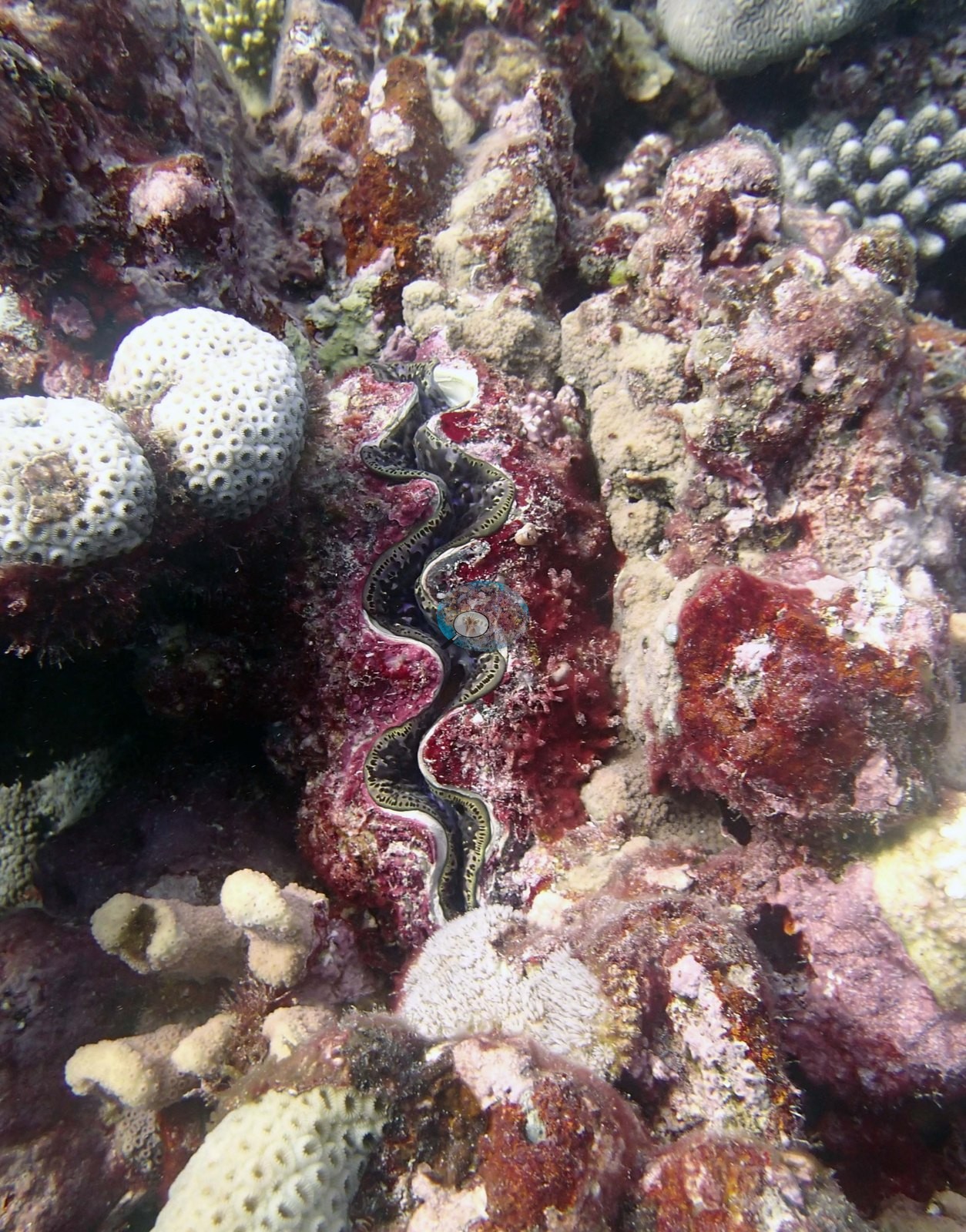
It looks along the floor of a narrow channel cut between two walls of reef framework, the walls rising steeply on both sides to within a few meters of the surface — bright surface light fills the gap between them — and the channel floor running away as pale sand strewn with rubble (e.g., Image 1). The walls themselves are a cemented, consolidated framework rather than a field of discrete colonies: coral rock crusted heavily in pink, mauve and maroon coralline algae, filmed with olive-green turf, carrying small branching, encrusting, and tabular colonies set into it. This one frame corroborates the surveyors' account of the site directly — narrow channels between spurs, sides built of cemented branching and massive coral framework, rubble bound together with thick crustose coralline algae — and it is the closest thing in the set to structural evidence. Image 2 is a close downward view of pavement with a thin taut line running across it: a small pale-green digitate colony has settled on and around the line, with olive-green encrusting colonies, a small brown massive, deep red and maroon coralline crusts, pale mauve CCA, and green turf around it. Image 3 is a giant clam with a fluted shell and a finely speckled mantle wedged into a crevice, surrounded by dark red coralline crusts, pale massive colonies with coarse corallites visible on their surface, and small branching colonies.
Deeper areas have scattered larger table acroporids that were not damaged.
Coral cover
- Reference: Khaled bin Sultan Living Oceans Foundation, Global Reef Expedition (in situ transect)
- 15%, from the Khaled bin Sultan Living Oceans Foundation's in situ transect survey (Global Reef Expedition site data); the photographs illustrate the site.
- Transect mean 15 ± 2.6% SE (n = 5 transects; Living Oceans Foundation per-transect benthic data); the headline above is this all-transect mean.
Stressors
Typical recent thermal stress: mean annual maximum of 5.2°C-weeks (Degree Heating Weeks, 2021-2025, NOAA CRW; Northern Tonga virtual station). The 2013 survey notes for this site record few snails and no Culcita cushion stars (Khaled bin Sultan Living Oceans Foundation expedition site notes, 2013; regional summary in Bruckner 2014). An old rope lies across the reef, colonized by a branching coral (e.g., Image 2). The survey notes record that the reef appears to have suffered extensive damage from the tsunami, with considerable rubble, most of it cemented together with thick crustose coralline algae; many of the Pocillopora colonies have disease, and there are large patches of cyanobacteria. The Living Oceans Foundation transects here recorded 0.6% cyanobacteria and no recently dead coral.
Fish and other fauna
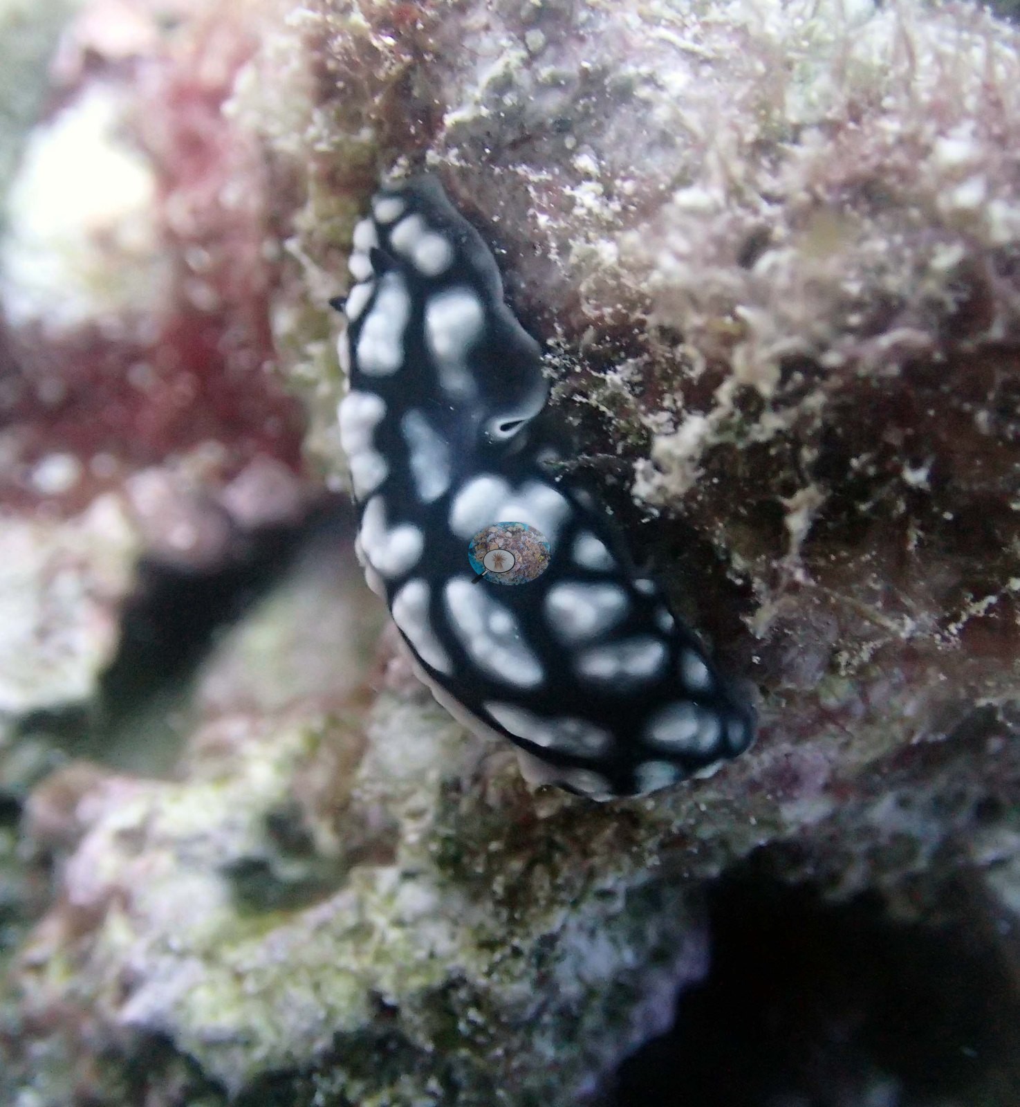
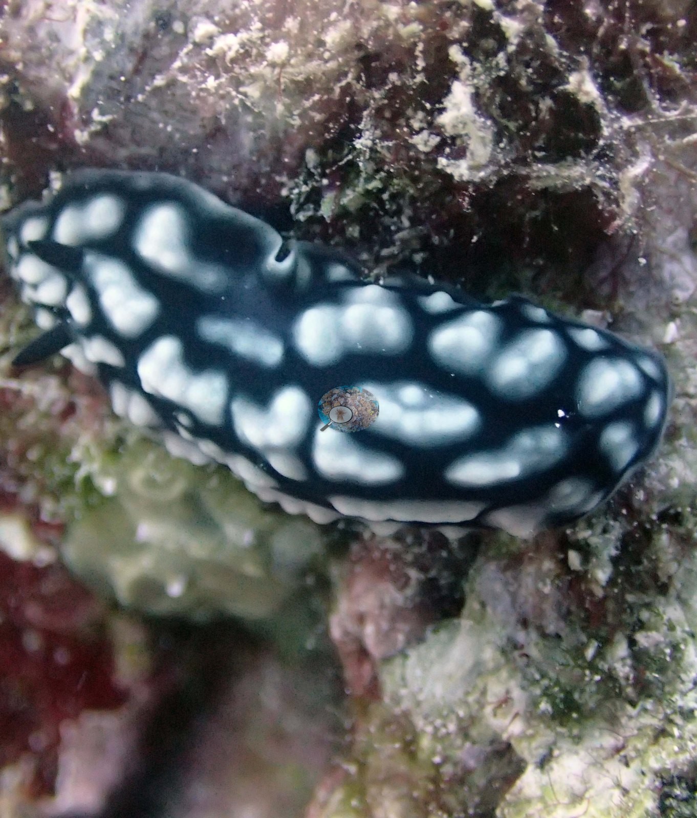
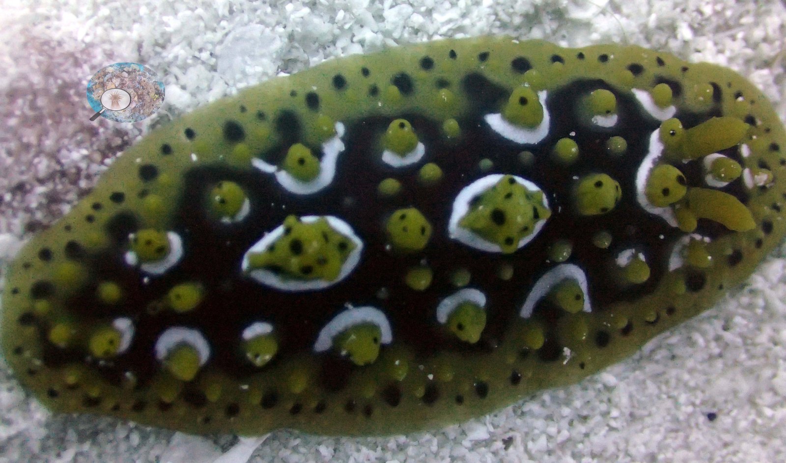
A few small fish hold in the channel. The remaining three frames are close portraits of two nudibranchs: a black one with raised white tubercles, photographed twice (e.g., Image 4, Image 5), and an olive-yellow one with dark-ringed white spots in Image 6.
Comparisons with other data
Apart from the Living Oceans Foundation survey shown here, we found no other surveys at this site as of 2026. If you would like to share yours, please email us.
Other notes
The photographic set is small and this account is short accordingly: six frames survive from the dive (Niuatoputapu, 30 Sep 2013), of which only three carry any habitat information at all — the other three are macro portraits of nudibranchs. There are no sampled-colony photographs: Anderson noted that none of the target Pocillopora ('no Pdam'), the expedition's sampling target, was found here. That is Anderson's record and it stands as one of the more informative things in the set. The nudibranch portraits are pretty and record what was on the reef that day, but they say nothing about habitat.
What these six frames cannot settle is most of the site. There is no photographic evidence here for the spur-and-groove profile below the shallows, the depth gradient from 3–4 m to 20 m, the tsunami rubble field, the thin plating Montipora recolonization, the diseased Pocillopora, or the cyanobacterial patches the surveyors recorded — the survey narrative retained below is the record of all of that. No scale reference, quadrat or transect line appears in any frame, so nothing here is measurable, and with no depth stamps the frames cannot be placed on the profile.
Survey team's site notes: The tops of the spurs have 10-30 cm microrelief, while the sides consist of a cemented branching and massive coral framework with 40-80 cm relief. This area has small table acroporids, Pocillopora, small Favia, Montastrea, and Montipora. At the base of the spurs are more digitate and table acroporids, some larger plates of Turbinaria, patches of staghorn coral, Merulina, Leptastrea, and other corals. At 20-30 m the spurs and mounds are much lower relief, with scattered coral bommies and rocks up to 1 m diameter, intermixed with some larger P. lobata and table acroporids. There is considerable recolonization, mostly thin plating sheets of Montipora at 5.0-30% cover.
Data & links
- Download benthic survey data (CSV) ↗
- Download all LOF-GRE data (933 sites) ↗
- Photo portfolio ↗
- Coral Reef Diagnostics region page ↗
All images from Coral Reef Diagnostics. Data are from the Khaled bin Sultan Living Oceans Foundation Global Reef Expedition and Coral Reef Diagnostics.
 © Coral Reef Diagnostics.