
Part of the regional summaries: Tonga - Niuatoputapu and Tonga
More info
Northern Tonga (NOAA CRW virtual station)A south-facing fringing reef off Niuatoputapu's southern tip, side-on to the trades (intermediate exposure), that slopes very gently to 25 m. About 30% of the corals were dead, including a large stand of staghorn at 15 m, and diseased Pocillopora were numerous. Surveyed on the Living Oceans Foundation's Global Reef Expedition.
Ko ha hakau matafonua (fringing reef) ʻoku hanga ki he tonga ʻi he muʻa tonga ʻo Niuatoputapu, ʻoku tuʻu fakatafaʻaki ki he matangi tokelau-hahake (trade winds) (fakaloto ʻa e fehangahangai mo e matangi), ʻoku hifo māmālie ʻaupito ki he 25 m. Naʻe mate ʻa e meimei 30% ʻo e ngaahi feo, kau ai ha vao staghorn lahi ʻi he 15 m, pea naʻe lahi ʻa e Pocillopora naʻe puke (diseased).
AI translation of this summary; the English is authoritative. (low confidence)
CTD-cast midpoint of 27.7–27.8°C
One other CRD dive site with its own page within 5 km shown in orange — click a marker to open its page.
Site description
Setting
South-facing fore reef at the southern tip, side-on to the trades (intermediate exposure).
General benthic features
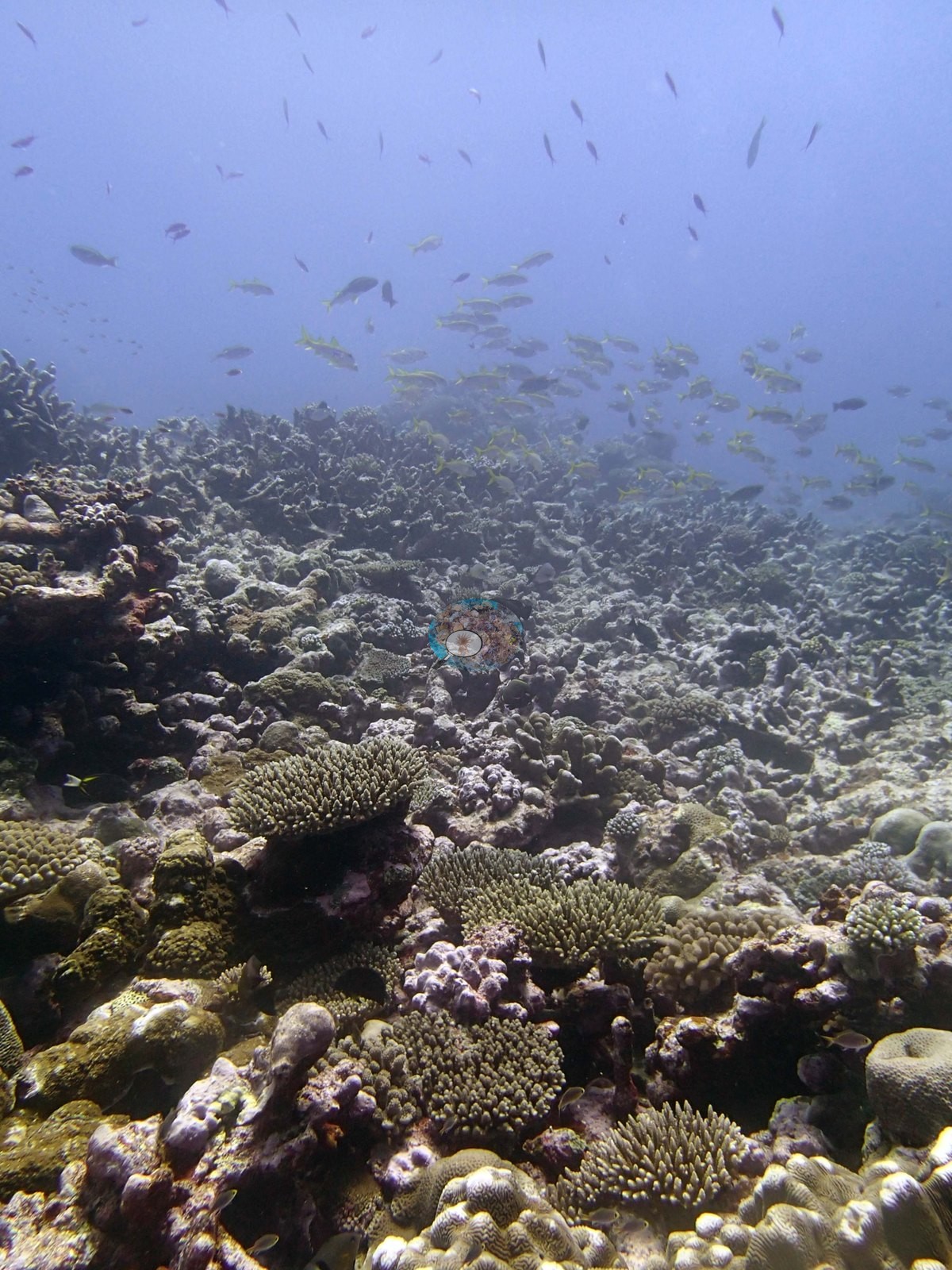
Across the usable wide views the reef reads as a broad, low-relief pavement rather than a structured spur system: Image 3, Image 6, Image 7, Image 14, and Image 16 all look out over gently rising ground carpeted almost continuously in small colonies, none of them large, with pale crustose-coralline and algal-filmed rock filling the spaces between.
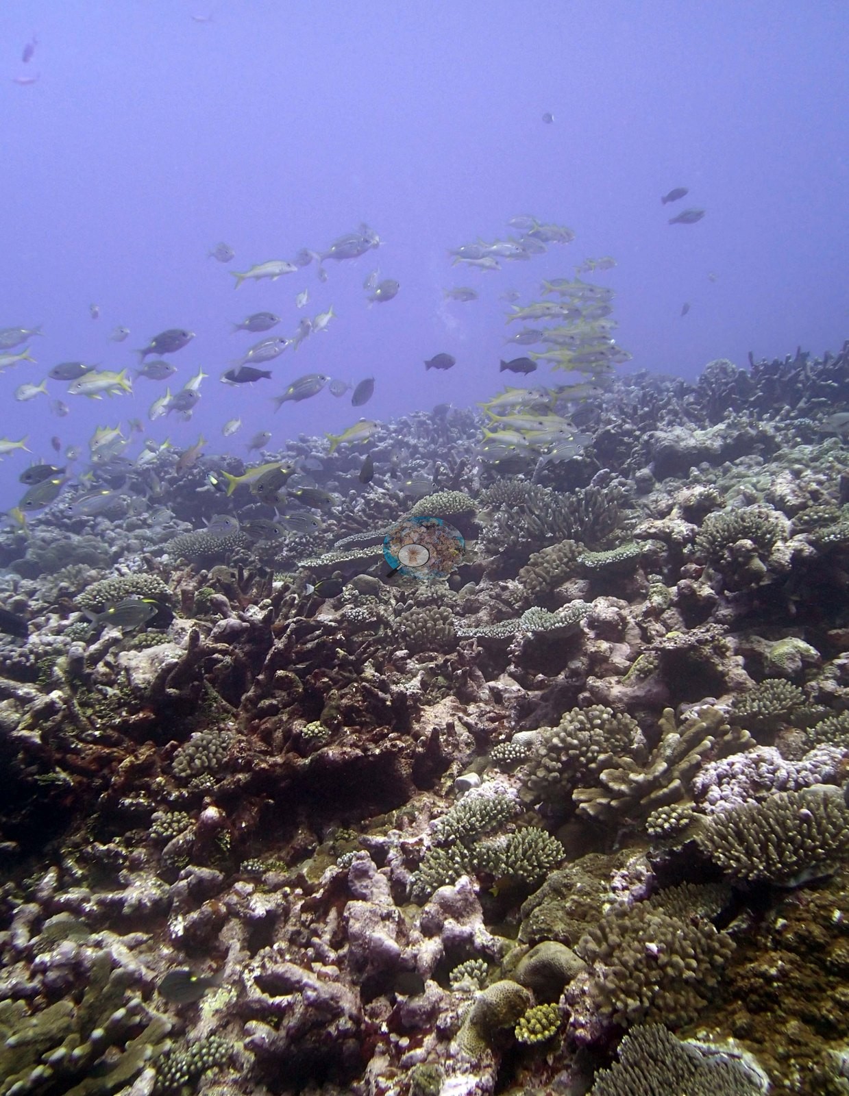
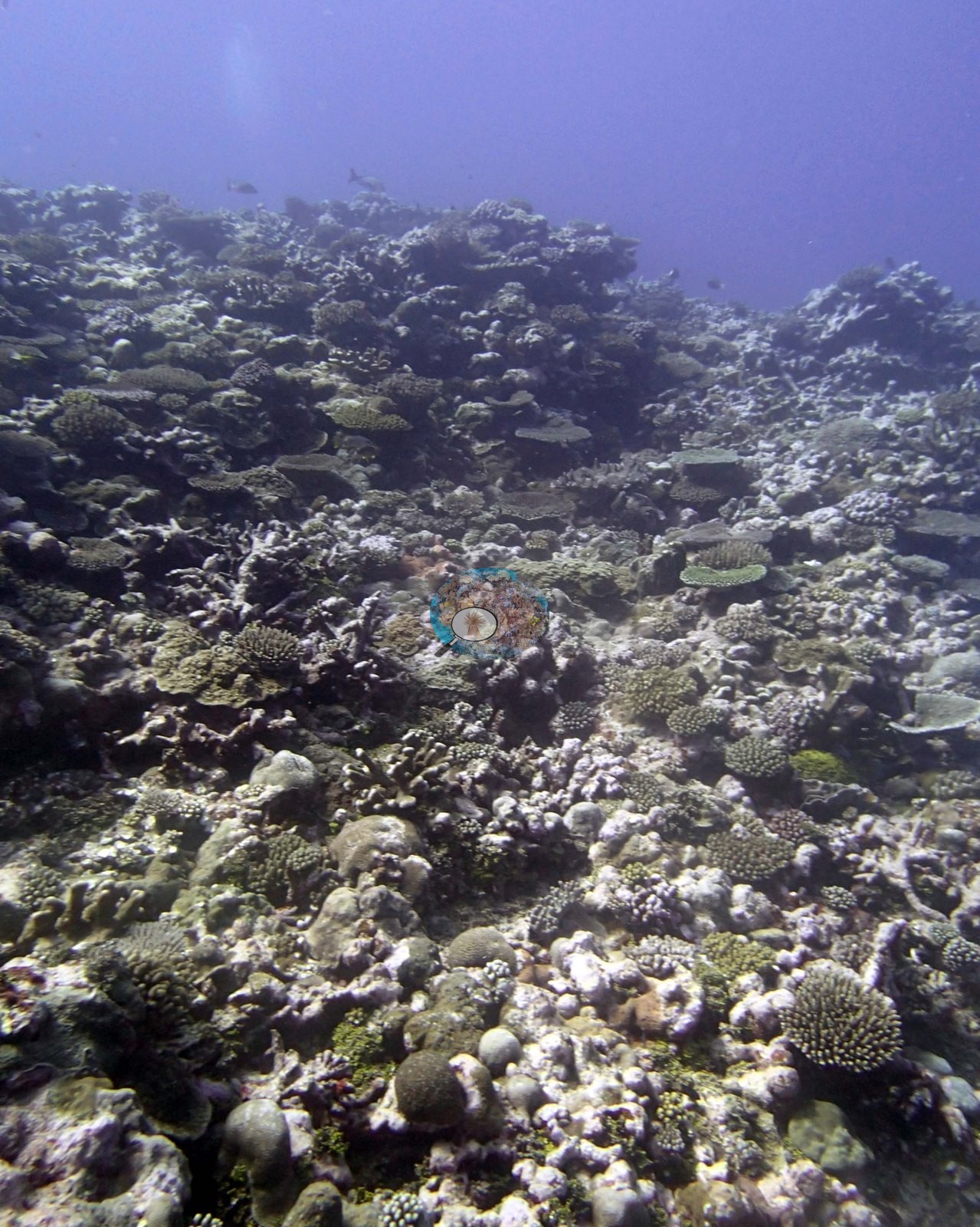
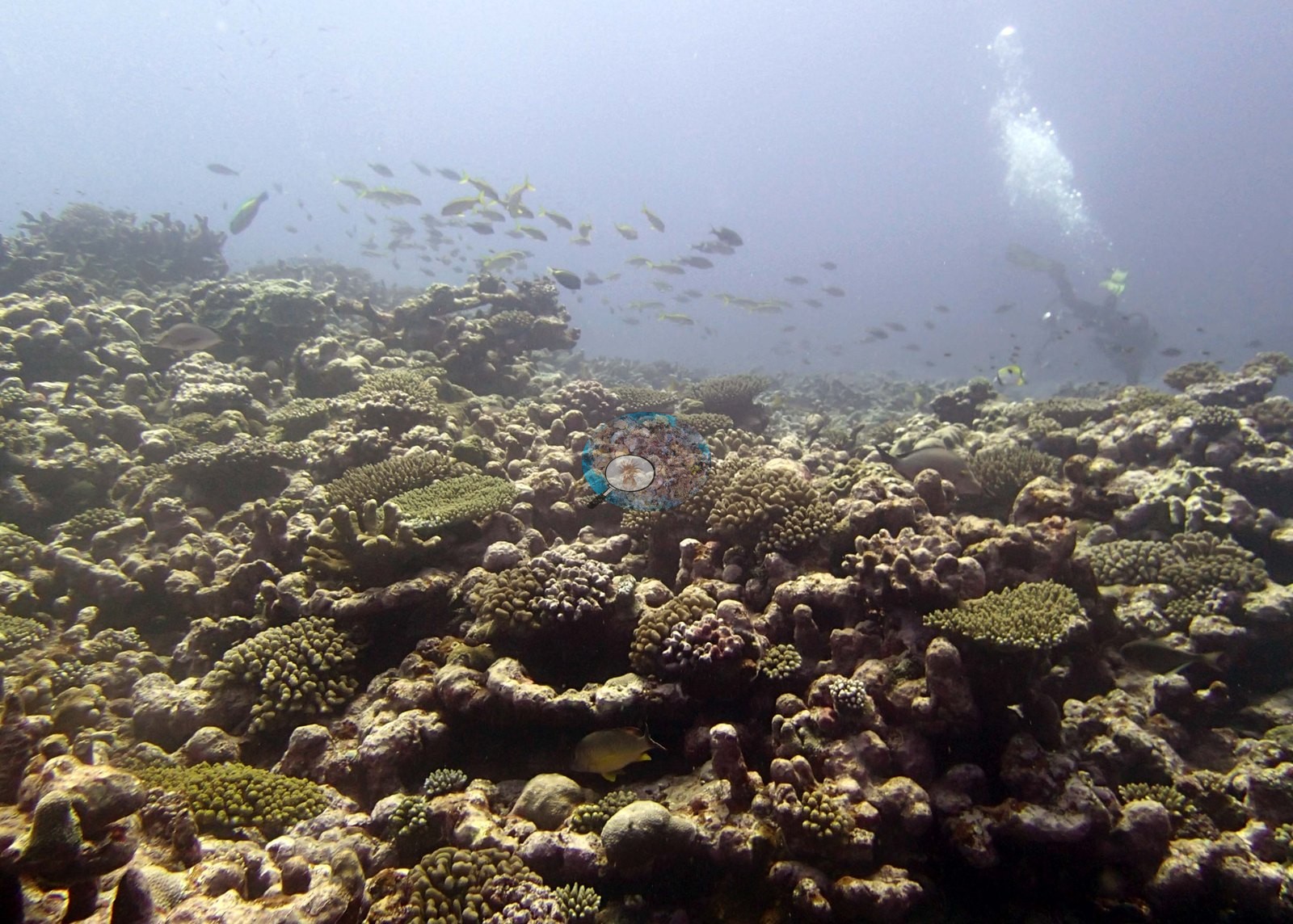
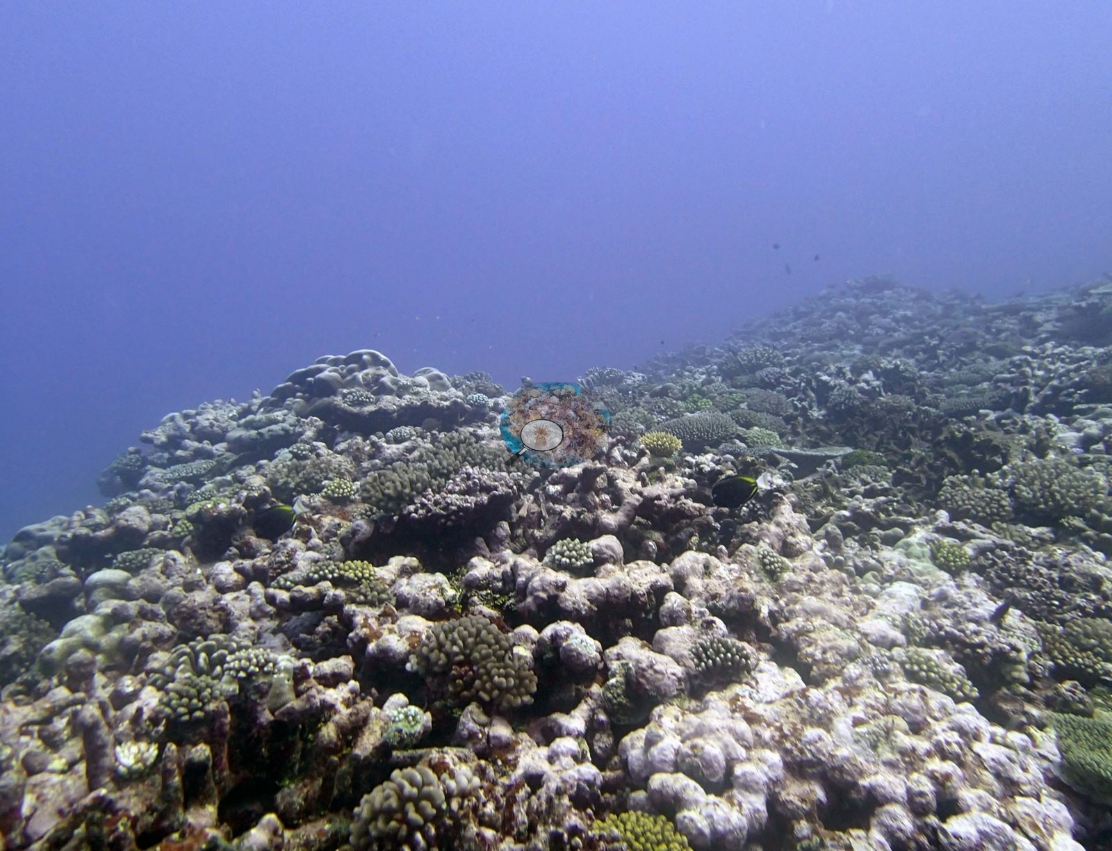
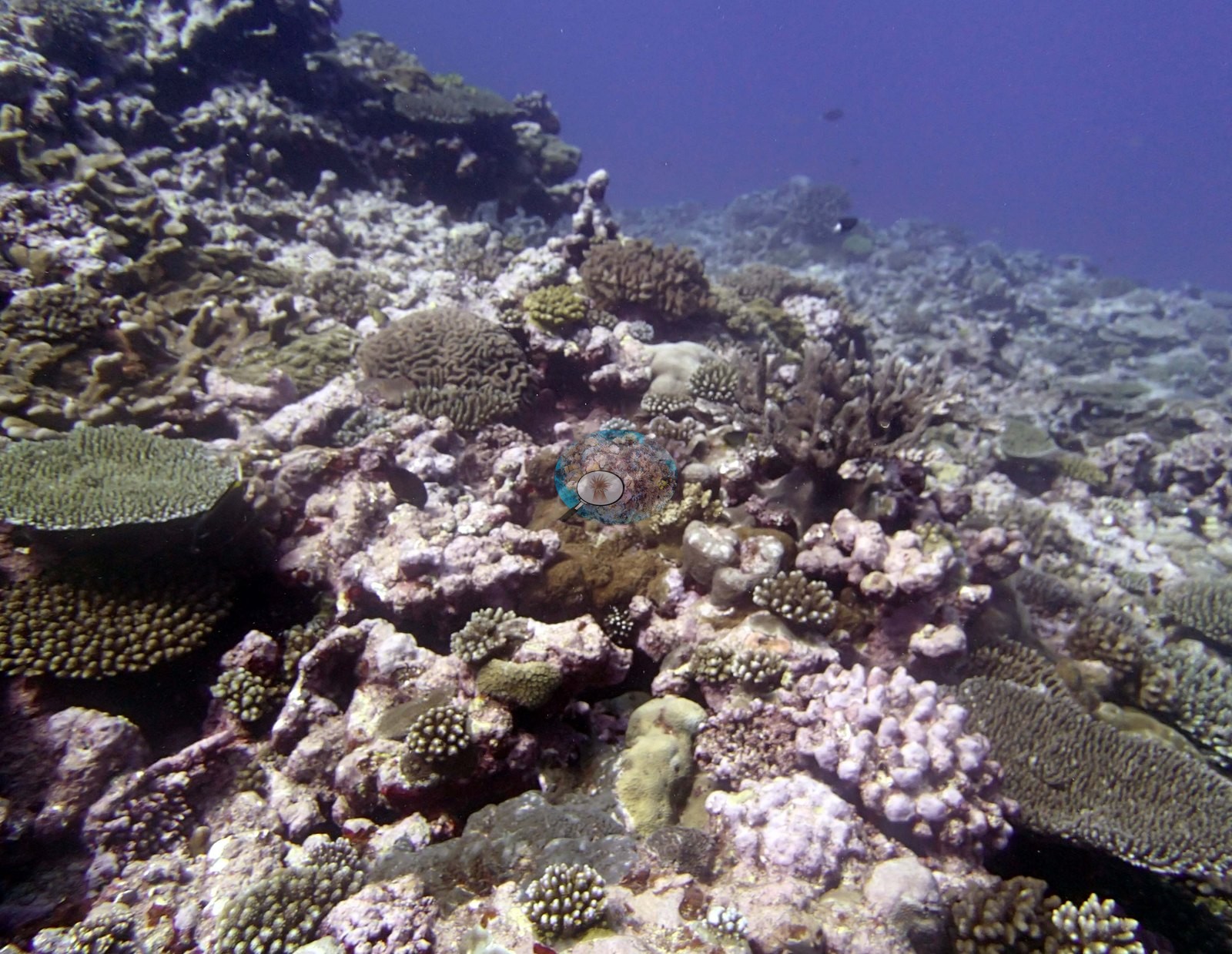
Growth forms are dominated by small branching and digitate corals — corymbose and digitate colonies, bushy branching colonies, and knobbly encrusting colonies — punctuated by modest tables (e.g., Image 2, Image 10, Image 16) and low massive and encrusting domes.
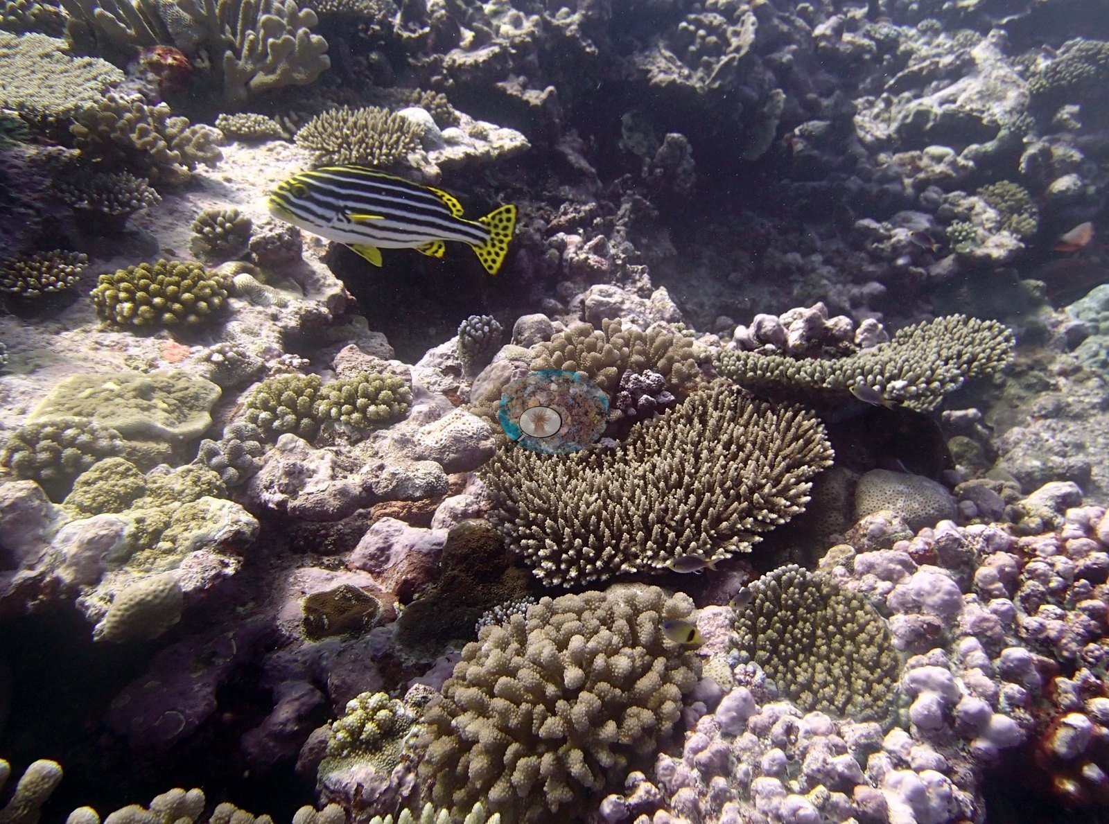
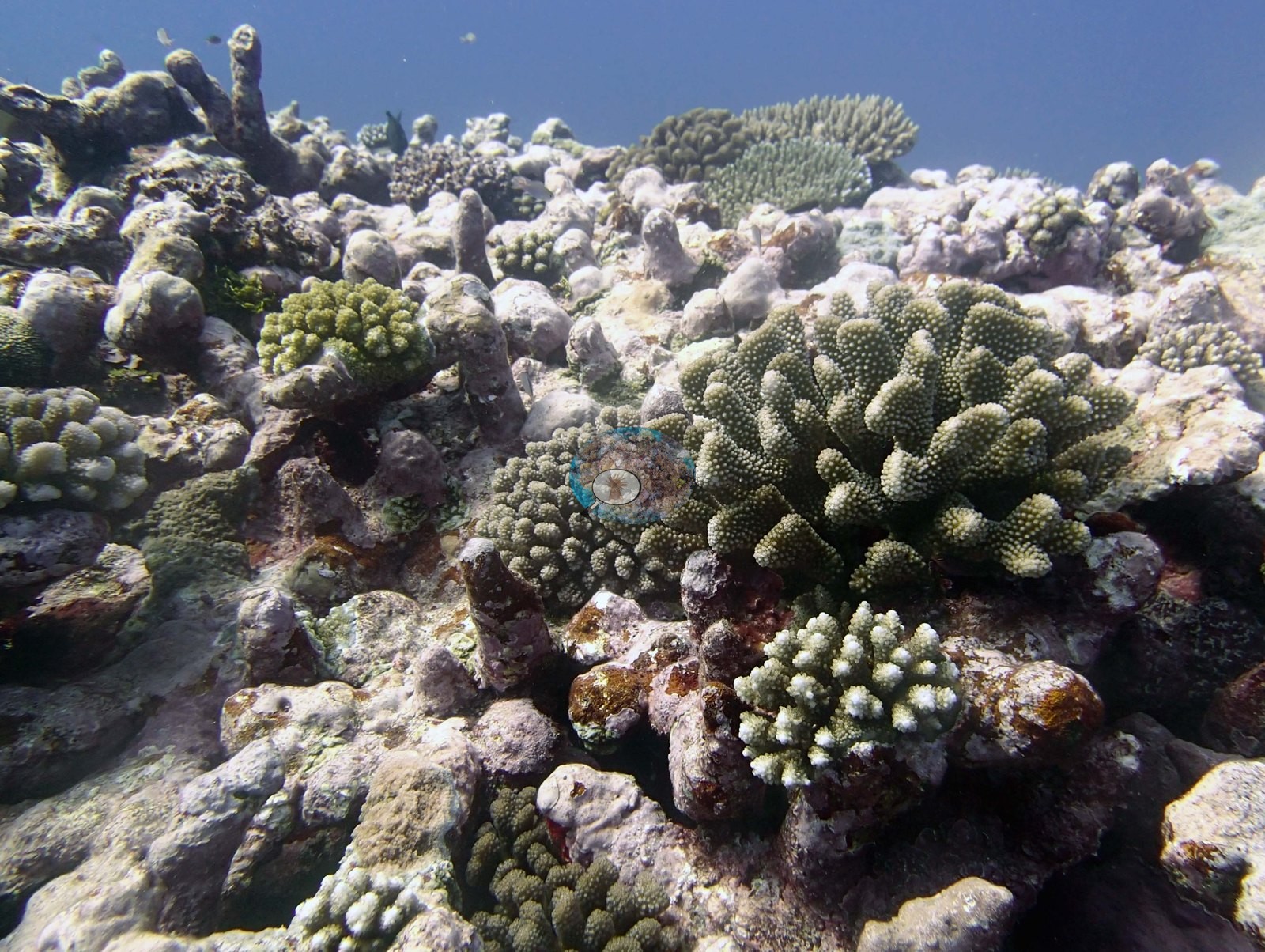
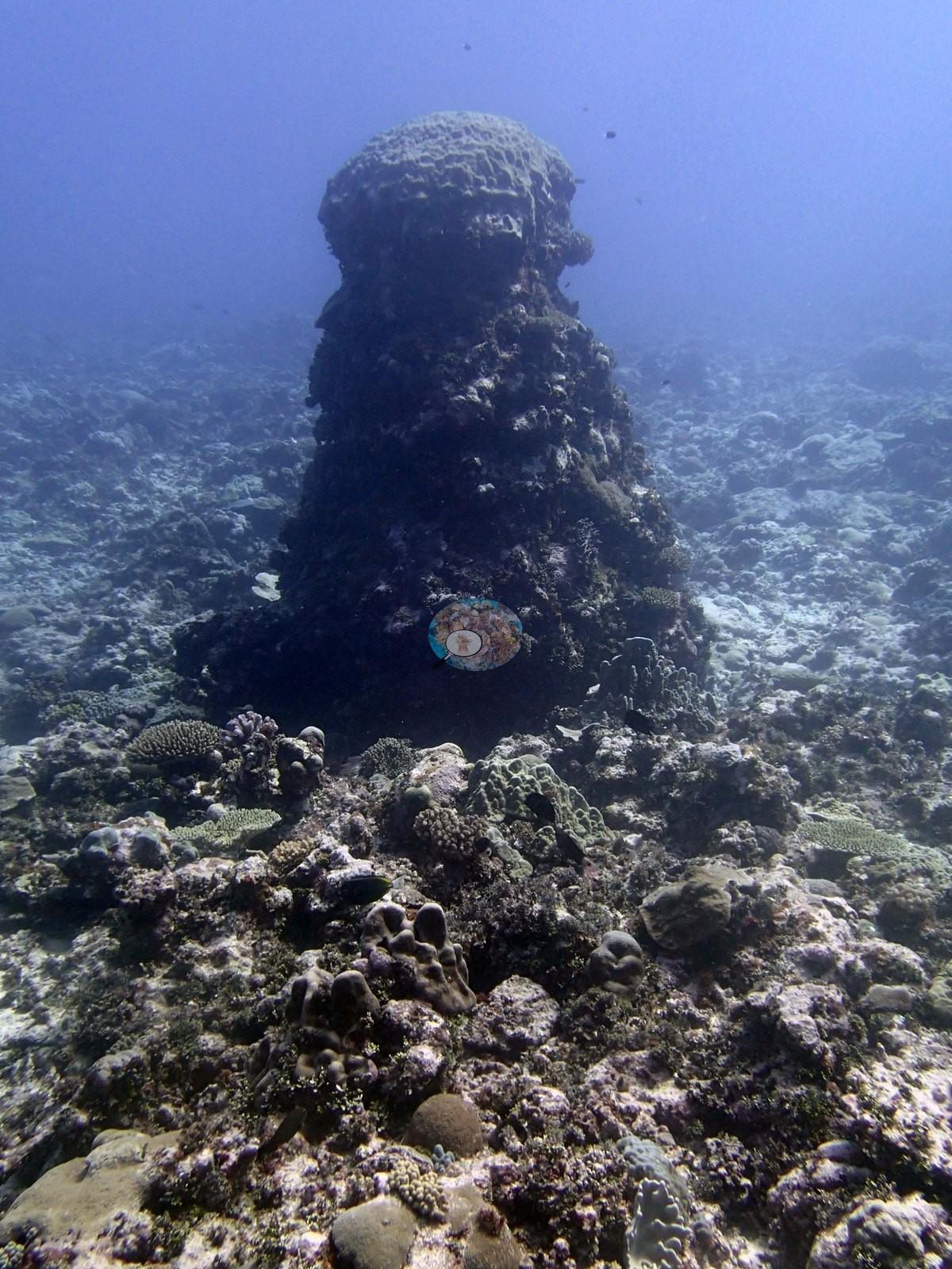
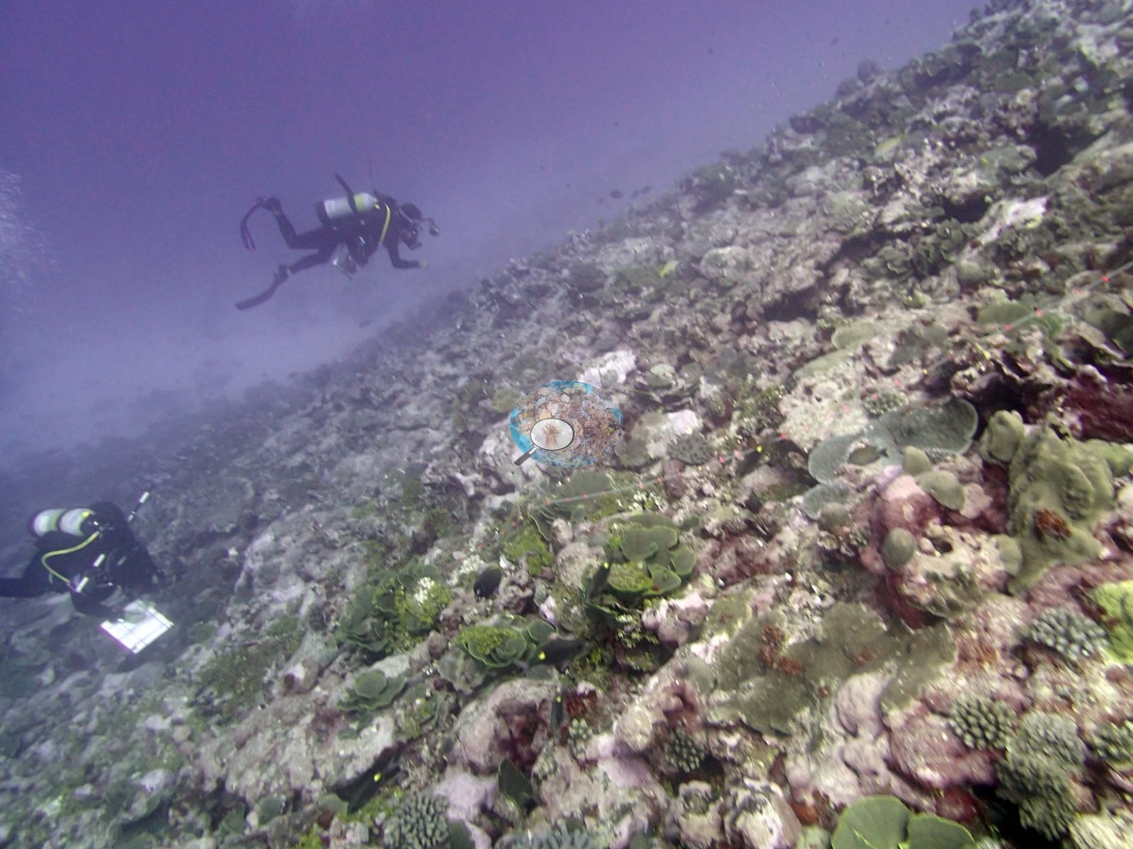
There is very little in these frames that resolves as a large colony; Image 2 and Image 10 are the closest, showing a meter-scale digitate stand and a small table respectively, and no frame carries a scale bar so even that is impression rather than measurement. Two frames stand apart. Image 4, which Anderson noted as a coral pillar, shows exactly that: a dark, stepped column of framework standing well clear of the surrounding pavement, algal-darkened on its flanks and capped with encrusting growth, the only pronounced vertical structure anywhere in the set. Image 13 looks along a steeper flank with two divers working above it, one with a slate; the ground there carries more foliose and plating growth and more bare pale rock than the shallower views.
Coral cover
- Reference: Khaled bin Sultan Living Oceans Foundation, Global Reef Expedition (in situ transect)
- Coral cover 34%, from LOF-GRE in situ transects (the mean of all of this station's own transects in the expedition's per-transect benthic data) — a diver transect measurement, not a photographic or computer-vision estimate.
- The transect figure is this site's authoritative cover value; the photographs above are qualitative only and no cover figure may be read off them.
- Transect mean 34 ± 3.5% SE (n = 6 transects; Living Oceans Foundation per-transect benthic data); the headline above is this all-transect mean.
Stressors
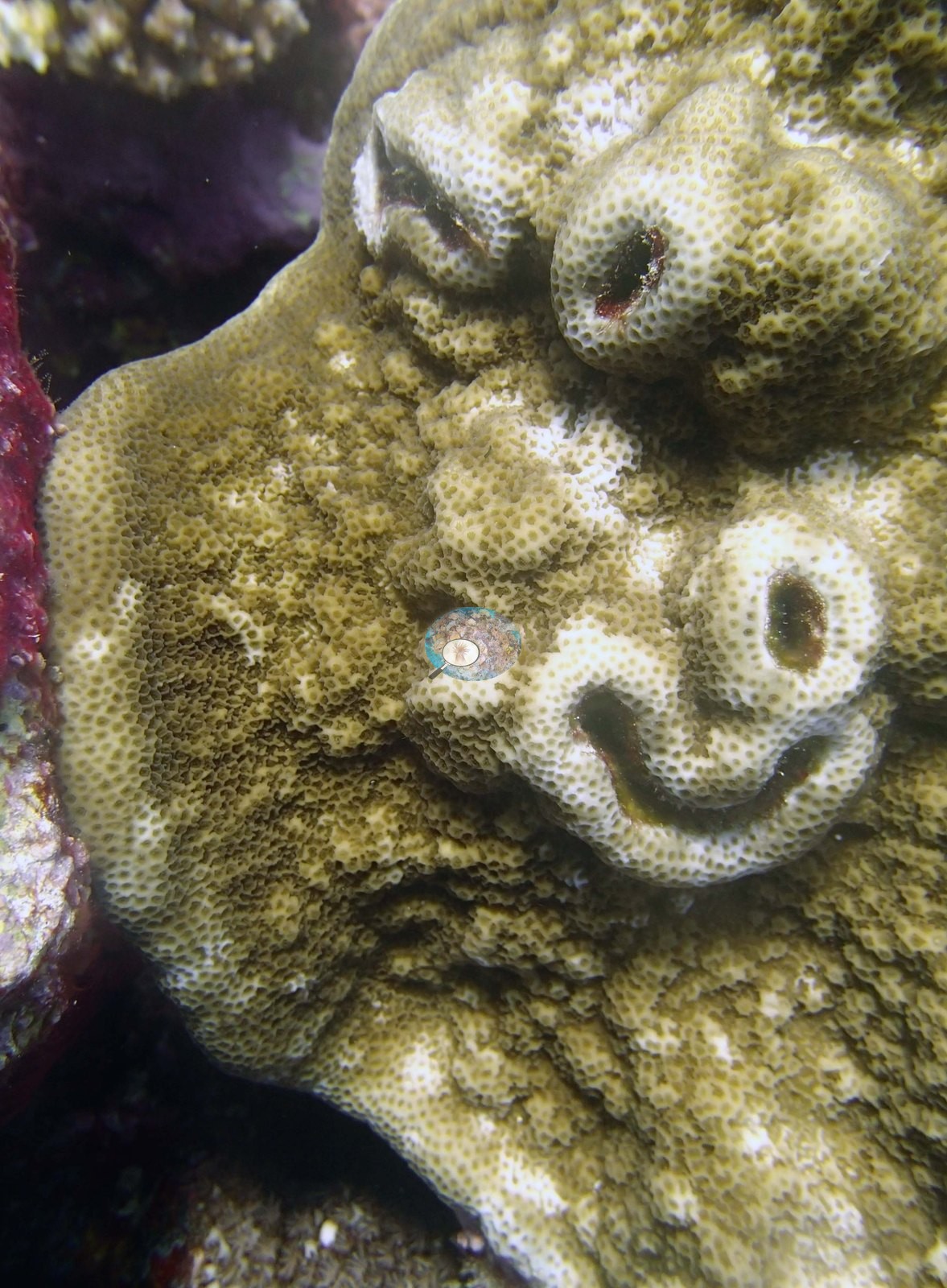
A great deal of pale, whitish substrate appears in Image 3, Image 6, Image 14, and Image 16. Read close, this is old carbonate rock and rubble crusted with coralline algae and turf, not bleached tissue — no frame in this set shows a bleached colony, and none is claimed. Image 5 is a massive-encrusting colony whose pale winding scars across an otherwise pigmented surface read as partial mortality or old damage that has been recolonized; it is a nice picture rather than a diagnosis.
The dense small-colony carpet and the amount of exposed, algal-covered rock are consistent with a reef carrying substantial old mortality, but the photographs cannot quantify that and no cover or mortality figure is read from them. There were about 30% dead corals including a large dead stand of thick branched staghorn coral at 15 m. The survey notes record high numbers of diseased Pocillopora. Typical recent thermal stress: mean annual maximum of 5.2°C-weeks (Degree Heating Weeks, 2021-2025, NOAA CRW; Northern Tonga virtual station). The Living Oceans Foundation transects here recorded 1.2% cyanobacteria and no recently dead coral.
Fish and other fauna
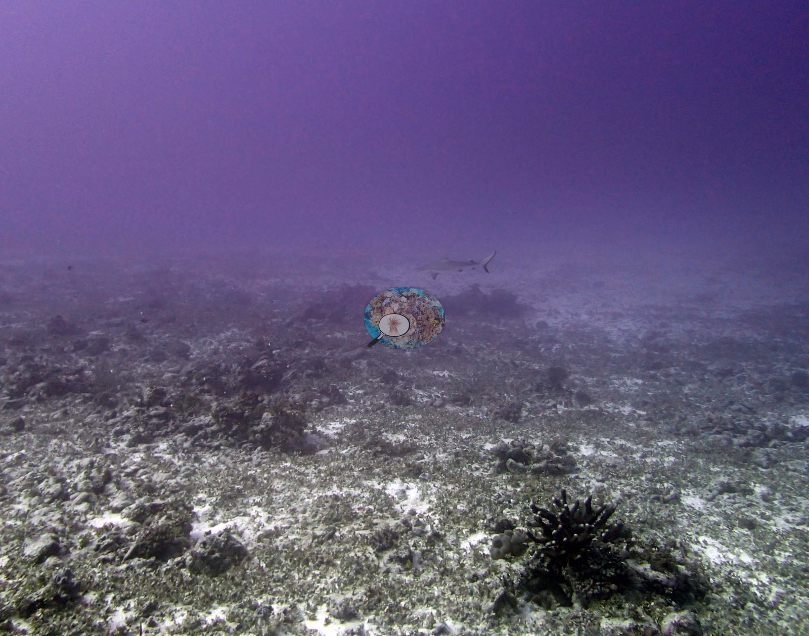
Fish are present without dominating: a loose school of yellow-striped snapper or goatfish hangs over the reef in Image 3, small planktivores drift through the water column in Image 1 and Image 7, a boldly striped yellow-and-black sweetlips sits among the corals in Image 2, and a distant shark is just resolvable in the murk of Image 20 above a rubble flat carrying a black spiny echinoderm in the foreground.
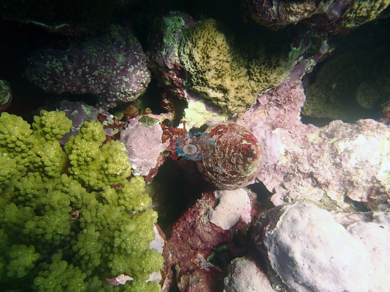
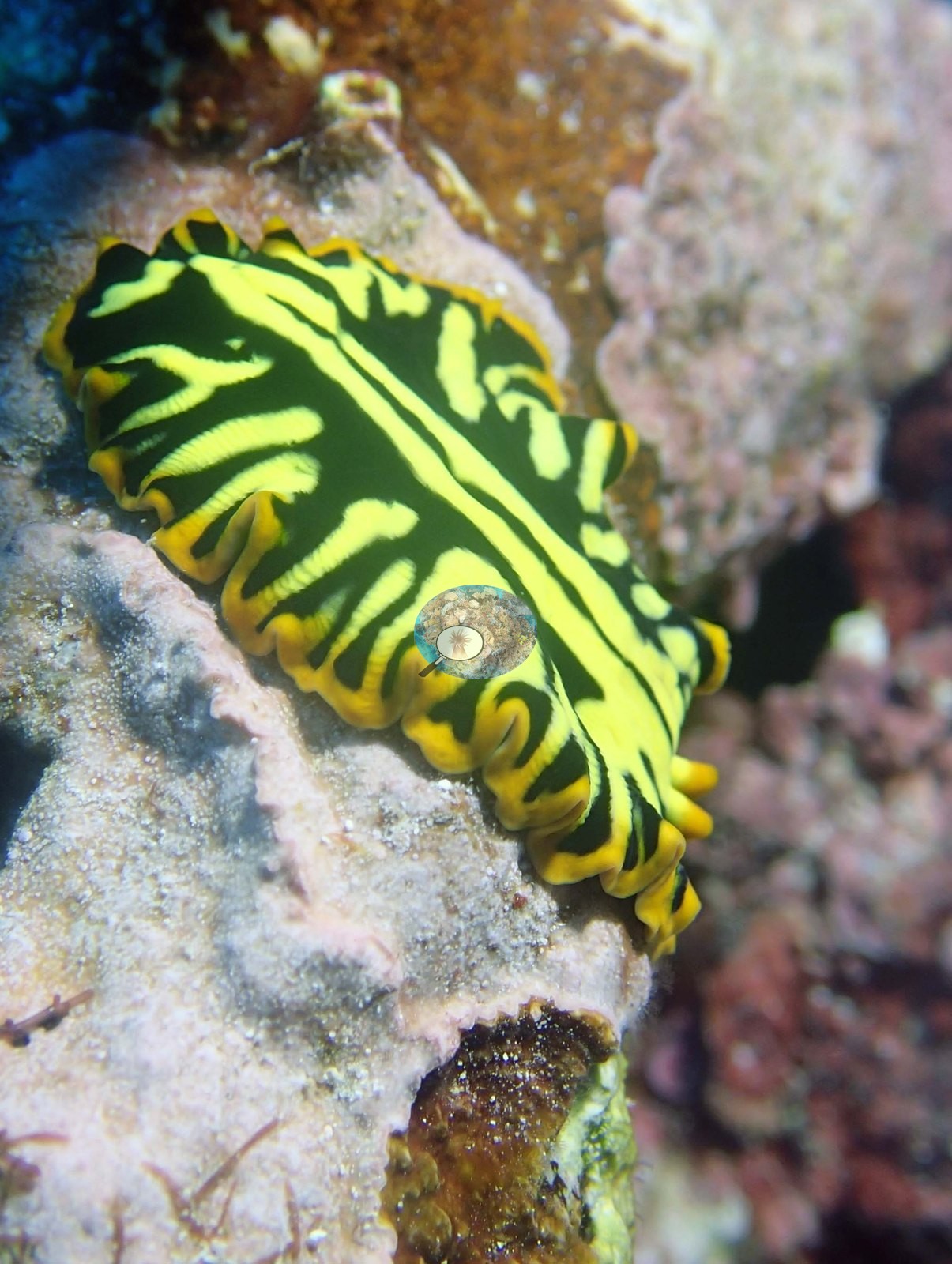
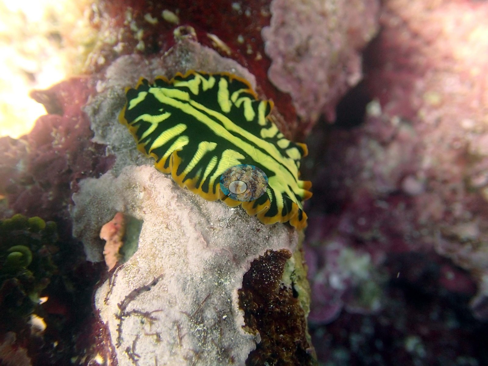
The macro frames are the memorable ones: Image 8 records a Trochus snail wedged against pink coralline crust beside a bright yellow-green digitate colony, and Image 12 and Image 17 are two views of a single spectacular black-and-lemon-yellow flatworm: a thin, flat body with a ruffled orange-edged margin and no gills or rhinophores, so Anderson's flatworm identification is the right one and the nudibranch identification for the other frame is not.
Comparisons with other data
TONI54 (Niuatoputapu) was surveyed during the Khaled bin Sultan Living Oceans Foundation Global Reef Expedition. Fringing reef that slopes very gradually from 4-5 m to 25 m depth. Small discontinuous spurs were surrounded by extensive low relief areas (up to 50 cm relief) of rubble with small corals. At 10 m was a larger spur with massive Astreopora, Montipora, Porites, and table acroporids at 30-50% cover. Cover remained fairly high at 15 m (30-50%) with more digitate and table acroporids, Montipora crusts, Montastrea, Echinopora, and staghorn coral. Slightly deeper are more Porites, Goniastrea and Coscinaraea forming large columns, stout branched Isopora, foliaceous Turbinaria, and many other species. The reef had large amounts of Halimeda (30% cover), 2.0–5.0% of the benthos had damselfish algal lawns.
Other notes
Twenty-six photographs were taken at Niuatoputapu on 29 September; Anderson noted that Pocillopora damicornis was not found here. Eleven of the twenty-six are tagged sampled-colony close-ups and are treated below purely as a record of what was collected; the account of the reef comes from the fifteen site frames only. Beyond that the photographs cannot settle this site's depth zonation or the deeper-slope community. Eleven colonies were tagged and photographed at this site in a separate series — numbered plastic tags, plus one whose number is obscured — each a small branching colony held or pointed to by a gloved hand, with sample vials in shot in Image 21.
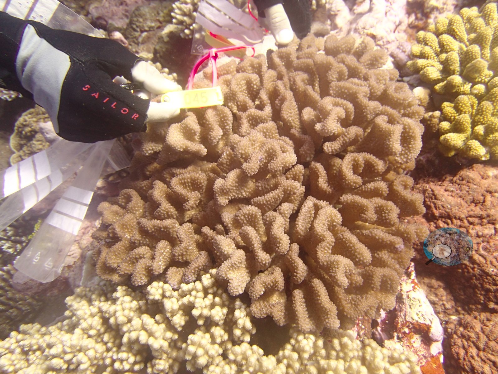
Colors run from gray-purple through cream to olive and pink. No species is assigned to them here; Anderson's own annotation for this site is that P. damicornis was absent. These frames describe the colonies, not the reef.
CV model comparison
More info
The photo row below is a methods comparison only: it is never averaged with the transect and never used as the headline. It compares site means only: the photos come from the same survey dive as the transect but not from the same points.| Method | Hard coral cover (%) | Photos (n) ± SE |
|---|---|---|
| Transect mean (all transects) | 34.3 | |
| CoralSCOP unedited | 34.2* | n = 13 · SE 5.9 |
| Claude | 20.1* | n = 13 · SE 6.3 |
The two computer-vision methods disagree by 14.1 pp (20.1–34.2%) and read 0.1–14.2 pp below the in-water transect (34.3%); the headline is the in-water transect because a diver transect is independent of the photographs.
*Based on only 13 reef-habitat photos; this estimate is less certain.
See a more detailed, stand-alone version of this table.
Data & links
- Download benthic survey data (CSV) ↗
- Download all LOF-GRE data (933 sites) ↗
- Photo portfolio ↗
- Coral Reef Diagnostics region page ↗
All images from Coral Reef Diagnostics. Data are from the Khaled bin Sultan Living Oceans Foundation Global Reef Expedition and Coral Reef Diagnostics.
 © Coral Reef Diagnostics.