
Part of the regional summaries: Tonga - Ha'apai and Tonga
More info
Southern Tonga (NOAA CRW virtual station)A densely built Ha'apai reef with almost no bare rock, crowded with lilac and violet Acropora, stacked table corals, and broad staghorn fields under schools of damselfish and surgeonfish. A boldly patterned giant clam anchors a crevice community, and no bleaching was visible. Surveyed on the Living Oceans Foundation's Global Reef Expedition.
Ko ha hakau ʻi Haʻapai kuo langa fefuʻifuʻi, ʻoku meimei ʻikai ha maka tukuʻau, ʻoku fonu ʻi he Acropora lanu laileki mo vaioleti, ngaahi feo fakatēpile (table corals) fakafuʻufuʻu, mo e ngaahi ngoue staghorn fālahi ʻi lalo ʻi he ngaahi kulupu ika damselfish mo e surgeonfish. ʻOku nofo ha vāsuva (giant clam) ʻoku ngali faka-tā hono lanu ʻi ha kiʻi ava mo e ngaahi meʻa moʻui ʻoku nofo ai, pea naʻe ʻikai hā ha hinehina (bleaching).
AI translation of this summary; the English is authoritative. (low confidence)
CTD-cast midpoint; the cast read 24.2°C throughout
One other CRD dive site with its own page within 5 km shown in orange — click a marker to open its page.
36 of the 38 photographs on this page are shown beside the text that discusses them under "Site description" below.
Site description
General benthic features
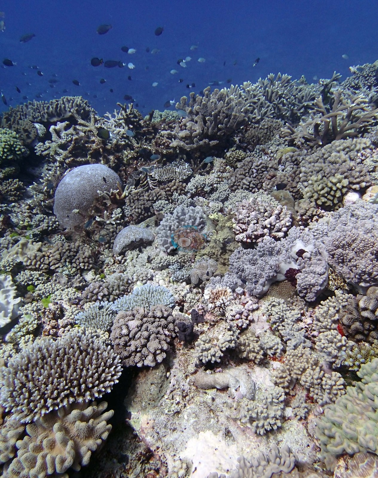
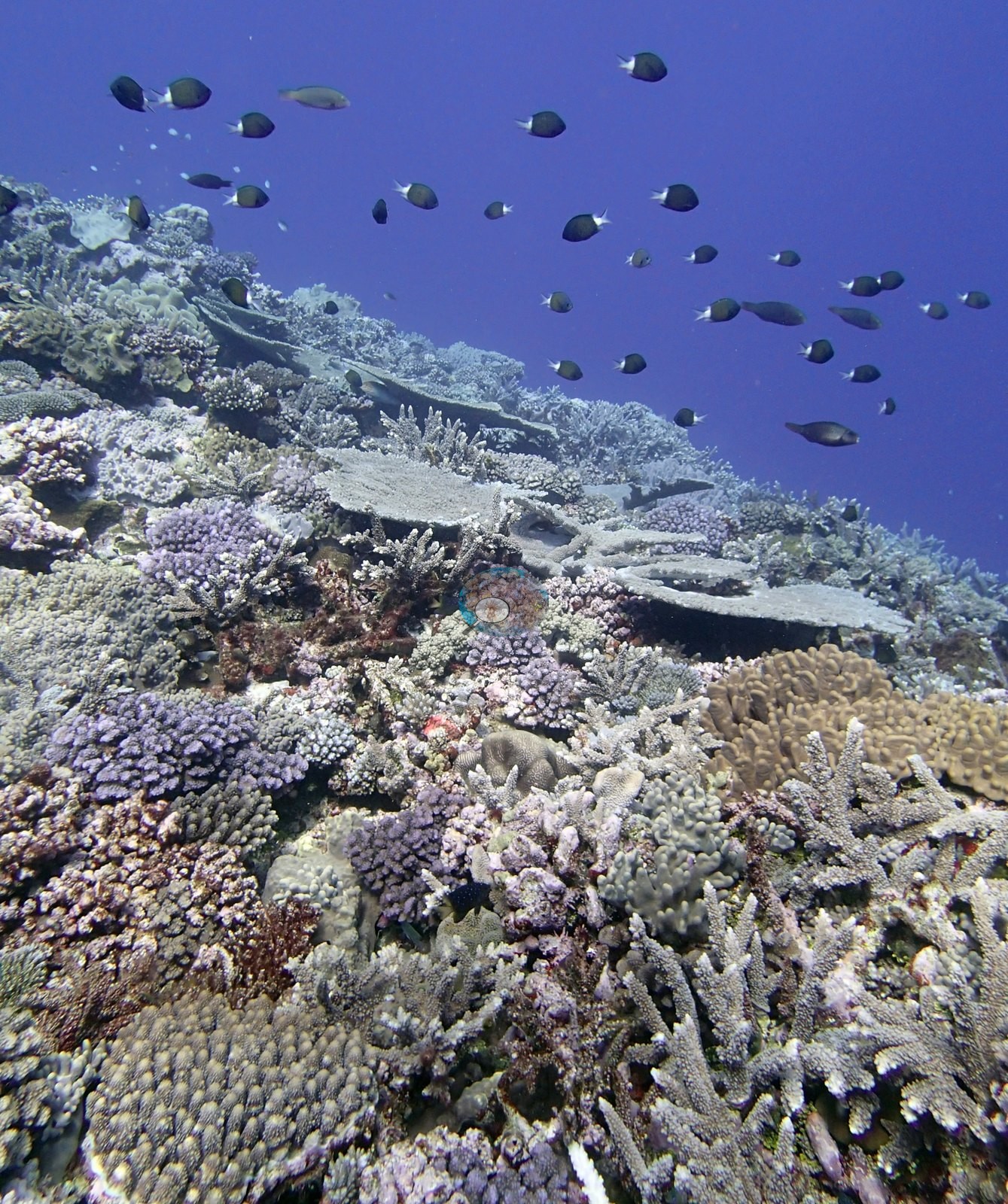
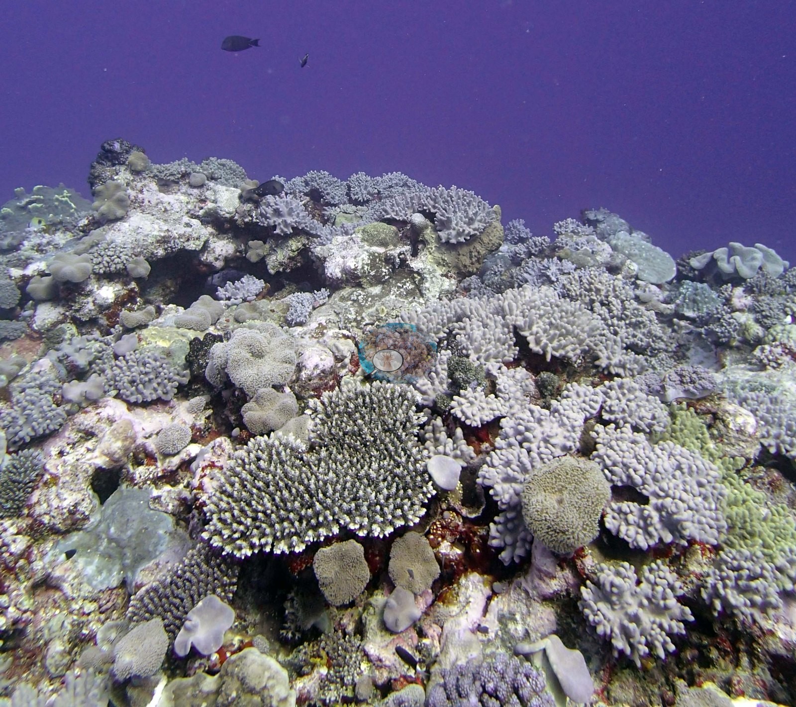
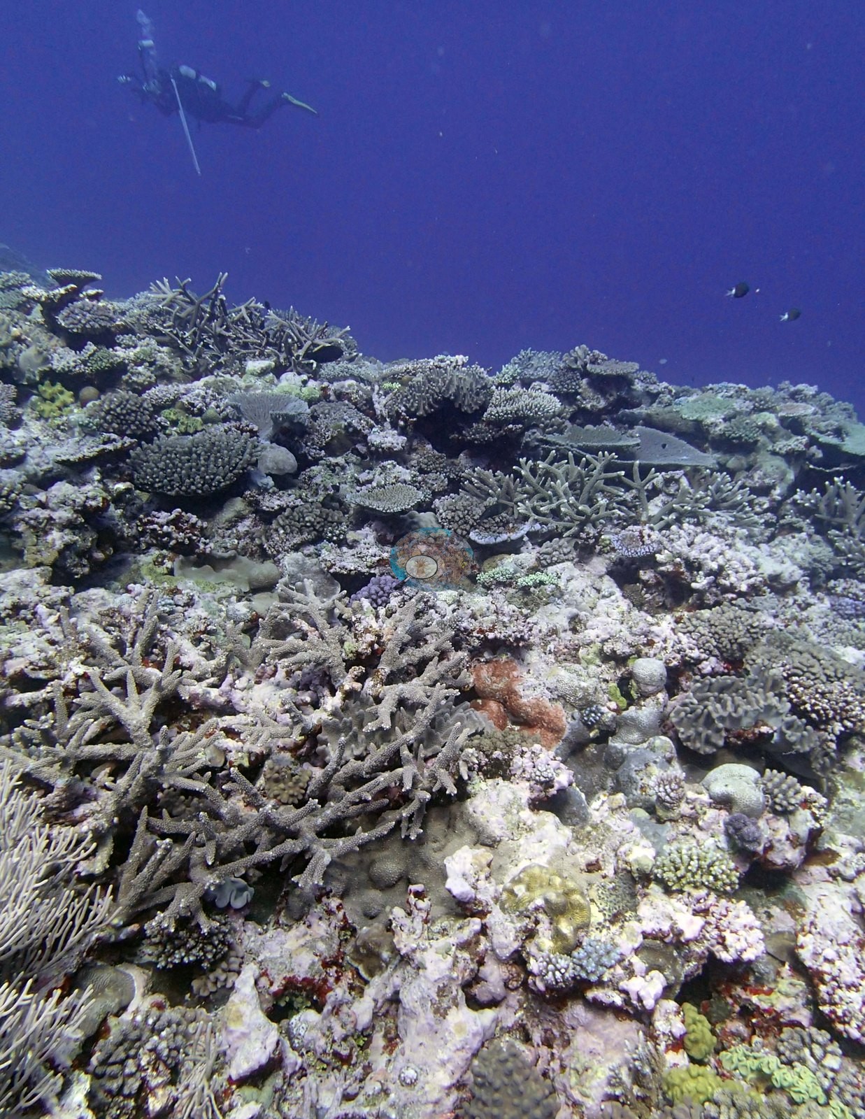
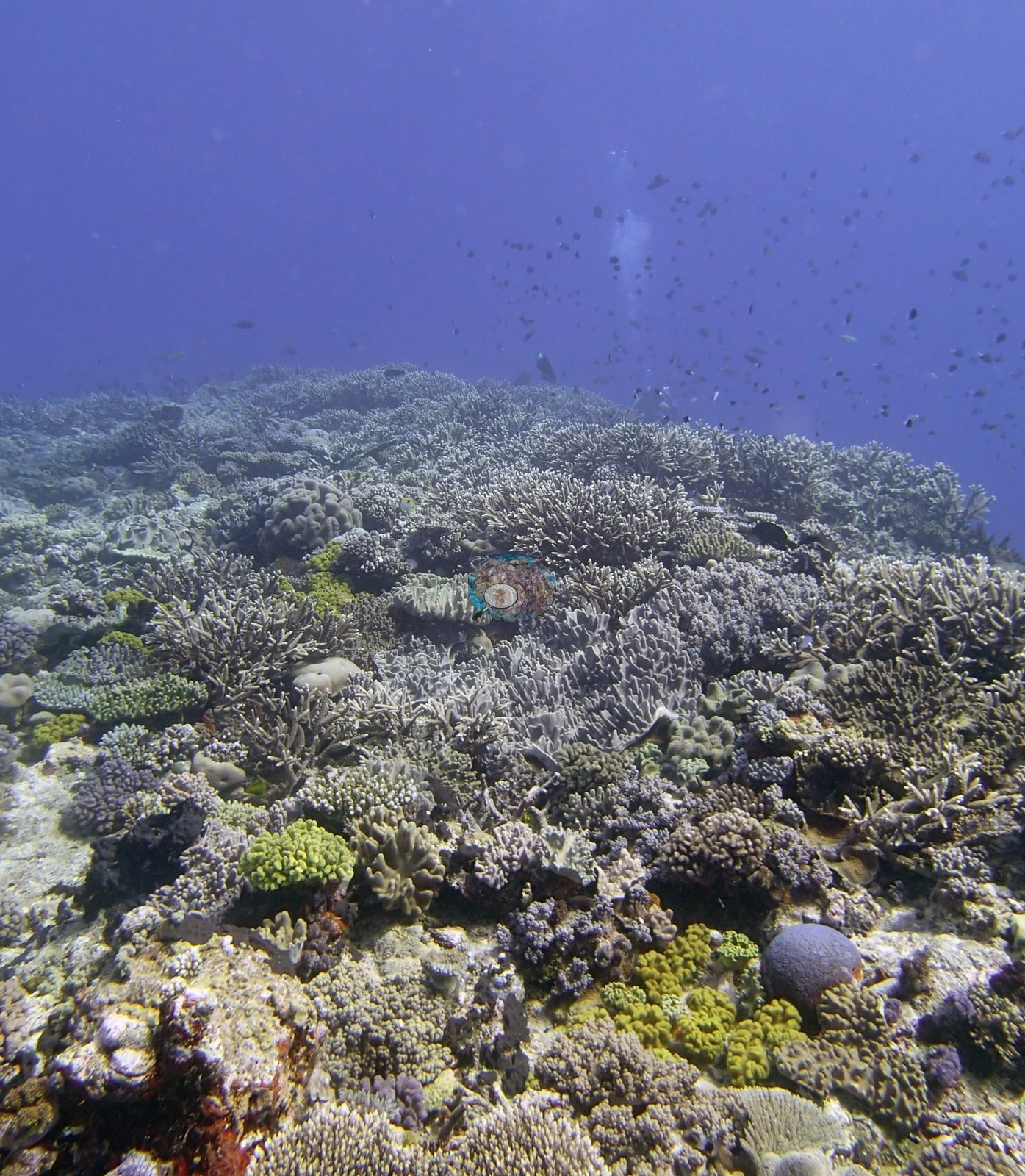
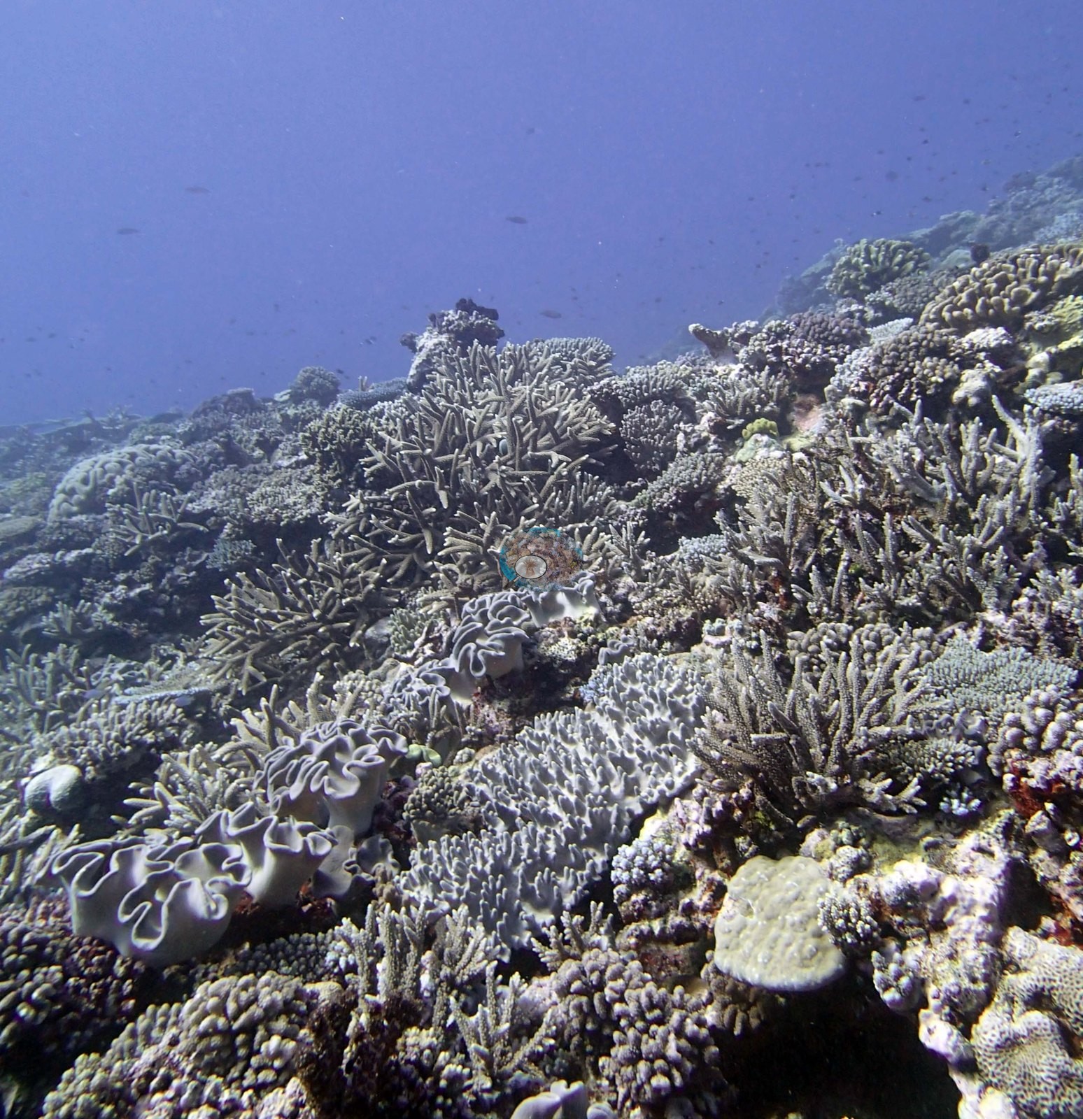
The wide frames show a densely built, high-cover reef. One looks up a slope crowded with lilac and violet corymbose Acropora, several stacked pale gray Acropora tables 40-80 cm across, staghorn branches, and a tan massive colony with meandering valleys on the right (a Platygyra or Leptoria growth form), while a loose school of dark white-tailed damselfish hangs in deep blue water above the crest (e.g., Image 21).
Another is a tight view of the reef top with essentially no bare substrate: gray-lilac corymbose acroporids packed edge to edge, a clean Acropora table around 50 cm at center, gray mushroom-shaped and lobate soft corals, pale plating colonies, and dark maroon encrusting growth filling the gaps (e.g., Image 2).
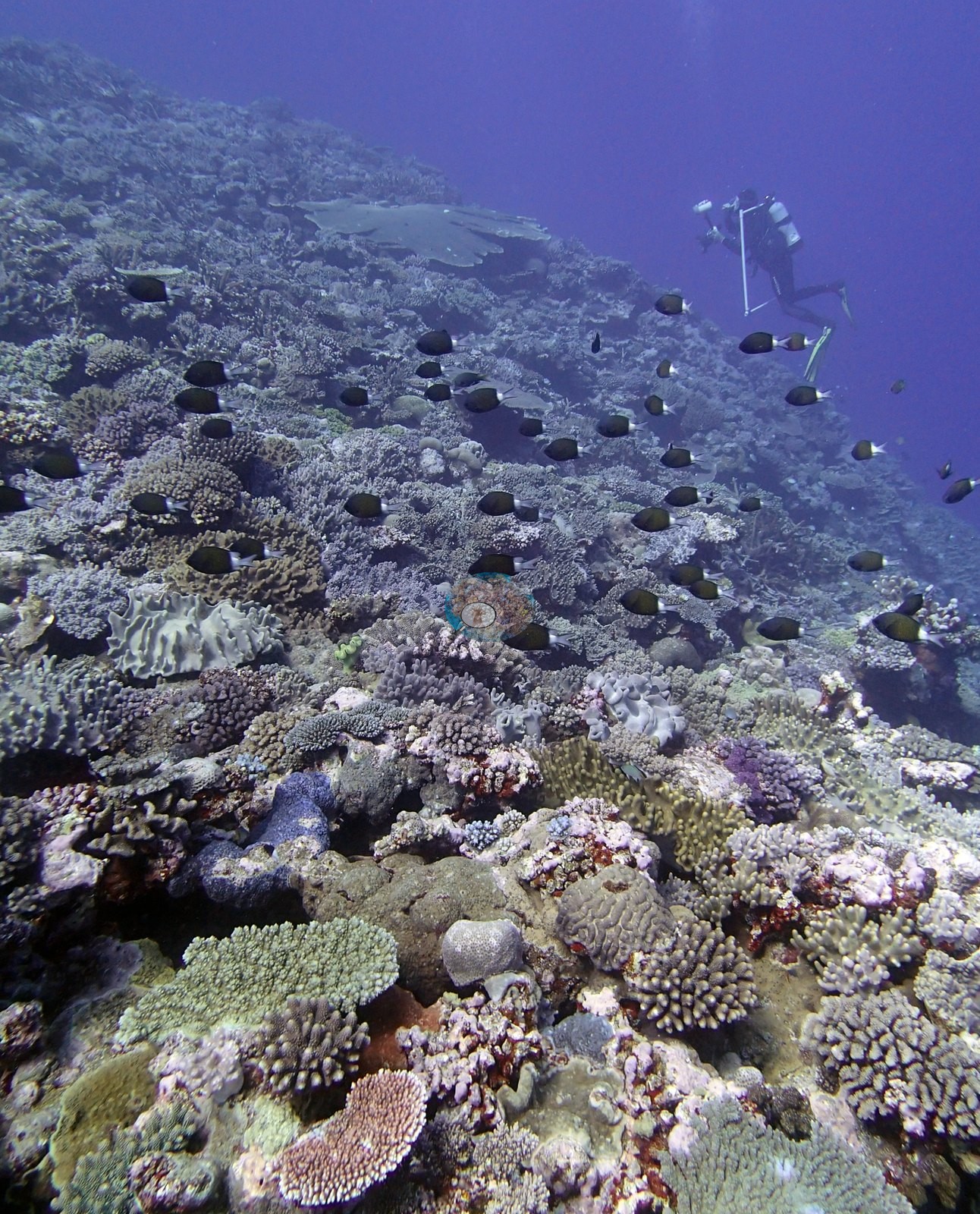
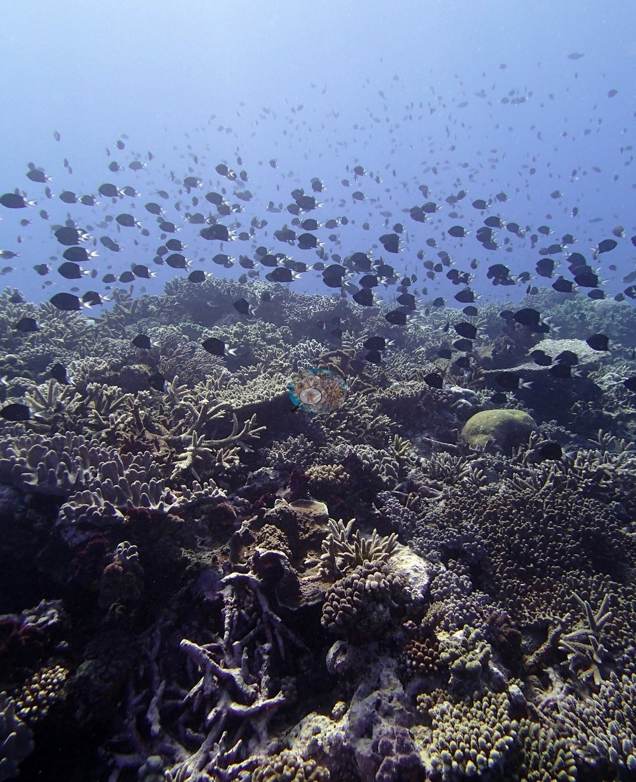
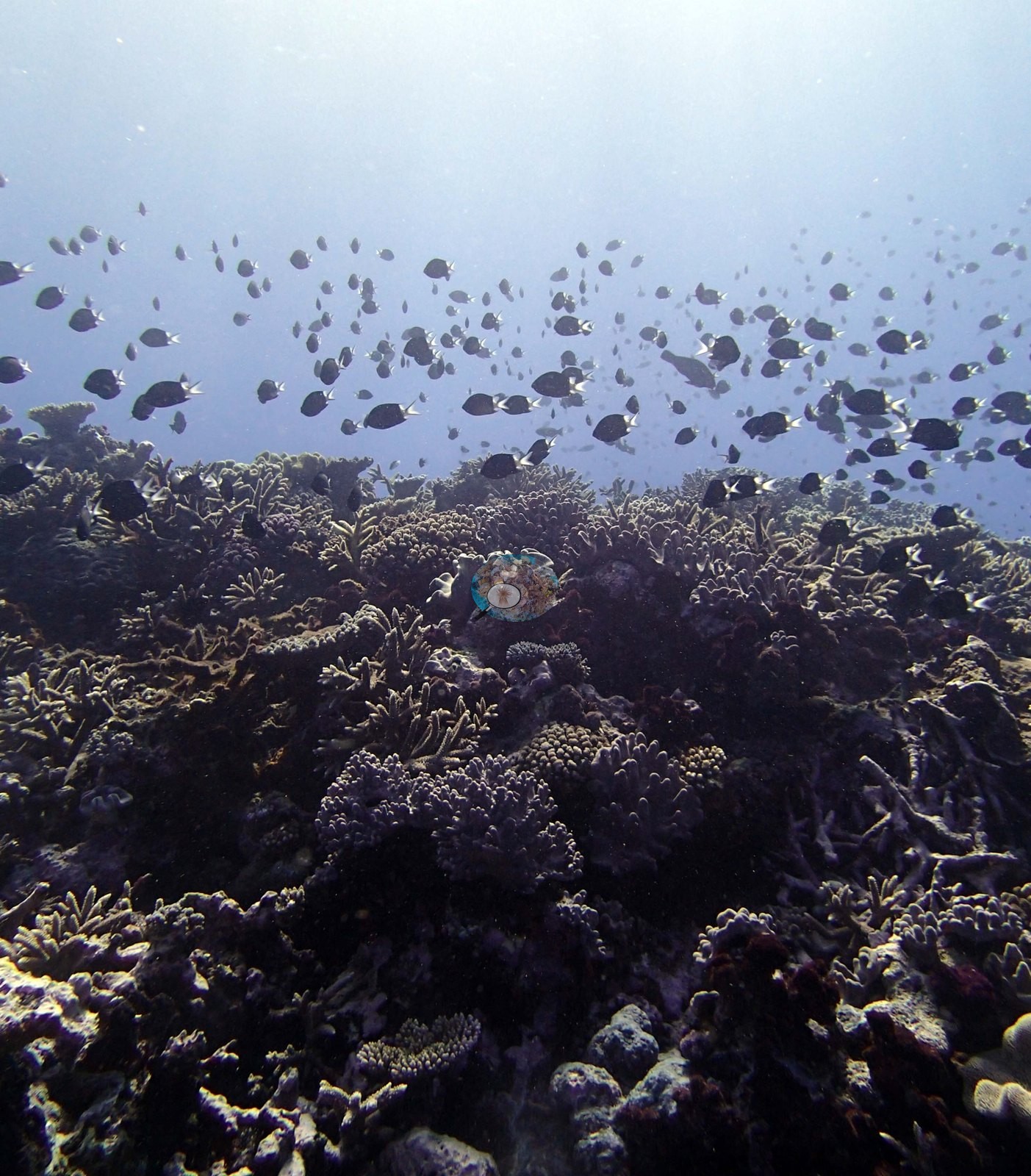
Another shows a broad staghorn-dominated field under a dense aggregation of dark damselfish and surgeonfish — a fitting image for a site where the survey specifically noted many damselfish algal lawns in the staghorn thickets — with pale and purple-brown branching Acropora, yellow-tipped corymbose colonies, and a tan massive Porites in the foreground, and turf-darkened dead branch sections mixed in Image 28.
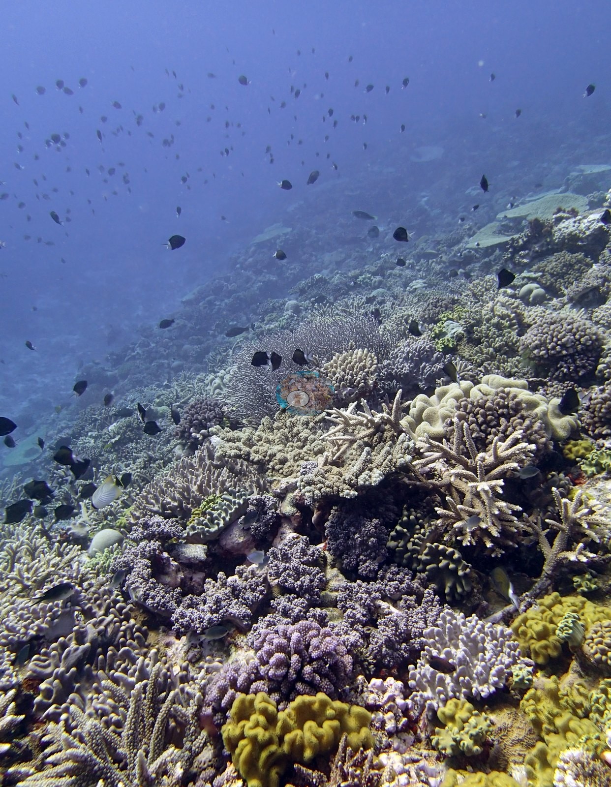
The dominant architecture across the set is corymbose and digitate Acropora in lilac, violet-gray, and pale tan, packed shoulder to shoulder over the reef top (e.g., Image 21, Image 10, Image 8, Image 5, Image 4, Image 24, Image 23, Image 11, Image 16, Image 32, Image 25, Image 17, Image 18, Image 35).
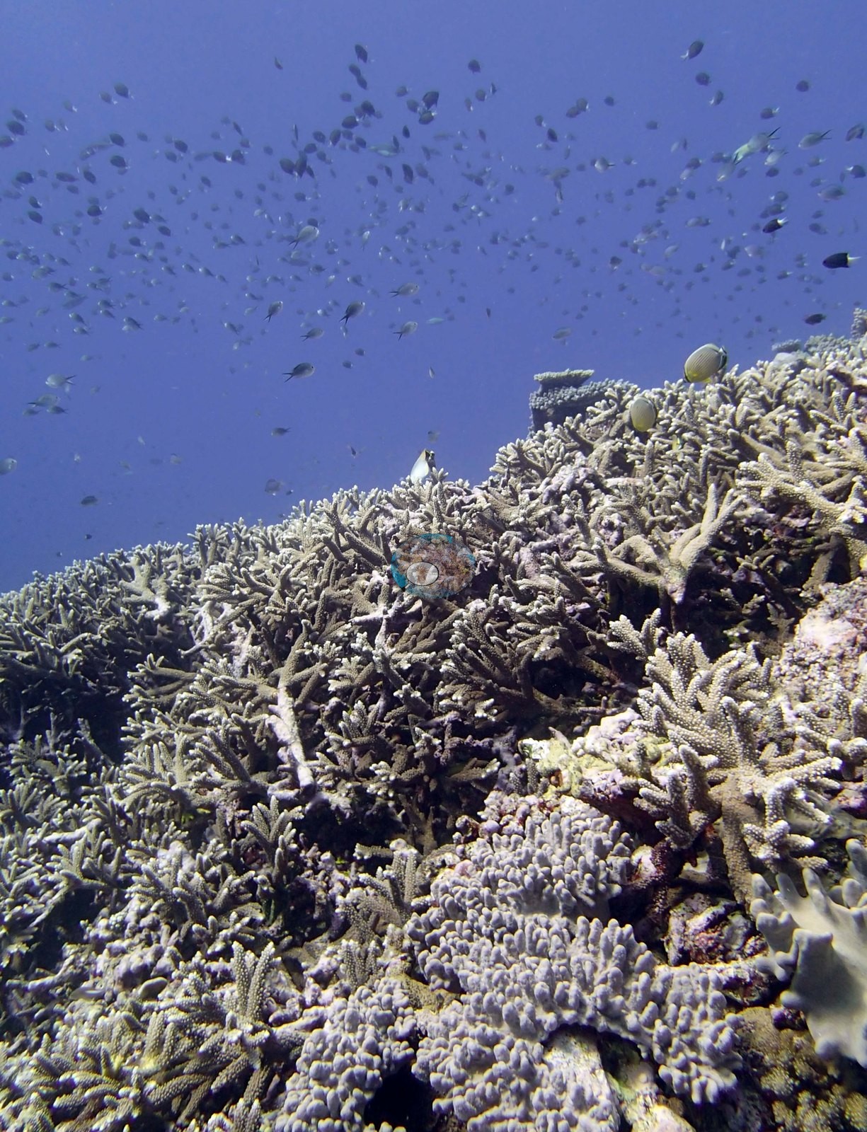
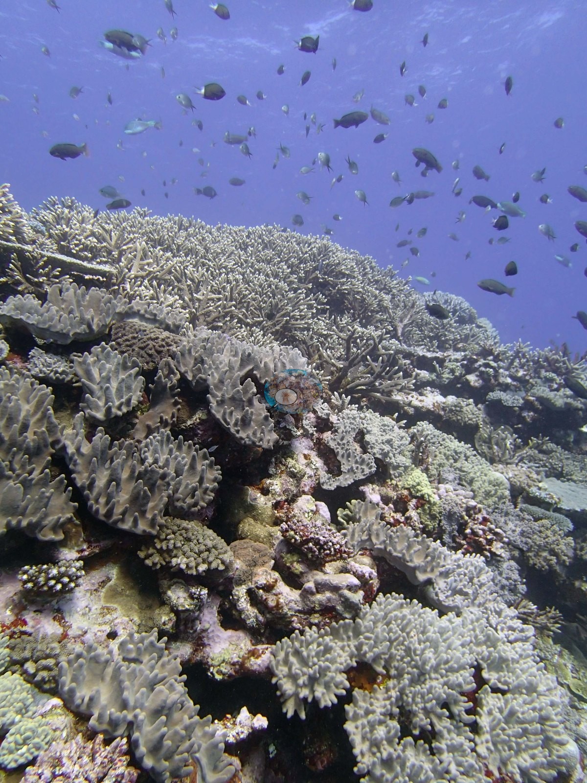
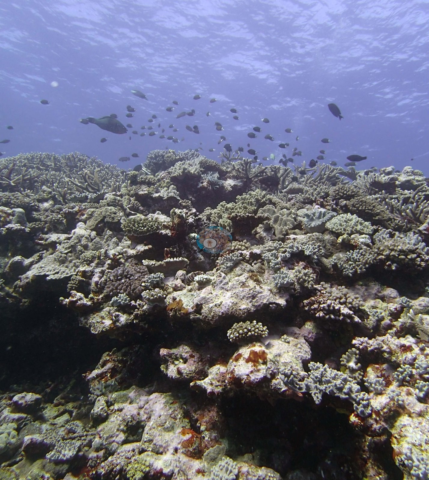
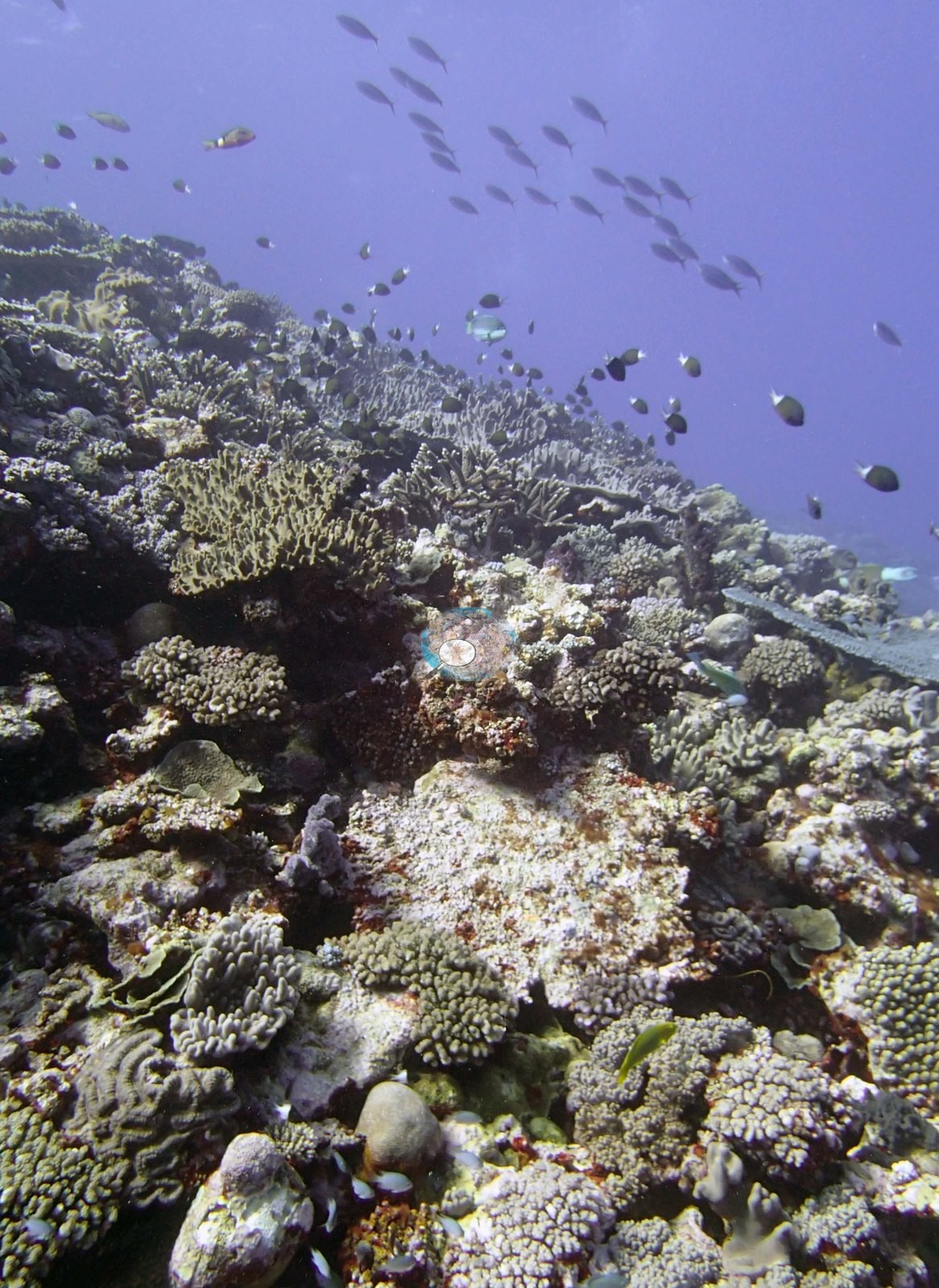
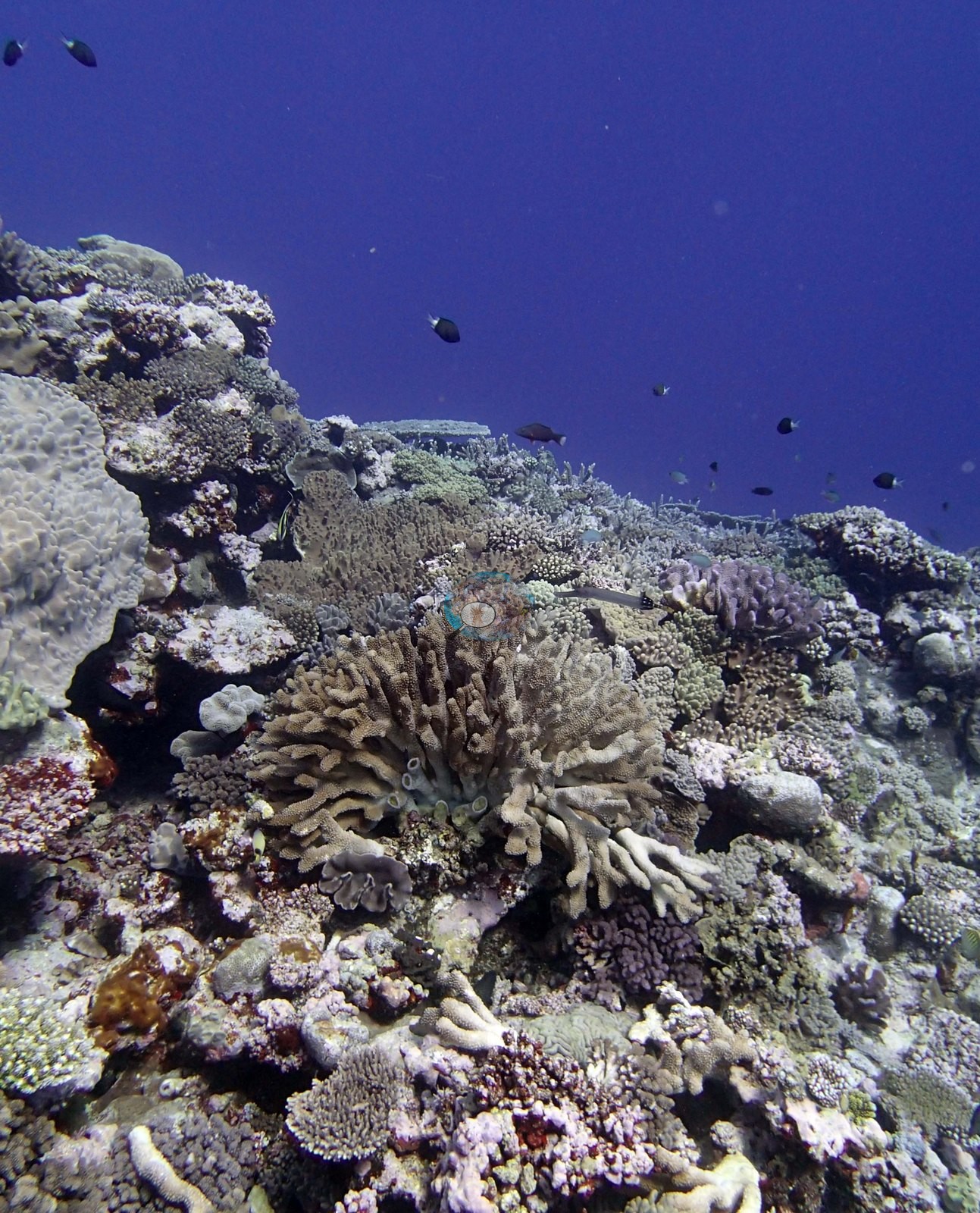
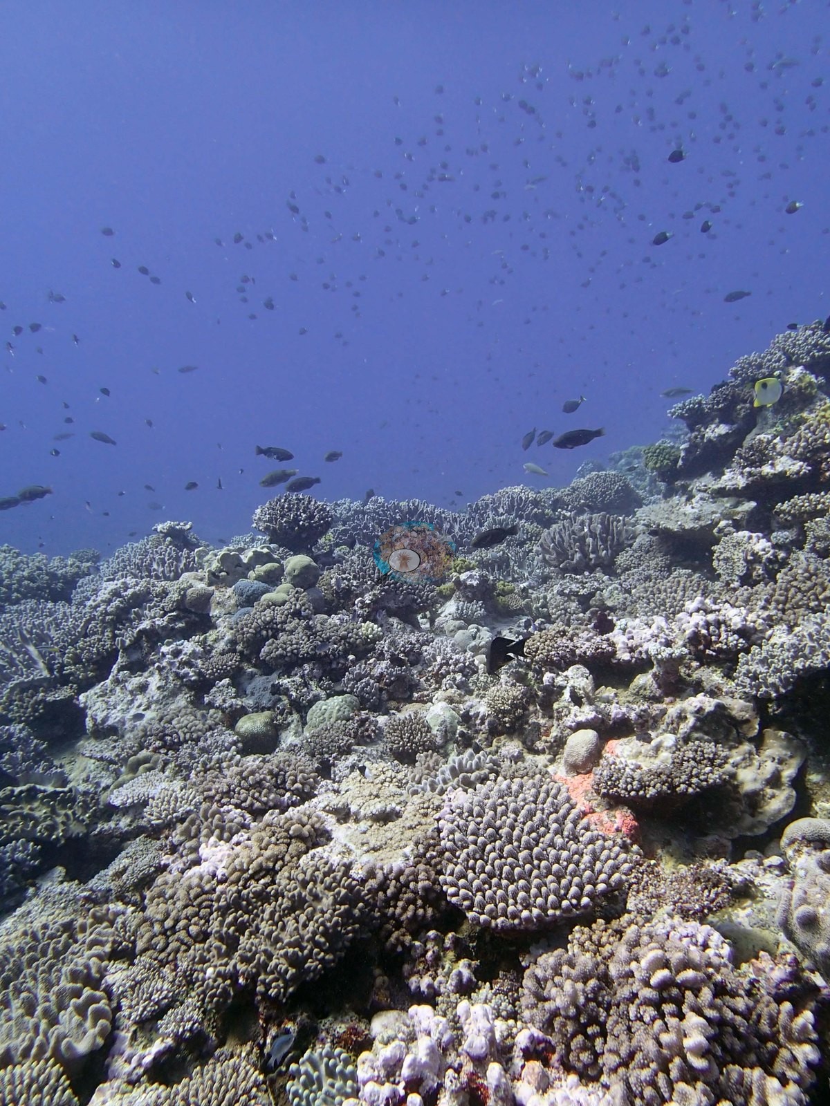
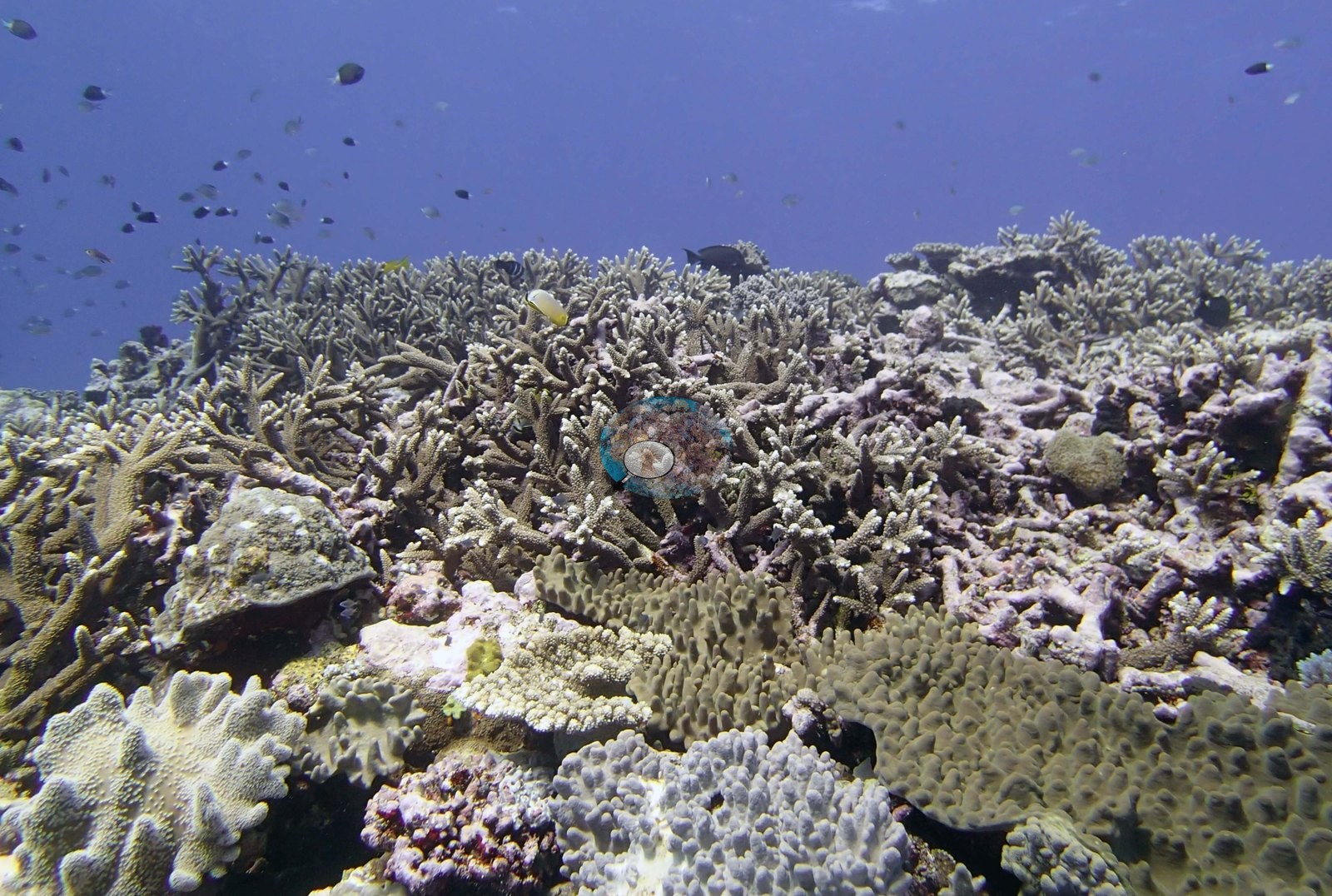
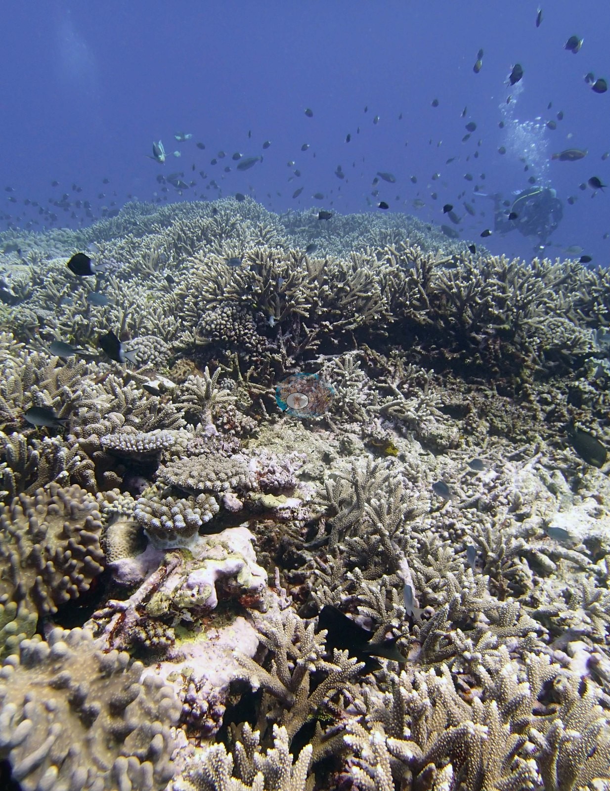
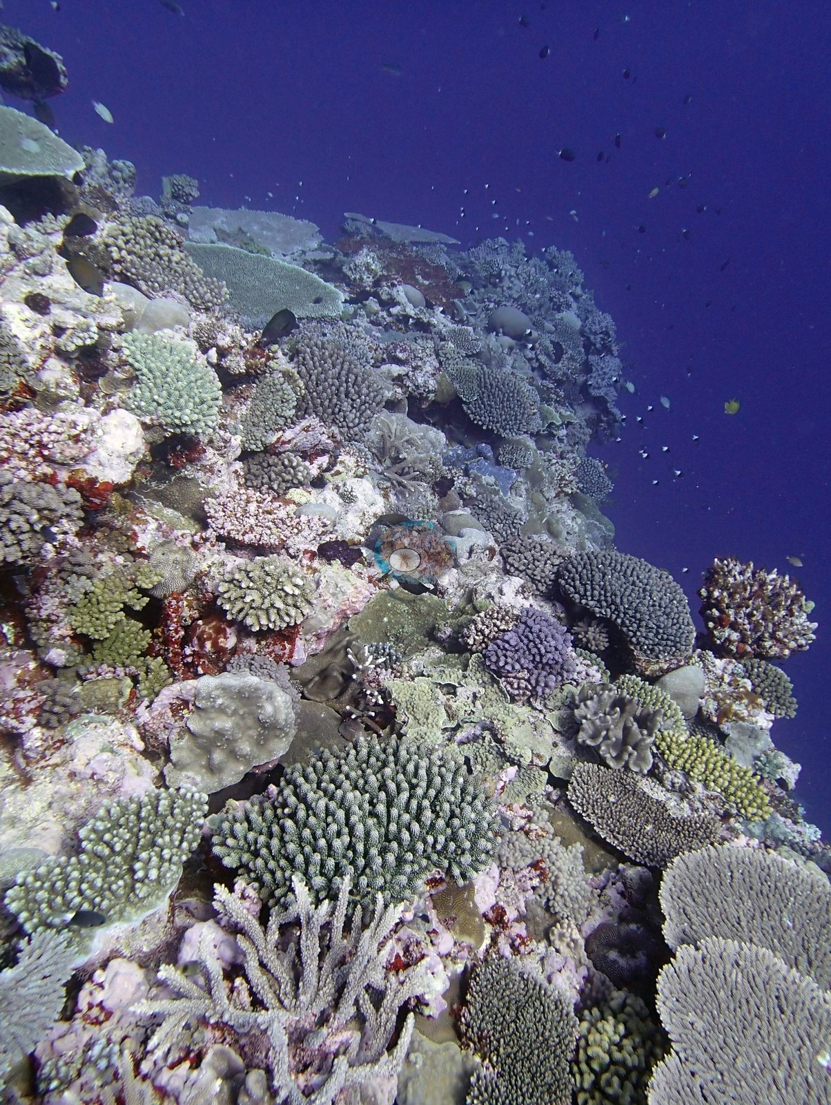
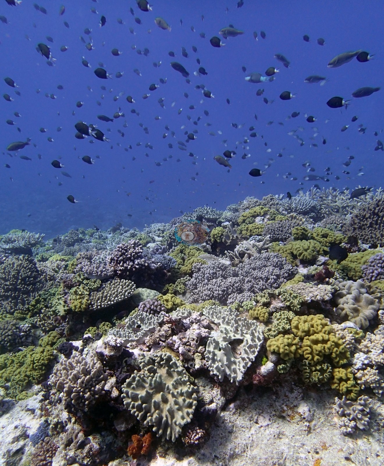
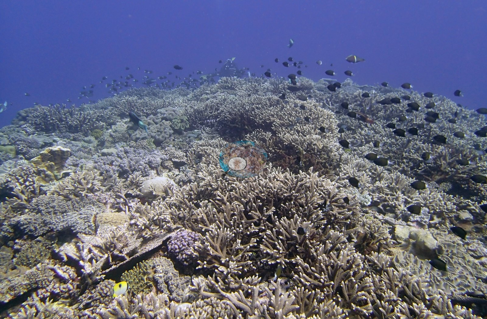
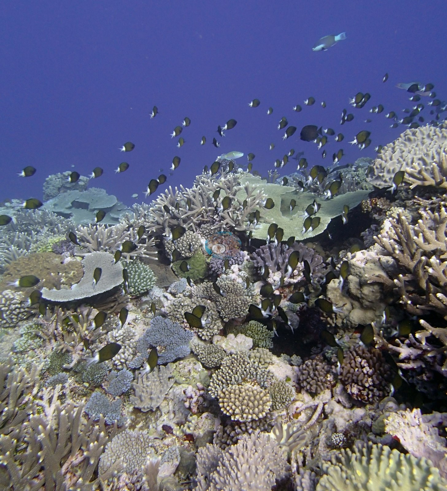
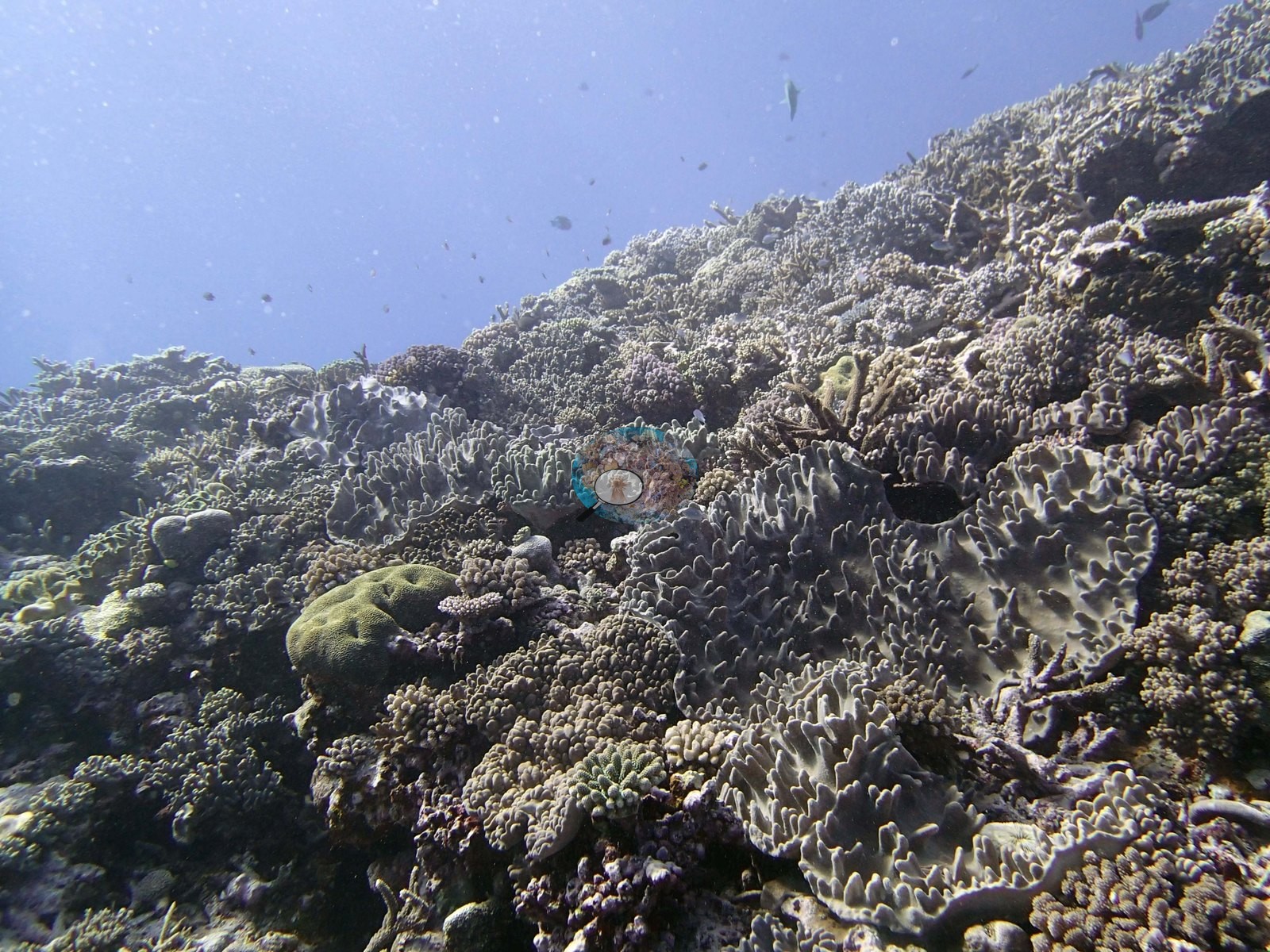
Staghorn-form Acropora thickets recur throughout, sometimes as clean pale branches (e.g., Image 36, Image 22, Image 19, Image 26) and sometimes as darker, turf-shaded branch fields with dead sections mixed among live tips (e.g., Image 7, Image 30, Image 22, Image 28).
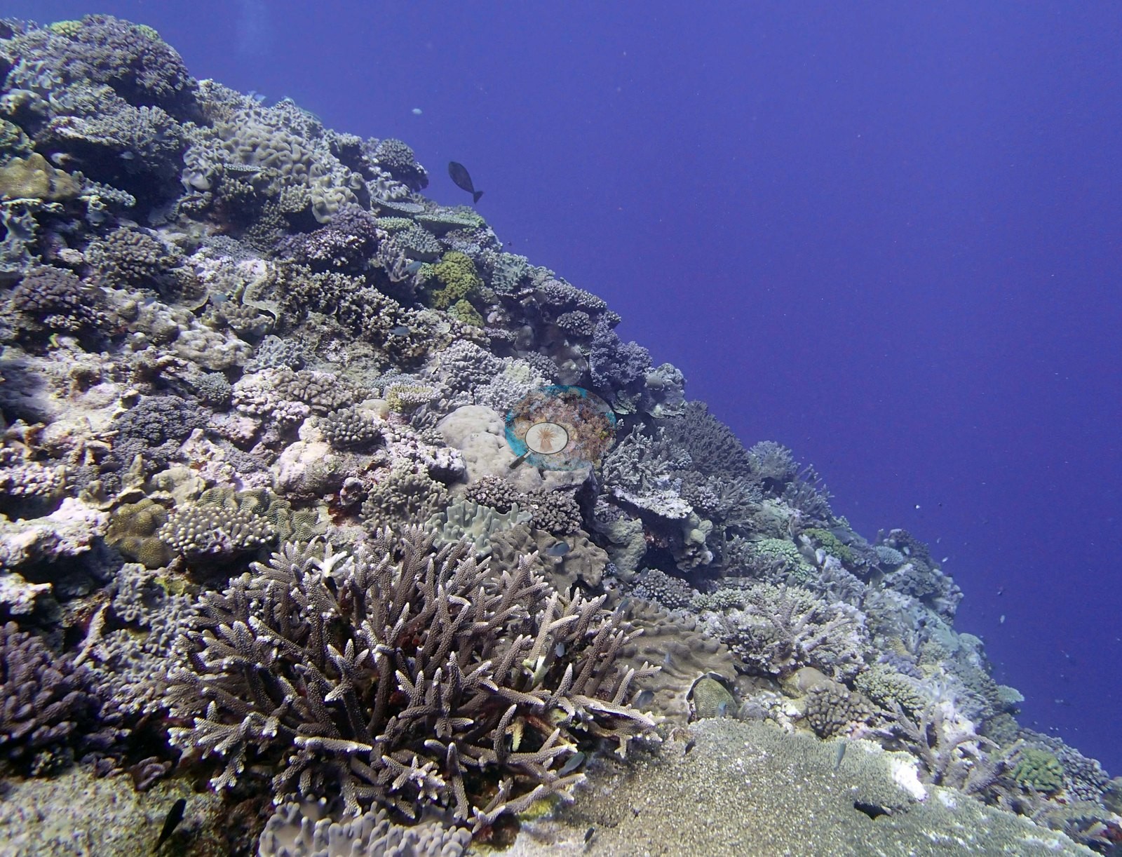
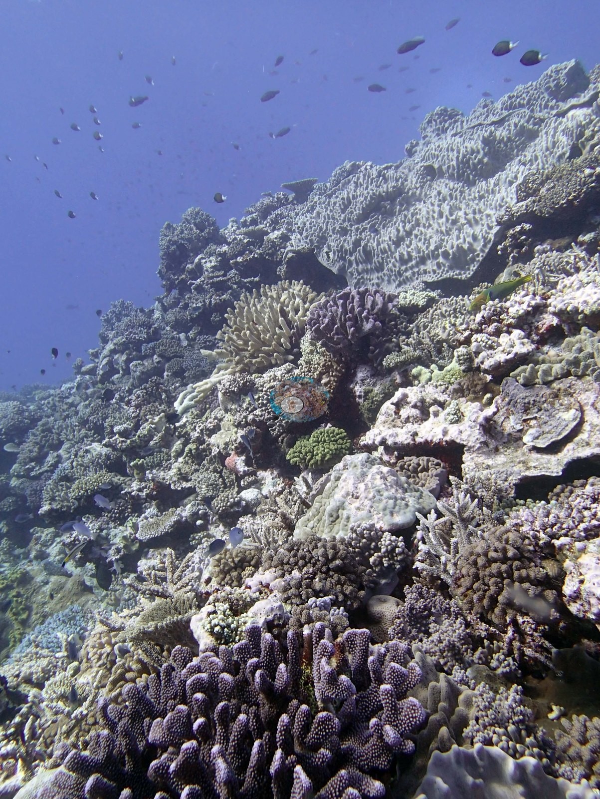
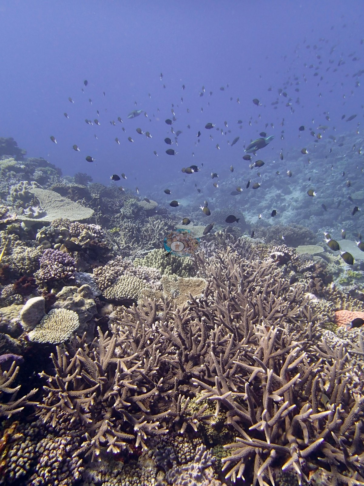
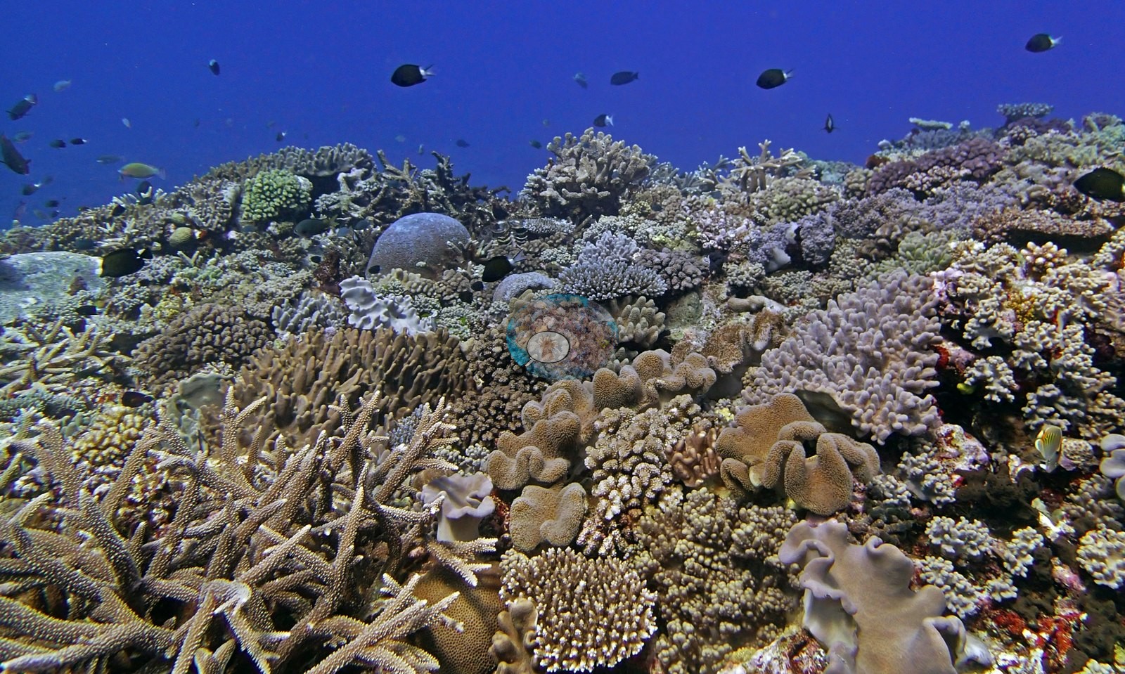
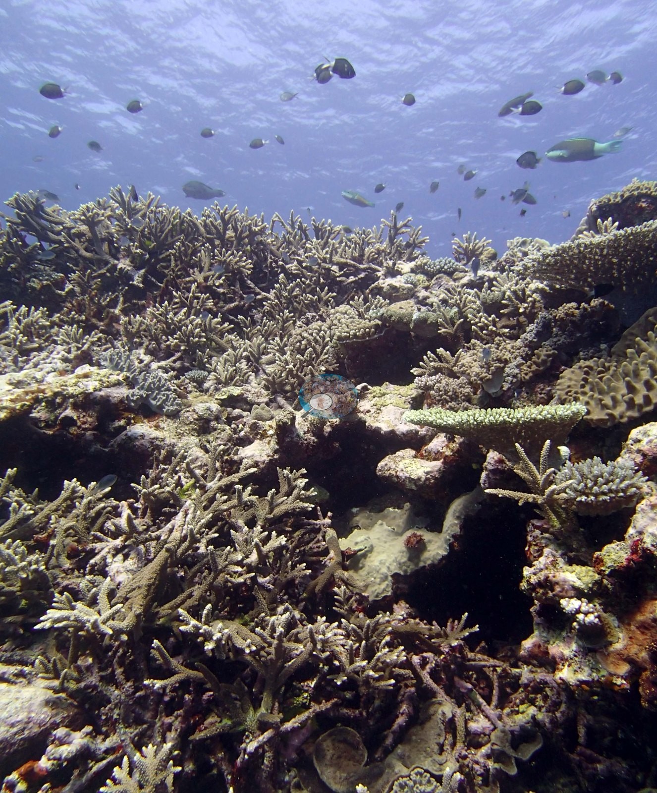
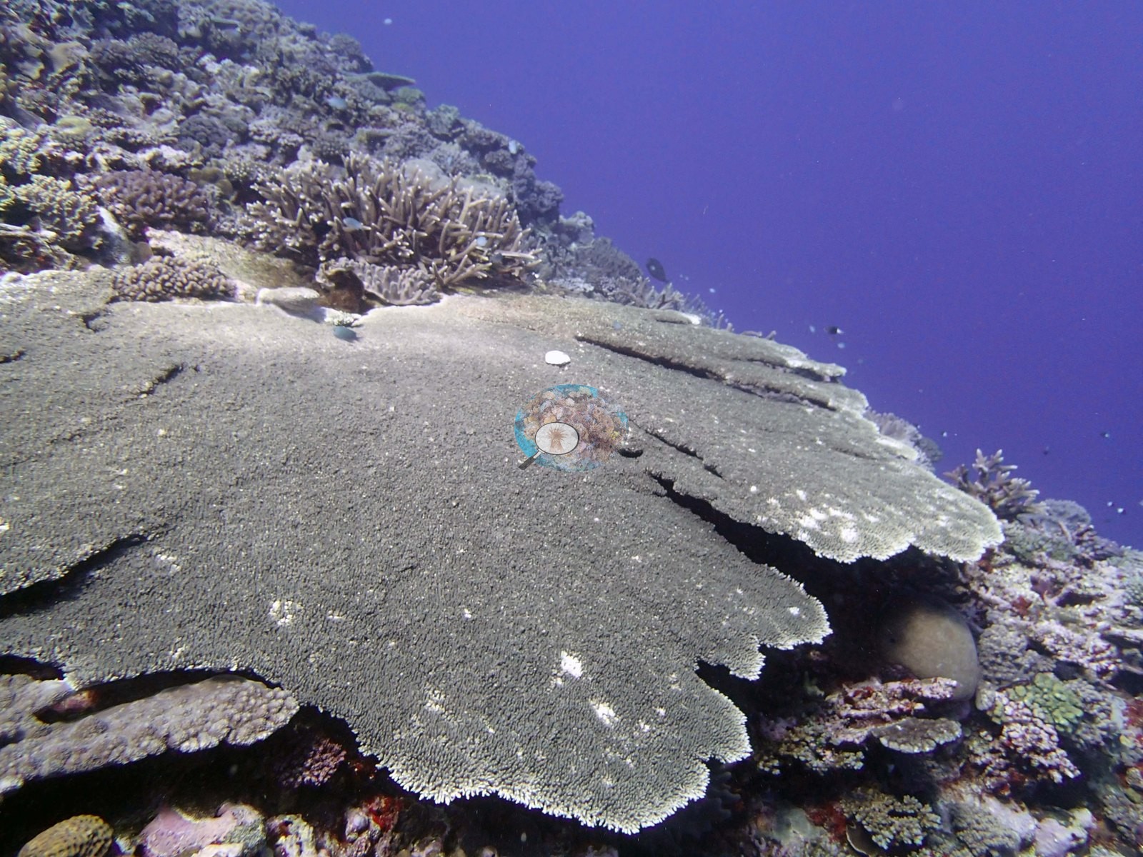
Tabulate acroporids appear repeatedly as discrete plates 40-80 cm across sitting proud of the corymbose matrix (e.g., Image 8, Image 2, Image 20, Image 32, Image 18); one frame (Image 34) holds a single smooth gray-brown table roughly a meter across with a clean growing margin, perched on the reef edge against open blue water, and a second broad plate of similar form dominates (e.g., Image 38).
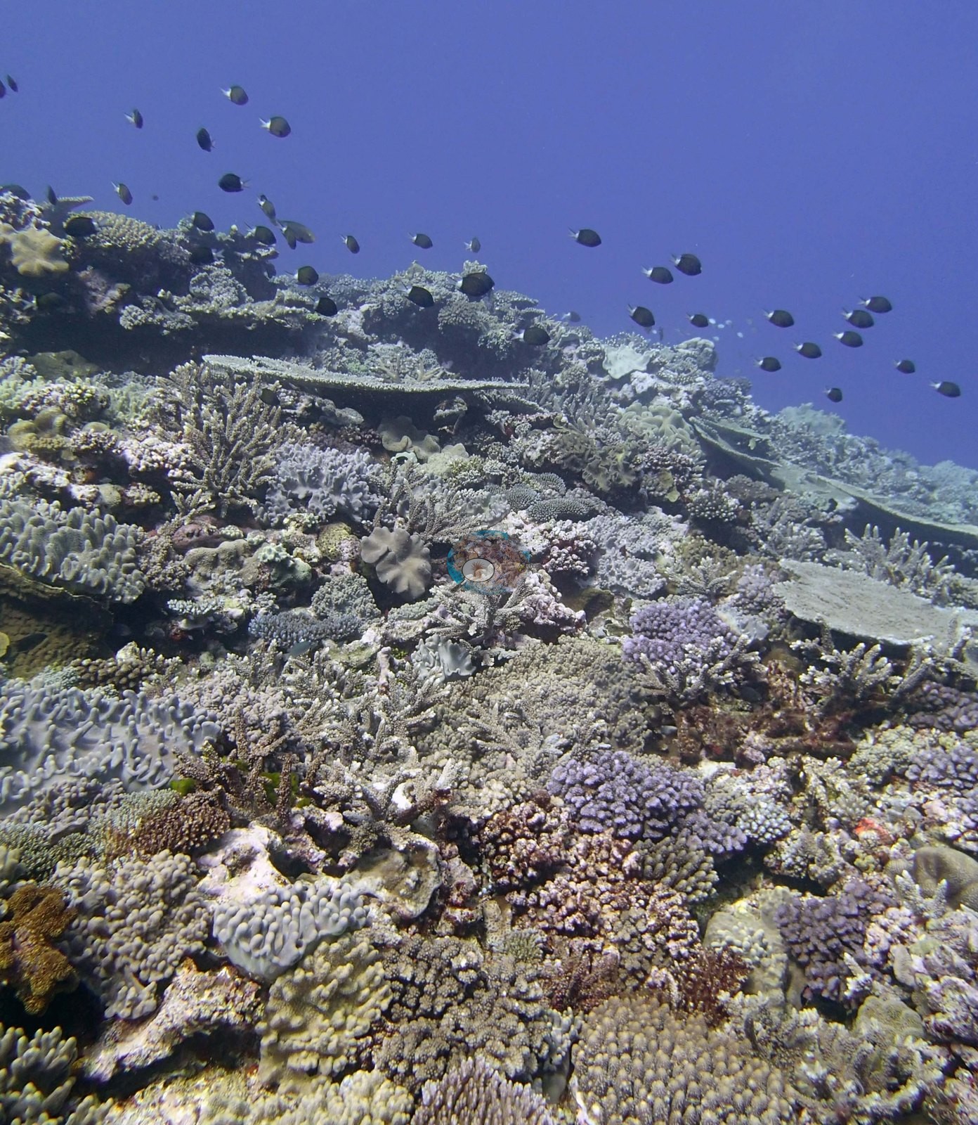
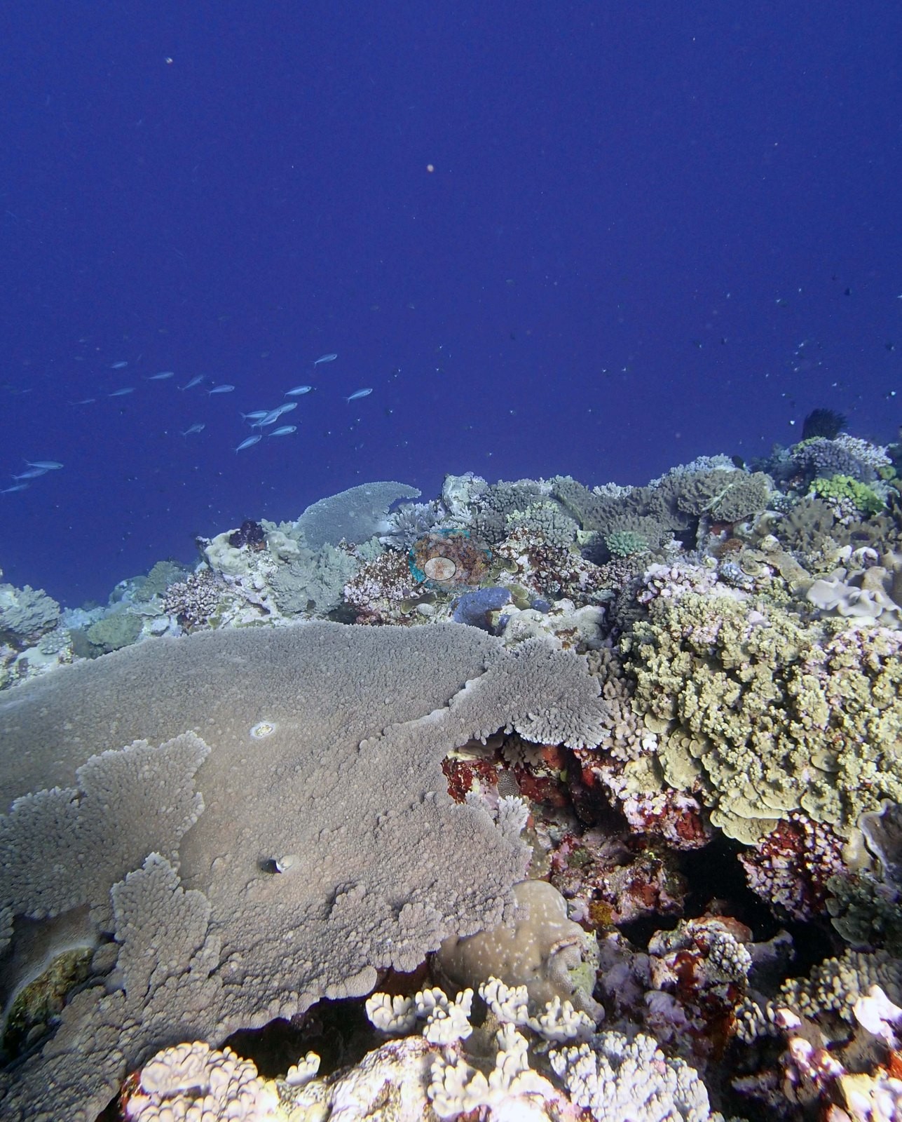
Massive corals are present but secondary: tan and olive-green domes with meandering or cerioid surfaces (a faviid-type growth form) and low submassive Porites-like mounds sit in the gaps (e.g., Image 9, Image 2, Image 11, Image 15, Image 32, Image 26).
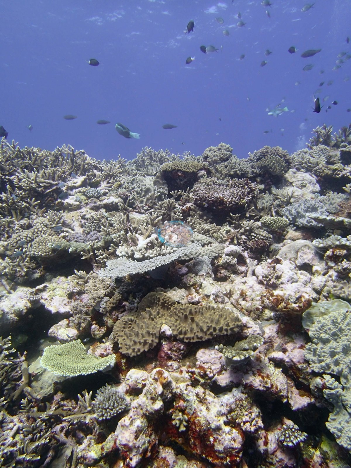
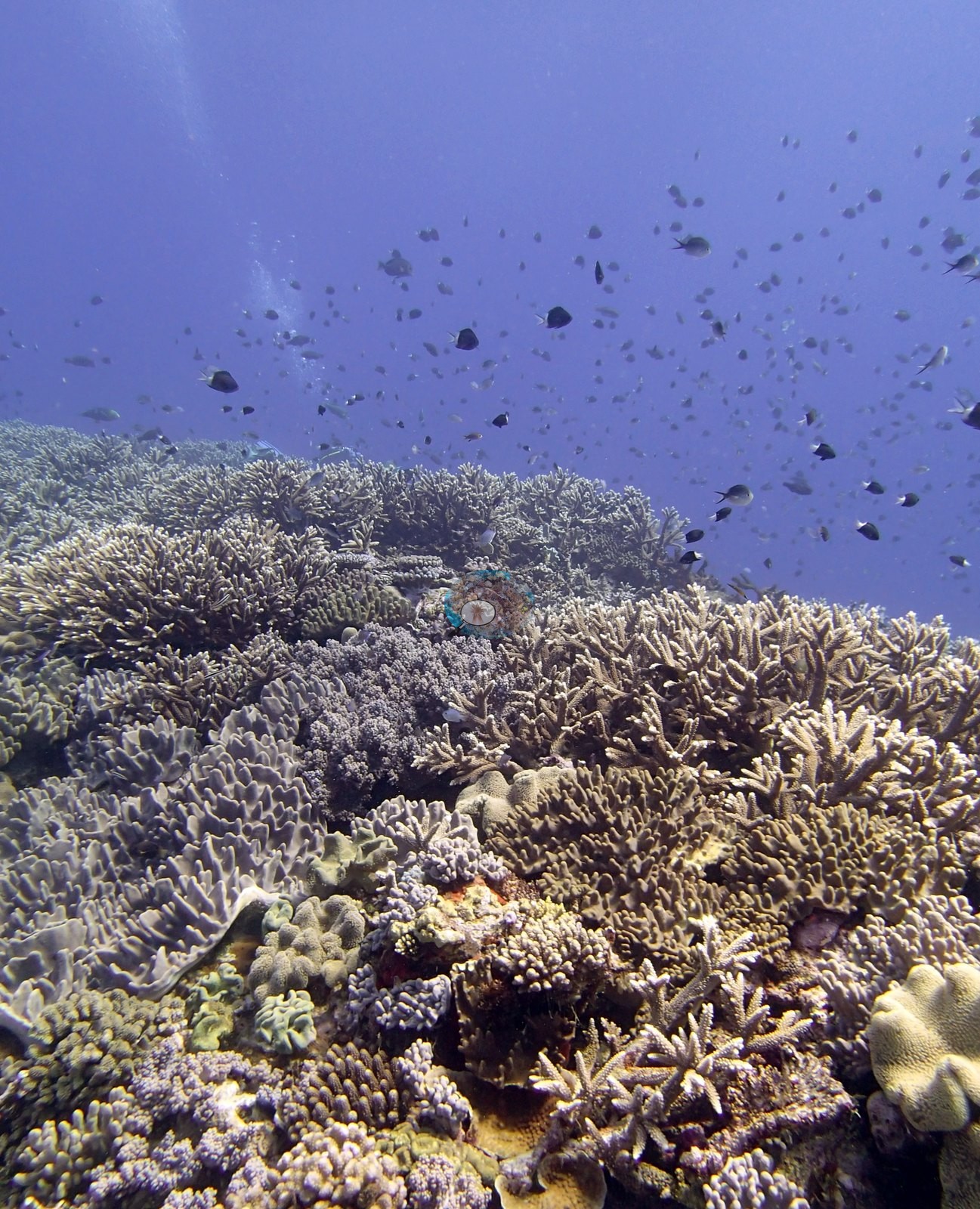
Encrusting and plating growth — ruffled gray-white plates, dark maroon-brown encrusting sheets, and pale foliose margins — fills much of the remaining substrate (e.g., Image 37, Image 2, Image 3, Image 32, Image 1).
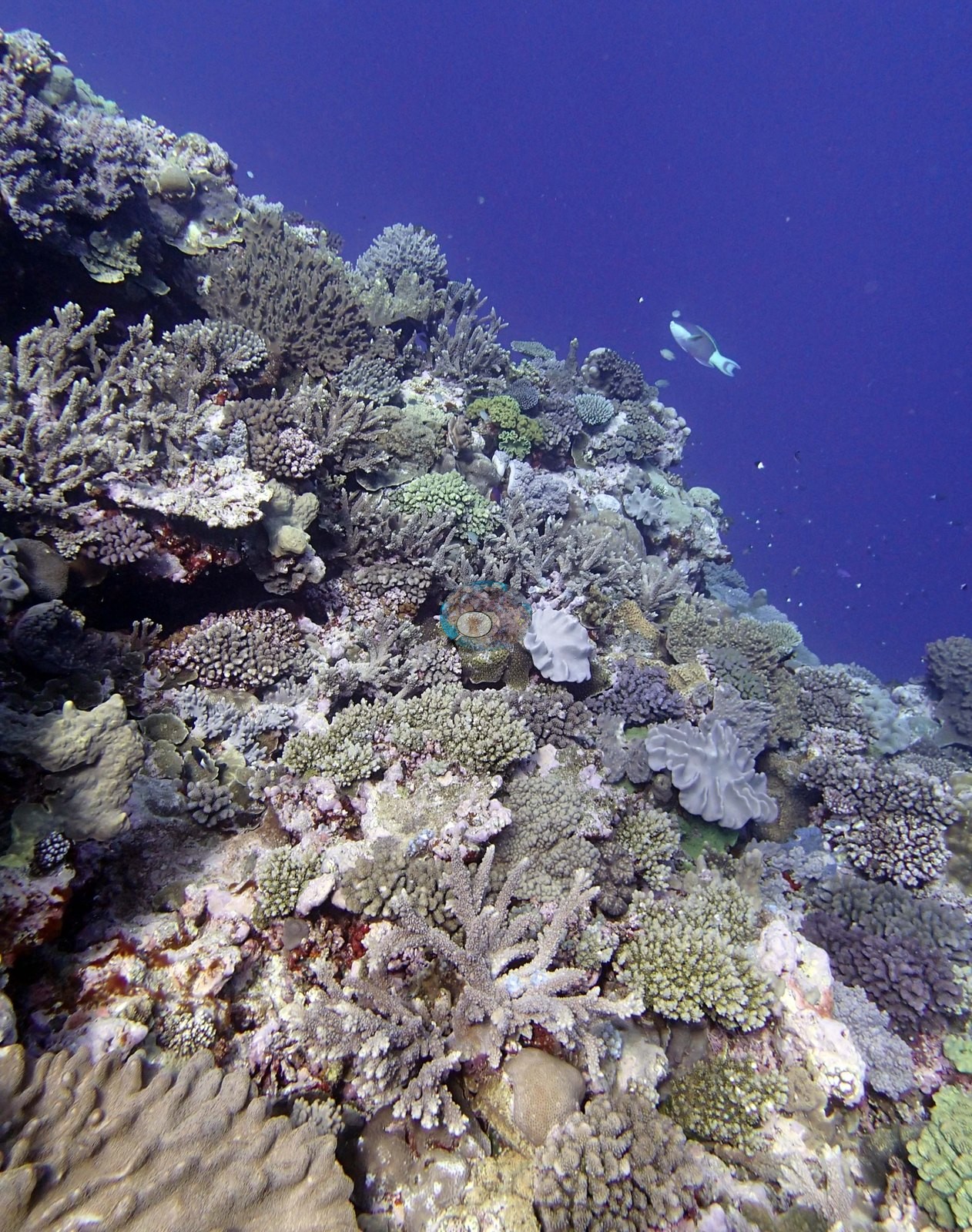
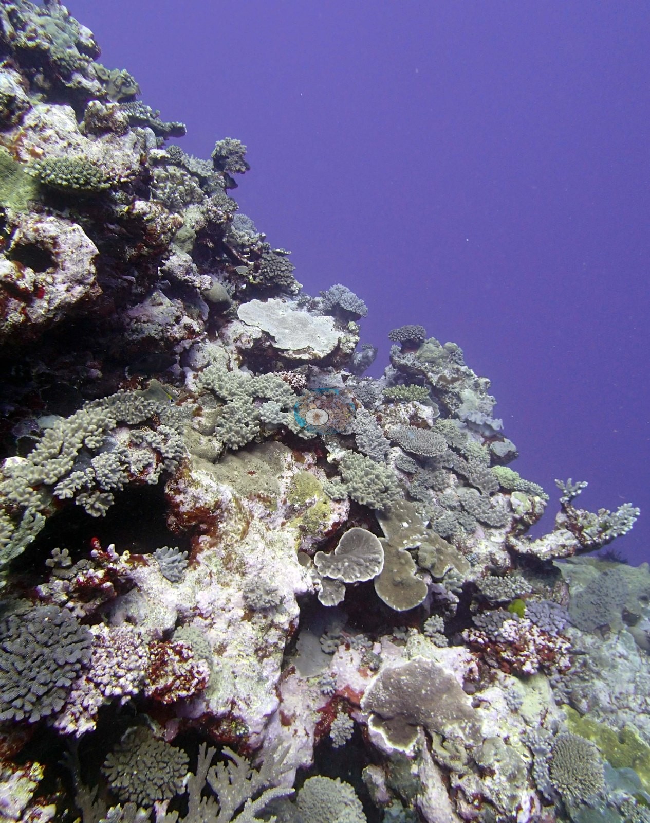
Two color elements recur. Mustard-yellow to chartreuse encrusting and short-branching growth appears in several frames on the terrace (e.g., Image 37, Image 14, Image 13, Image 25), and low gray and gray-green lobate and mushroom-form soft corals with tan lobed capitula are scattered through the reef top (e.g., Image 37, Image 2, Image 32, Image 26).
The photographs cannot separate the yellow growth from a hydrocoral (Millepora) versus a yellow Porites or encrusting Montipora, and this station's survey notes do record extensive Millepora deeper on the wall — so the identification is left open here rather than asserted.
Coral cover
- Reference: Khaled bin Sultan Living Oceans Foundation, Global Reef Expedition (in situ transect)
- Coral cover 44%, from LOF-GRE in situ transects (the mean of all of this station's own transects in the expedition's per-transect benthic data) — a diver transect measurement, not a photographic or computer vision estimate.
- Almost all frames are shot obliquely upward from below the crest, so they exaggerate the vertical profile of the reef and are not planar views — none of them can be used to derive a cover figure, so the computer-vision rows below are a methods comparison, not a cover estimate.
- Transect mean 44 ± 5.7% SE (n = 6 transects; Living Oceans Foundation per-transect benthic data); the headline above is this all-transect mean.
Stressors
No bleaching is visible in any frame. What the frames do not settle: no frame shows an obviously bleached, stark-white colony, and no recent mortality scar large enough to read is visible, but many colonies photograph pale gray-lilac under flat ambient light and that is a color-rendering effect, not a health call. Dead, turf-covered branch sections inside the staghorn thickets (e.g., Image 7, Image 28) indicate normal background branch loss, not an event. A large table, which Anderson identified as an acroporid, is healthy but carries about a dozen small white spots, possibly feeding scars; no corallivore is visible (e.g., Image 34). The Living Oceans Foundation transects here recorded 2.3% cyanobacteria and no recently dead coral; they carry no bleaching, disease, or crown-of-thorns field.
Fish and other fauna
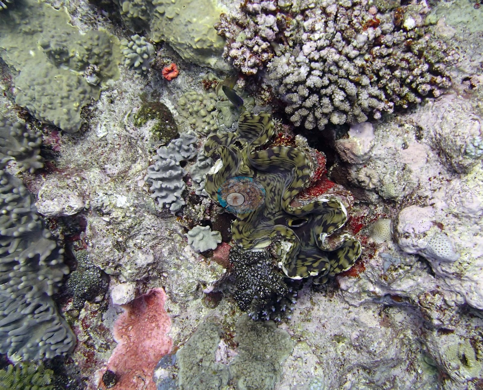
The detail shot is the best single record here: a giant clam (Tridacna) with a boldly mottled olive and black zigzag mantle wedged into the framework, a second dark speckled clam mantle just below it, salmon-pink encrusting sponge, an orange encrusting invertebrate, ruffled gray plates, and small blue-gray soft corals — a crowded, healthy-looking cryptic community (e.g., Image 1).
Fish are conspicuous in most frames: loose to dense clouds of dark, white-tailed damselfish and small planktivores hang over the crest in the majority of the wide shots (e.g., Image 21, Image 14, Image 13, Image 12, Image 9, Image 8, Image 4, Image 30, Image 6, Image 25, Image 17, Image 29), with the heaviest aggregations in two frames where Anderson noted large numbers of fish over the reef (e.g., Image 29, Image 6).
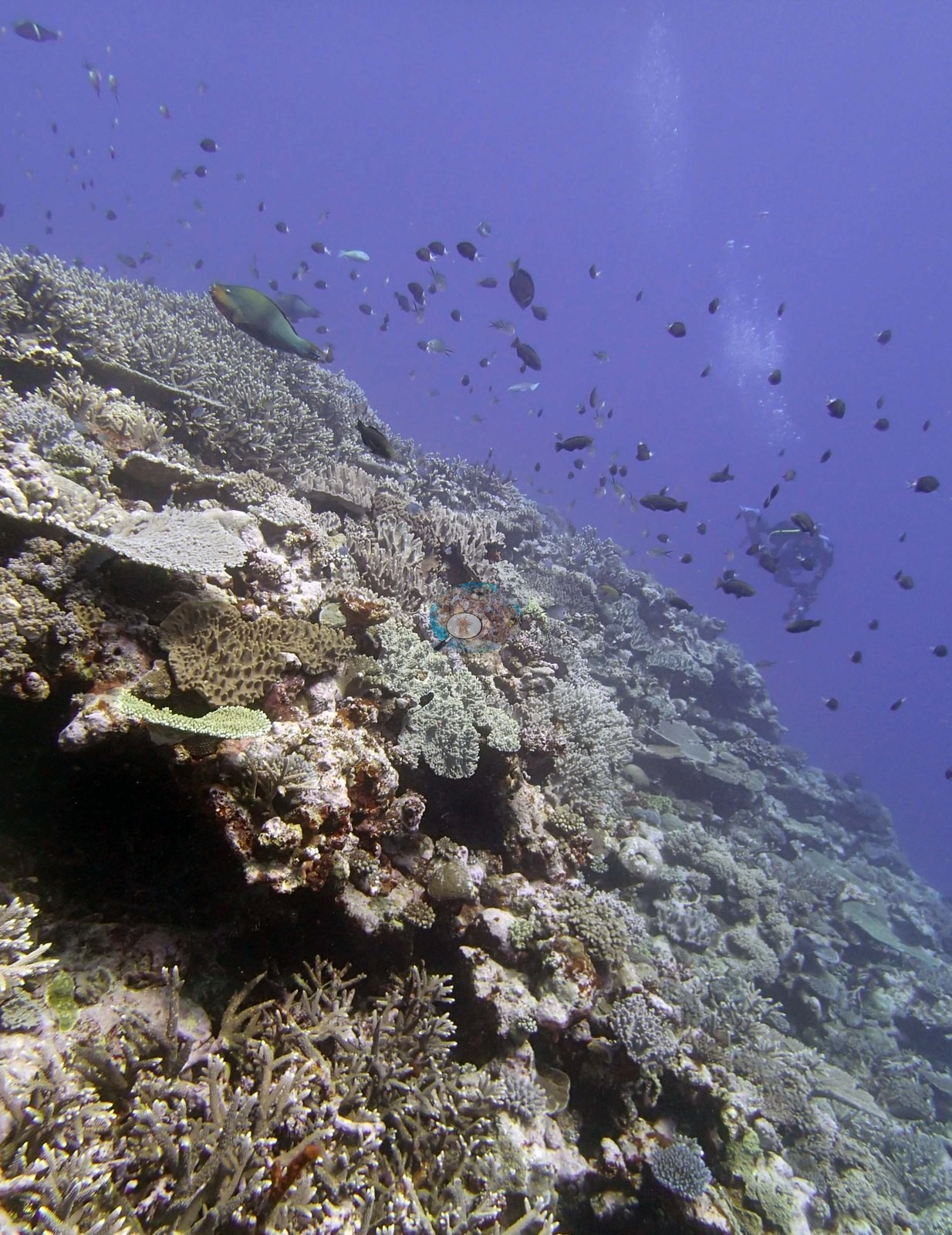
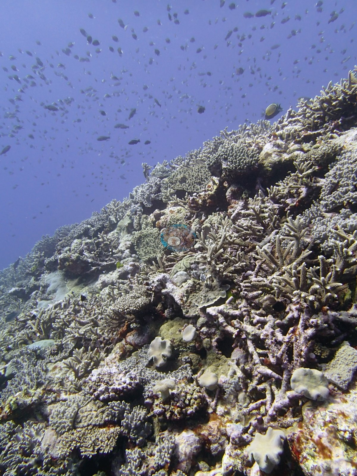
Larger-bodied fish (surgeonfish- and wrasse-sized silhouettes) pass over the reef in a few frames (e.g., Image 5, Image 25).
Comparisons with other data
Apart from the Living Oceans Foundation survey shown here, we found no other surveys at this site as of 2026. If you would like to share yours, please email us.
Other notes
This is not a sampled selection: almost every frame is a wide reef-scale view of the same shallow terrace and crest, shot upward or along the slope into blue water, and the set is remarkably consistent from frame to frame — a continuously built reef surface with very little exposed pavement or rubble anywhere in it. Anderson's field verdict on the site was very high coral cover on an exceptionally beautiful reef (e.g., Image 21, Image 31, Image 19, Image 17, Image 18, Image 28).
Four frames are overexposed or backlit: Image 35, the strongly backlit fish silhouette (e.g., Image 29), and the surface-glare frames over the crest (e.g., Image 7, Image 5). Depth is not resolvable from the imagery; all of it reads as shallow terrace and crest rather than the deeper wall described in the survey notes.
Survey team's site notes: Reef has a shallow terrace that slopes gradually from 4-8 m over about 100 m, then a steep slope from 10 m to 25 m, then a more gradual slope. At 5 m the dominant corals are staghorn thickets 1-5 m wide, digitate acroporids, small to medium table acroporids, Pocillopora, and numerous small to medium massives. The wall also had large areas of Millepora that was 20-40% cover at 15 m declining to 10% at 20-25 m. Coral cover dropped significantly below 15 m to about 10% dominated by small to medium Acropora, Pocillopora, with larger plates of Mycedium, Echinophyllia, and Montipora (50-200 cm), submassive Porites lobata (up to 1 m across).
CV model comparison
More info
The photo row below is a methods comparison only: it is never averaged with the transect and never used as the headline. It compares site means only: the photos come from the same survey dive as the transect but not from the same points.| Method | Hard coral cover (%) | Photos (n) ± SE |
|---|---|---|
| Transect mean (all transects) | 44.0 | |
| CoralSCOP unedited | 33.1 | n = 38 · SE 2.0 |
| Claude (edited photos) | 36.2 | n = 38 · SE 3.1 |
The two computer-vision methods agree within 3.1 pp (33.1–36.2%) and read 7.8–10.9 pp below the in-water transect (44.0%); the headline is the in-water transect because a diver transect is independent of the photographs.
See a more detailed, stand-alone version of this table.
Data & links
- Download benthic survey data (CSV) ↗
- Download all LOF-GRE data (933 sites) ↗
- Photo portfolio ↗
- Coral Reef Diagnostics region page ↗
All images from Coral Reef Diagnostics. Data are from the Khaled bin Sultan Living Oceans Foundation Global Reef Expedition and Coral Reef Diagnostics.
 © Coral Reef Diagnostics.