
Part of the regional summaries: Tonga - Ha'apai and Tonga

More info
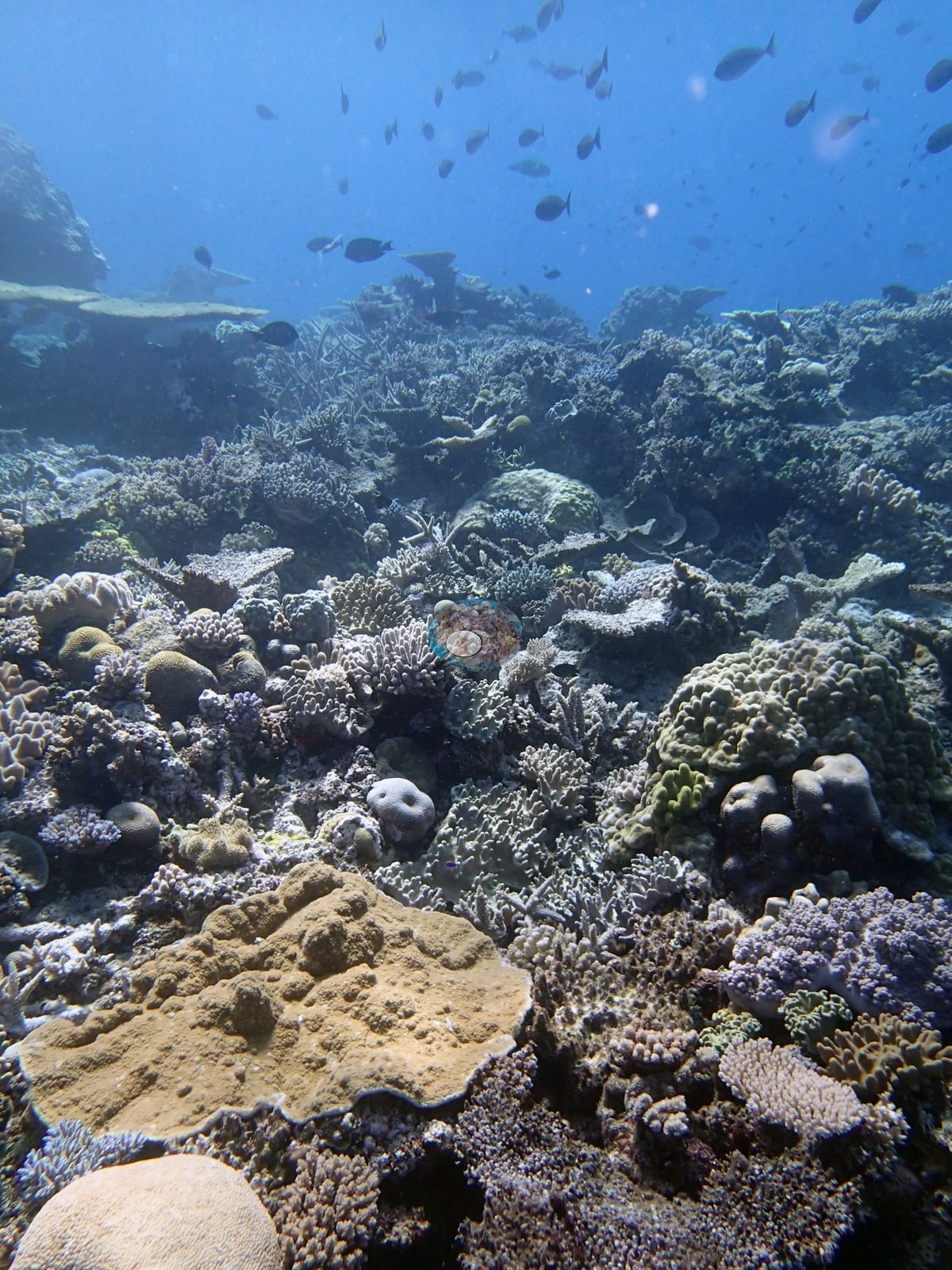
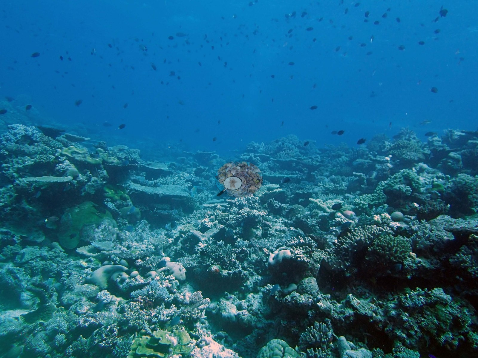
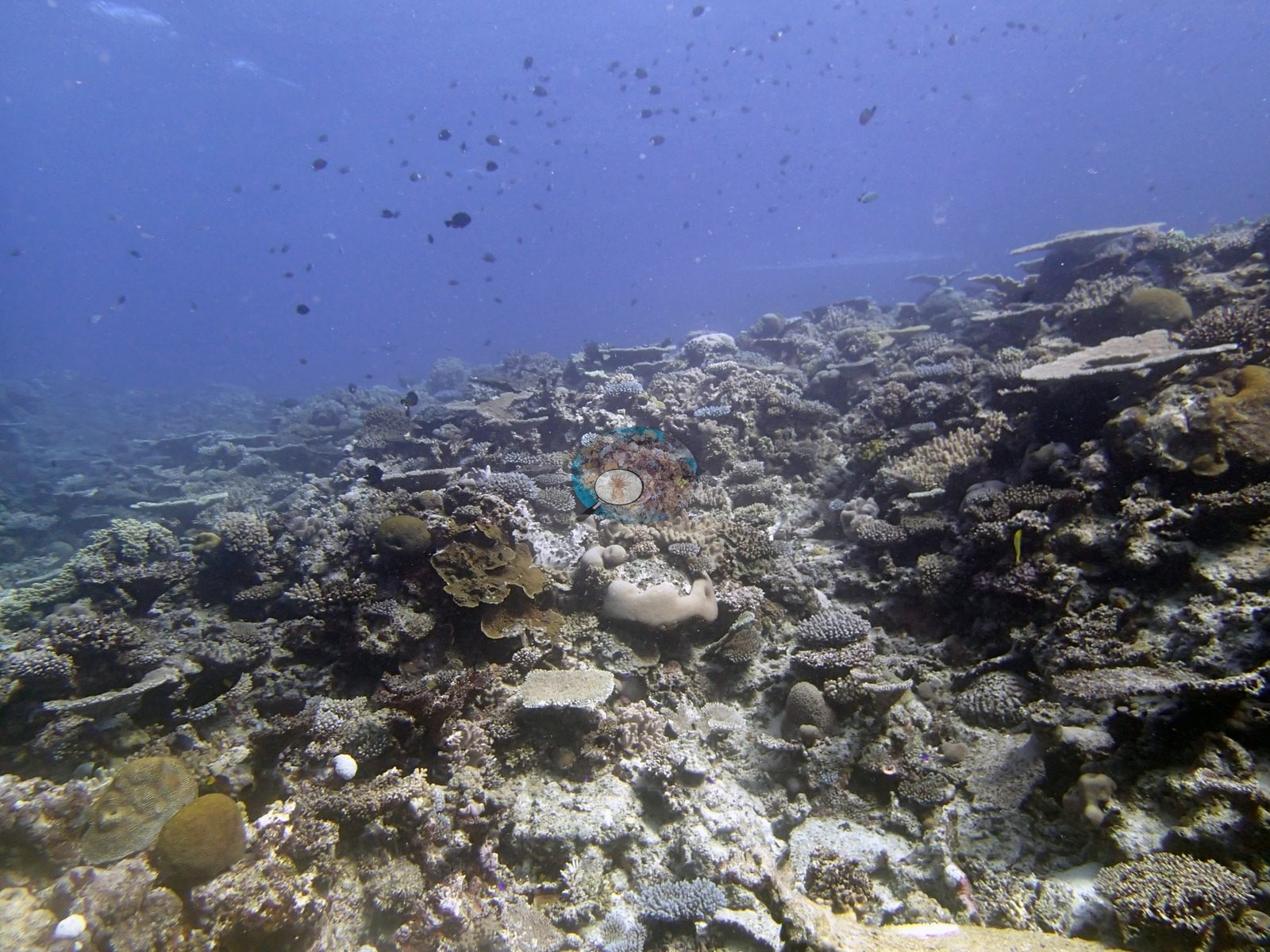
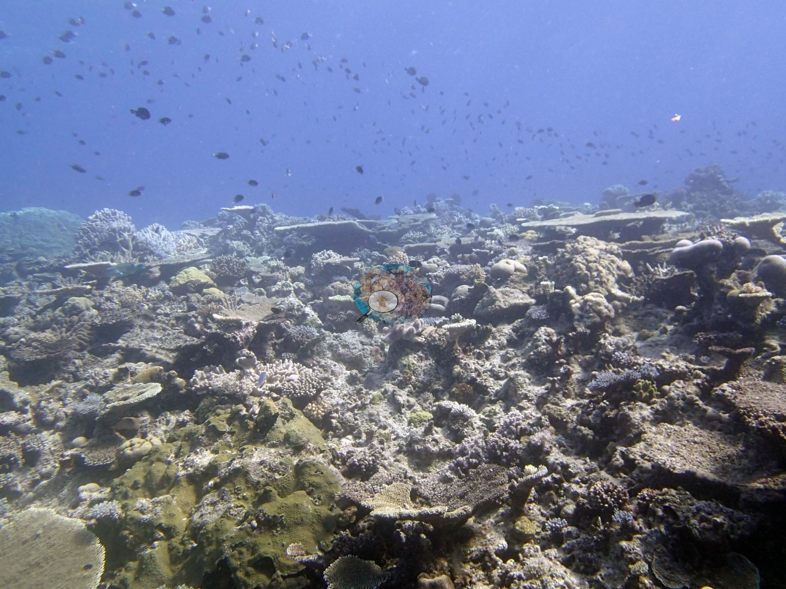
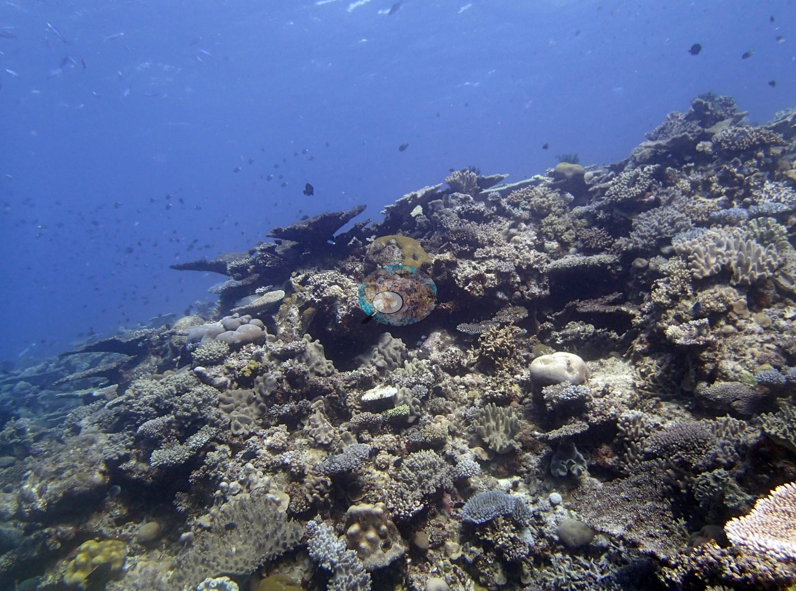
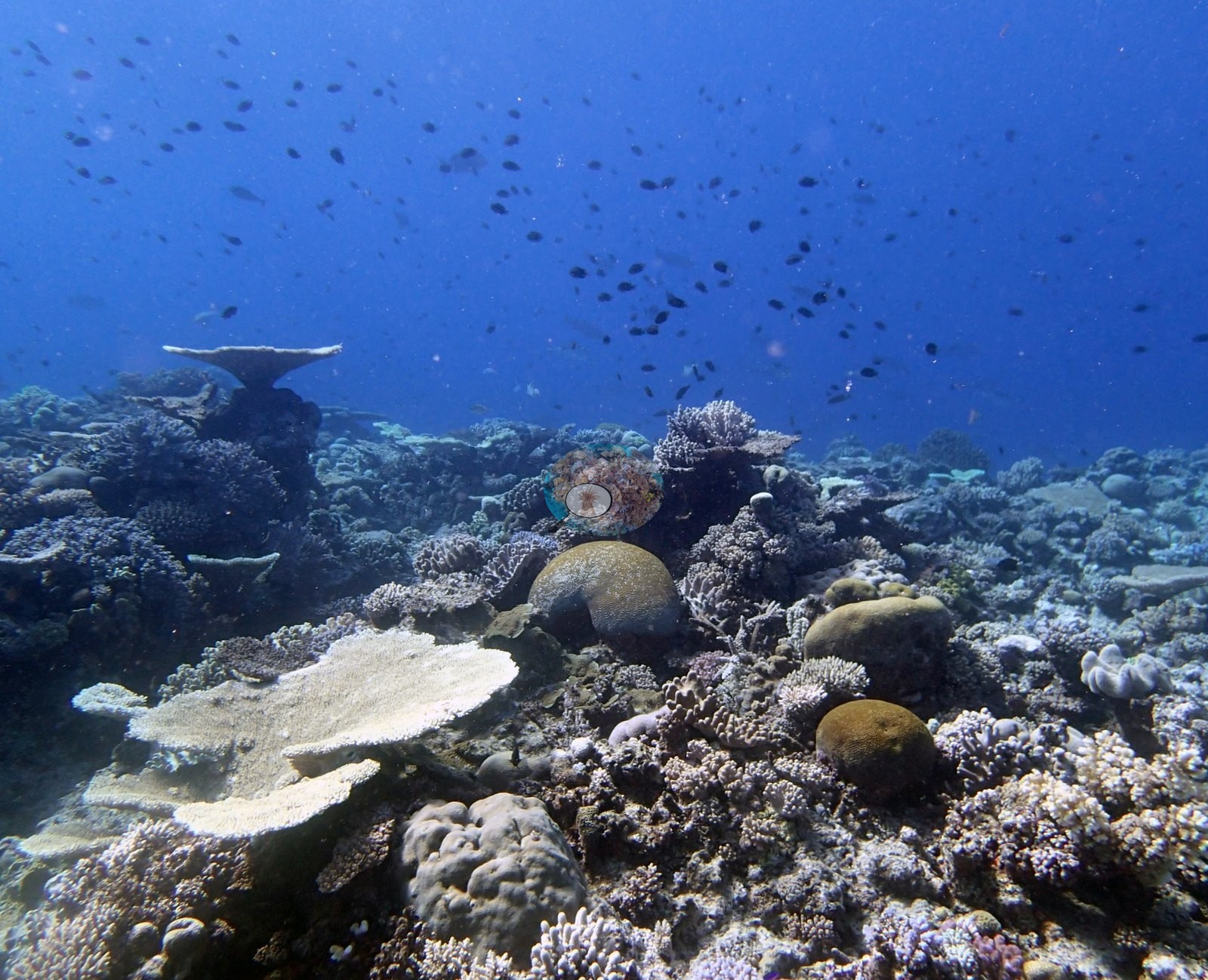
Southern Tonga (NOAA CRW virtual station)A gently rising platform of densely packed table, staghorn, and massive corals that Anderson described as a spectacular coral garden, running out to a coral wall and pillar with clouds of fish throughout. Stark white colonies appear in two of the photographs, and one of the three colonies Anderson sampled here was 20-30% bleached. Surveyed on the Living Oceans Foundation's Global Reef Expedition.
Ko ha tuʻunga ʻoku hake māmālie ʻo e ngaahi feo fakatēpile, staghorn, mo e feo lalahi (massive) ʻoku fefuʻifuʻi, ʻa ia naʻe ui ʻe Anderson ko ha ngoue feo fakaofo (spectacular coral garden), ʻo aʻu atu ki ha holisi feo (coral wall) mo ha pou, mo e ngaahi ʻao ika ʻi he feituʻu kotoa. ʻOku hā ʻa e ngaahi kulupu hinehina ʻaupito ʻi he tā ʻe ua, pea ko e taha ʻo e kulupu ʻe tolu naʻe toʻo sīpinga mei ai ʻe Anderson heni naʻe hinehina ʻaki ʻa e 20-30%.
AI translation of this summary; the English is authoritative. (low confidence)
CTD-cast midpoint of 24.4–24.5°C
Two other CRD dive sites with their own pages within 5 km shown in orange — click a marker to open its page.
34 of the 39 photographs on this page are shown beside the text that discusses them under "Site description" below.
Site description
Setting
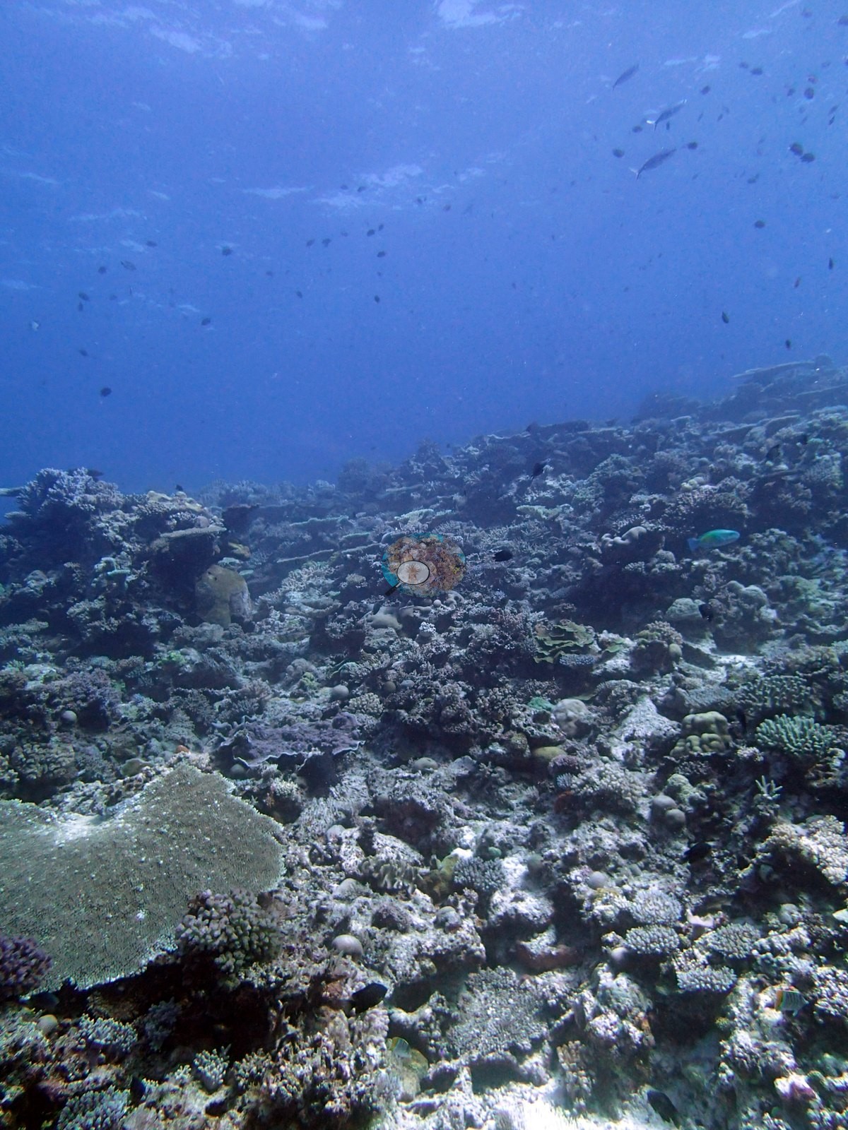
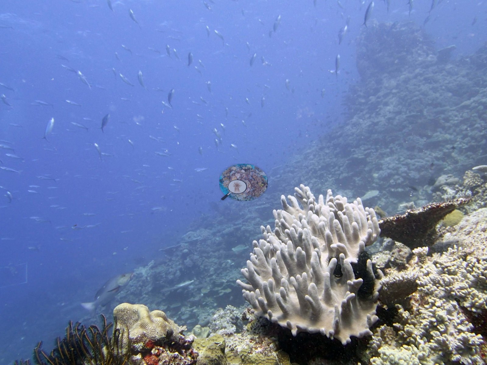
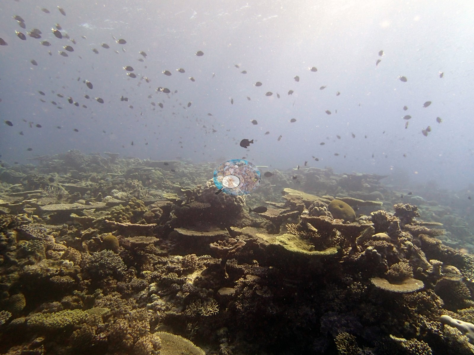
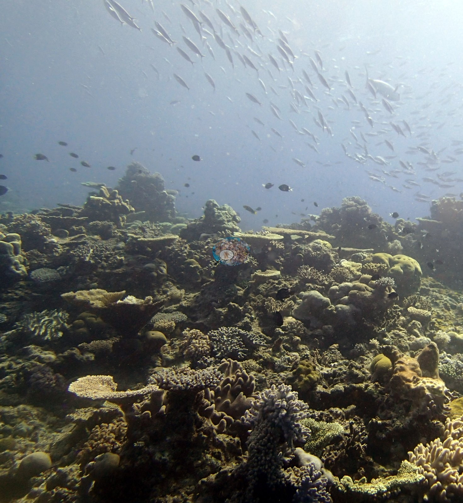
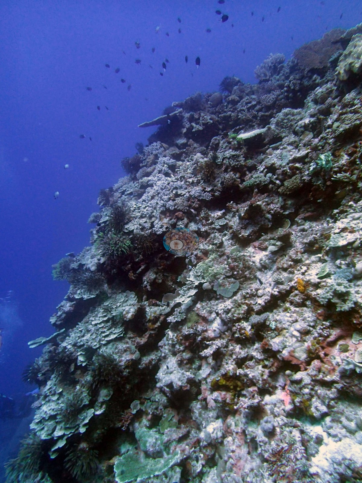
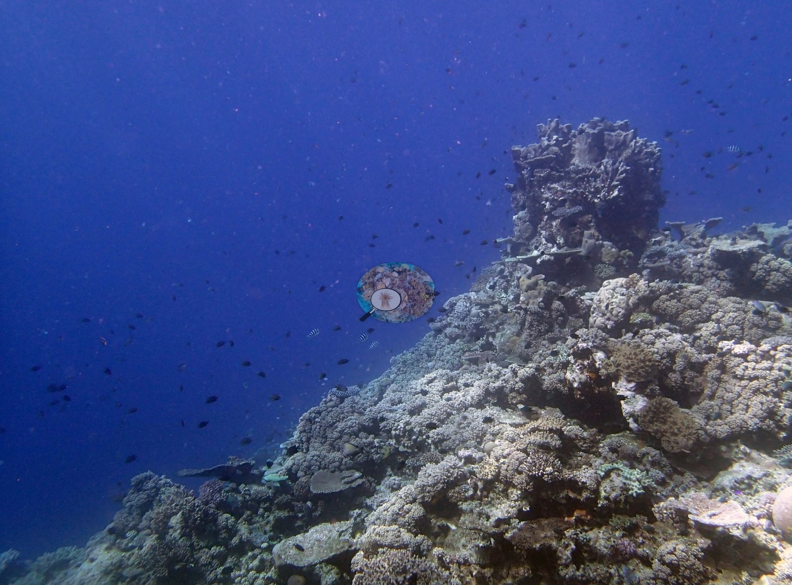
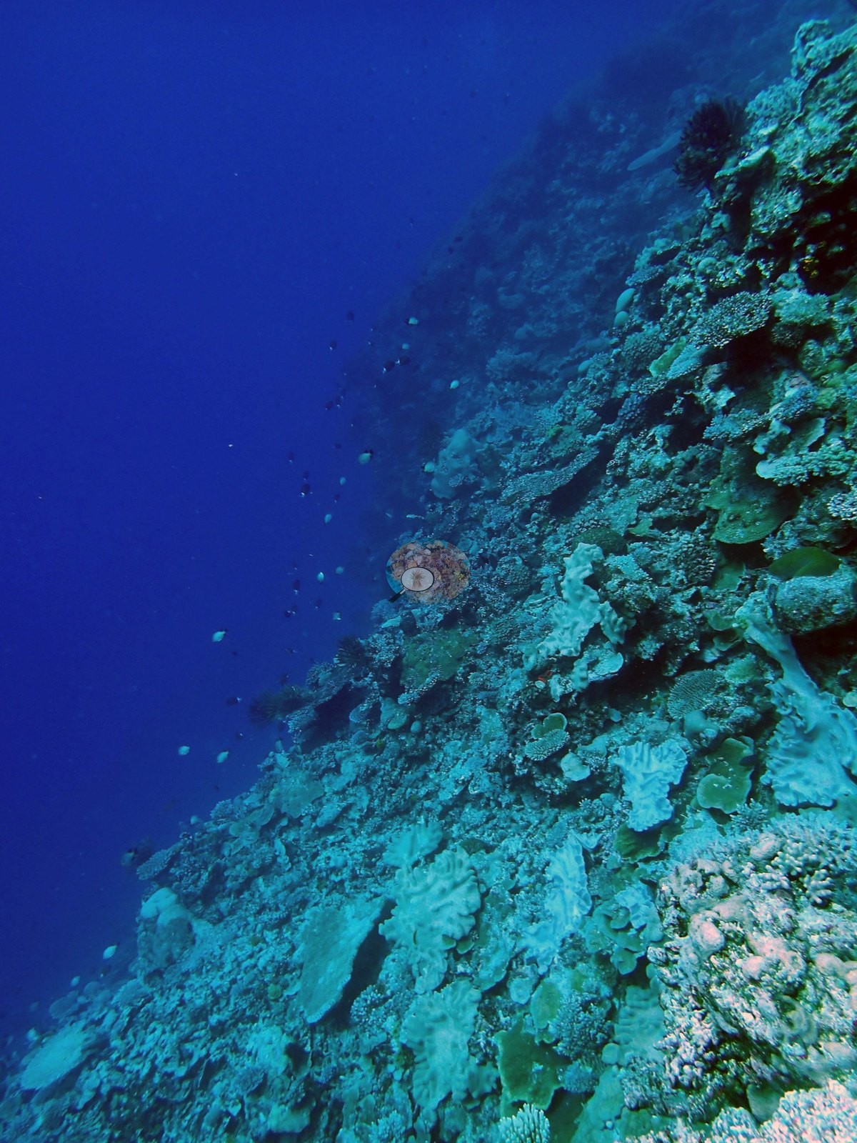
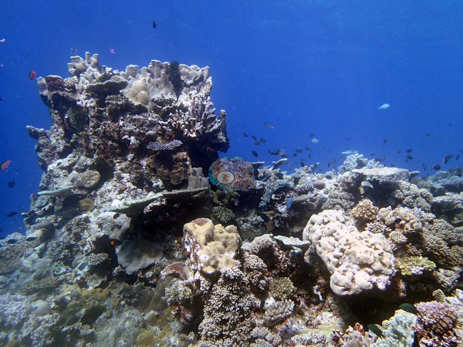
The reef in these photographs is a gently rising platform of coral that runs out to a drop-off: Anderson's notes take the sequence from a coral platform (e.g., Image 18) and a spectacular coral garden (e.g., Image 1) across to the approach to the drop-off (e.g., Image 5), down a coral wall (e.g., Image 33) and a coral pillar (e.g., Image 6), then back up the slope (e.g., Image 17).
General benthic features
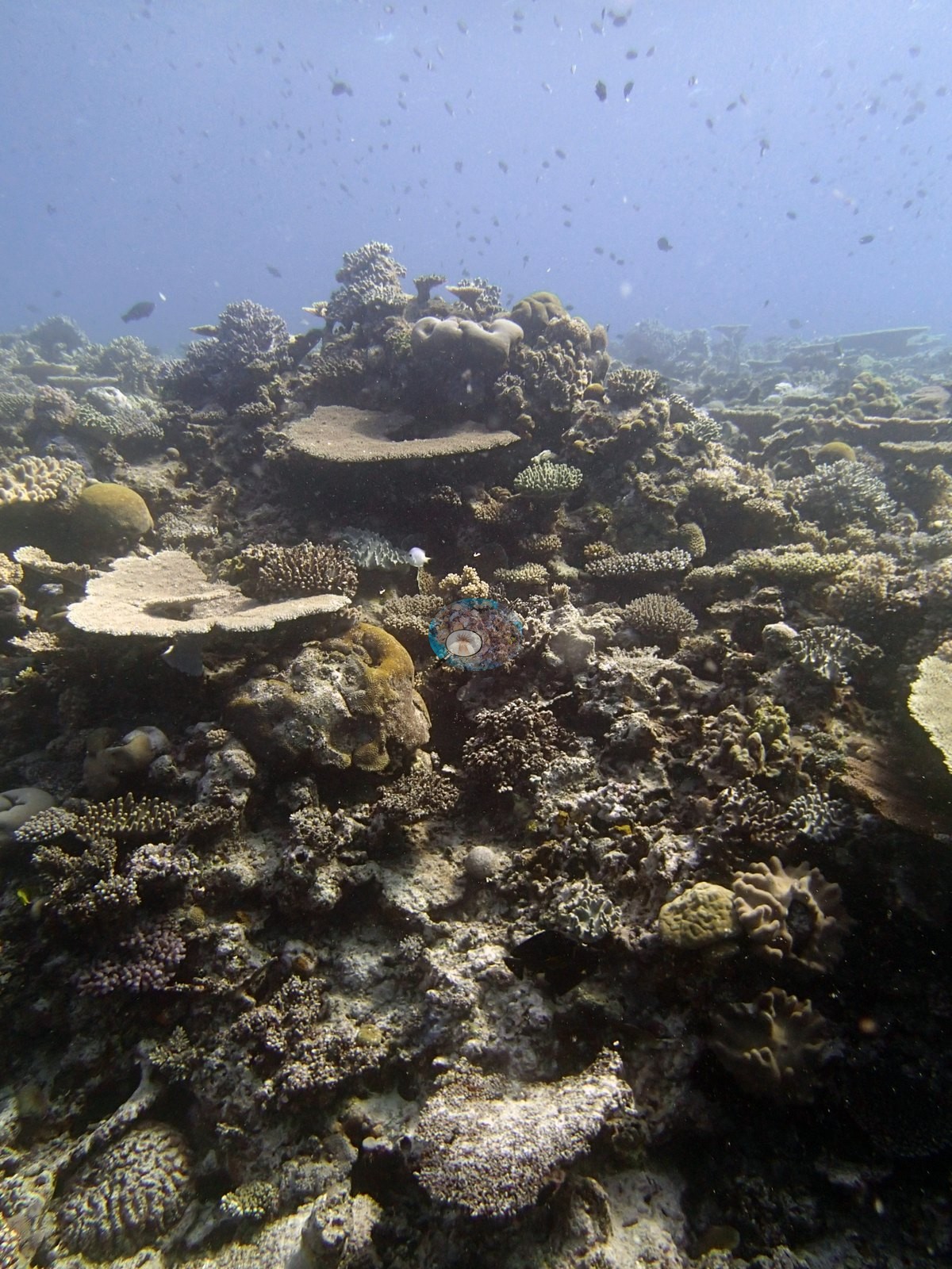
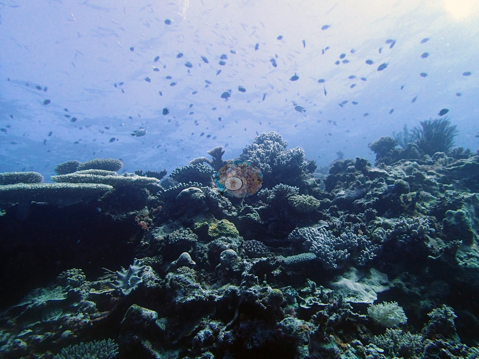
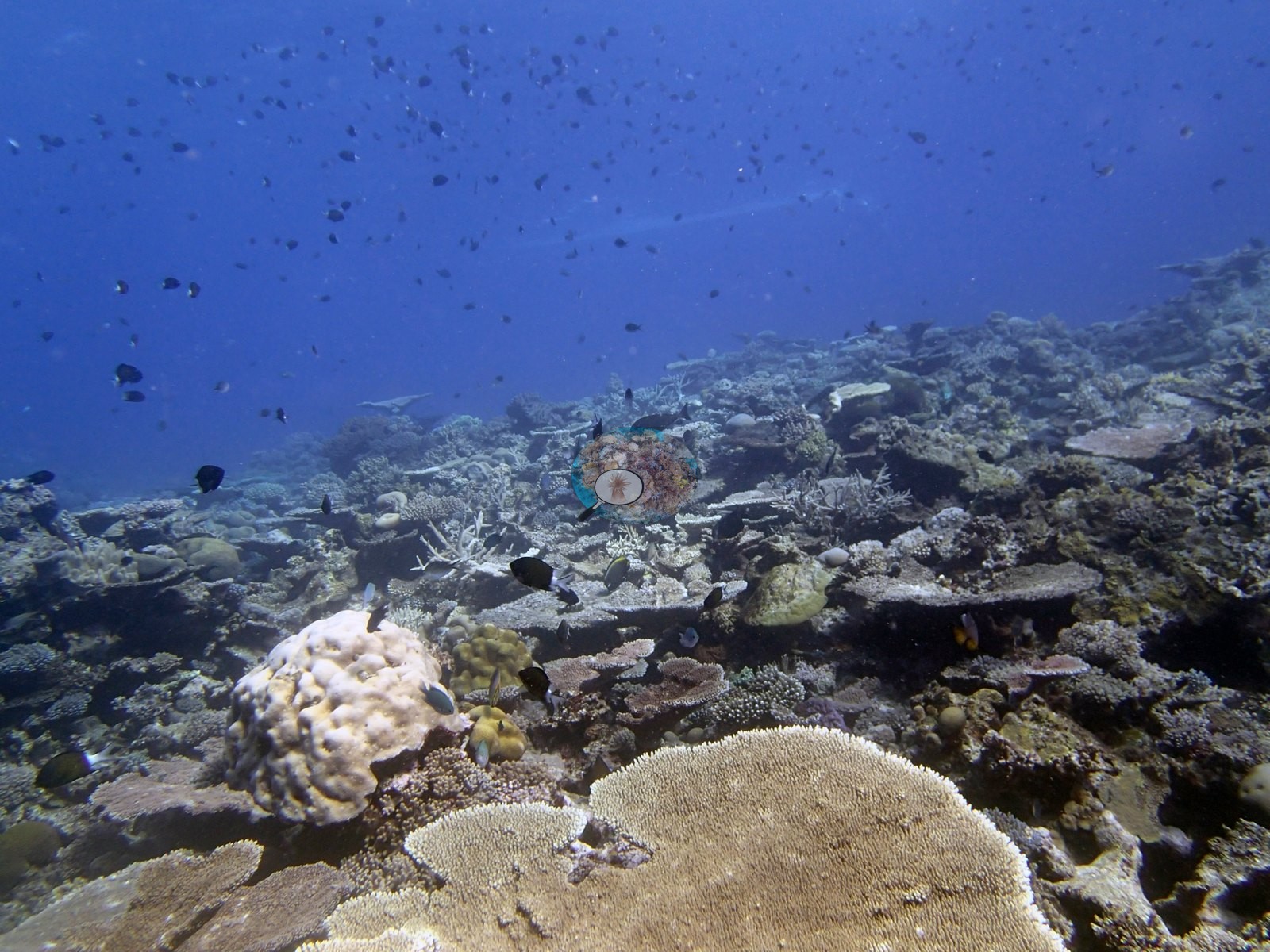
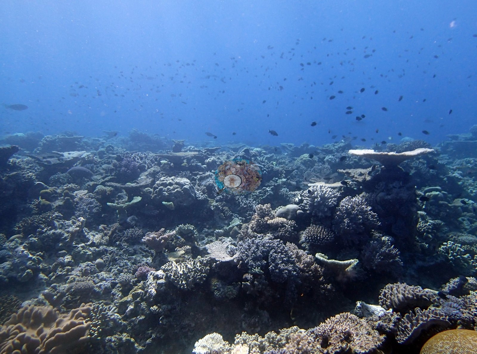
The platform frames (e.g., Image 1, Image 2, Image 18, Image 25, Image 34, Image 38, Image 39, Image 41) show a densely packed, structurally complex assemblage — tabulate and corymbose Acropora, staghorn and robust branching colonies, tan and cream massive Porites domes, plating and foliose colonies, low faviid mounds — with very little bare framework showing between them.
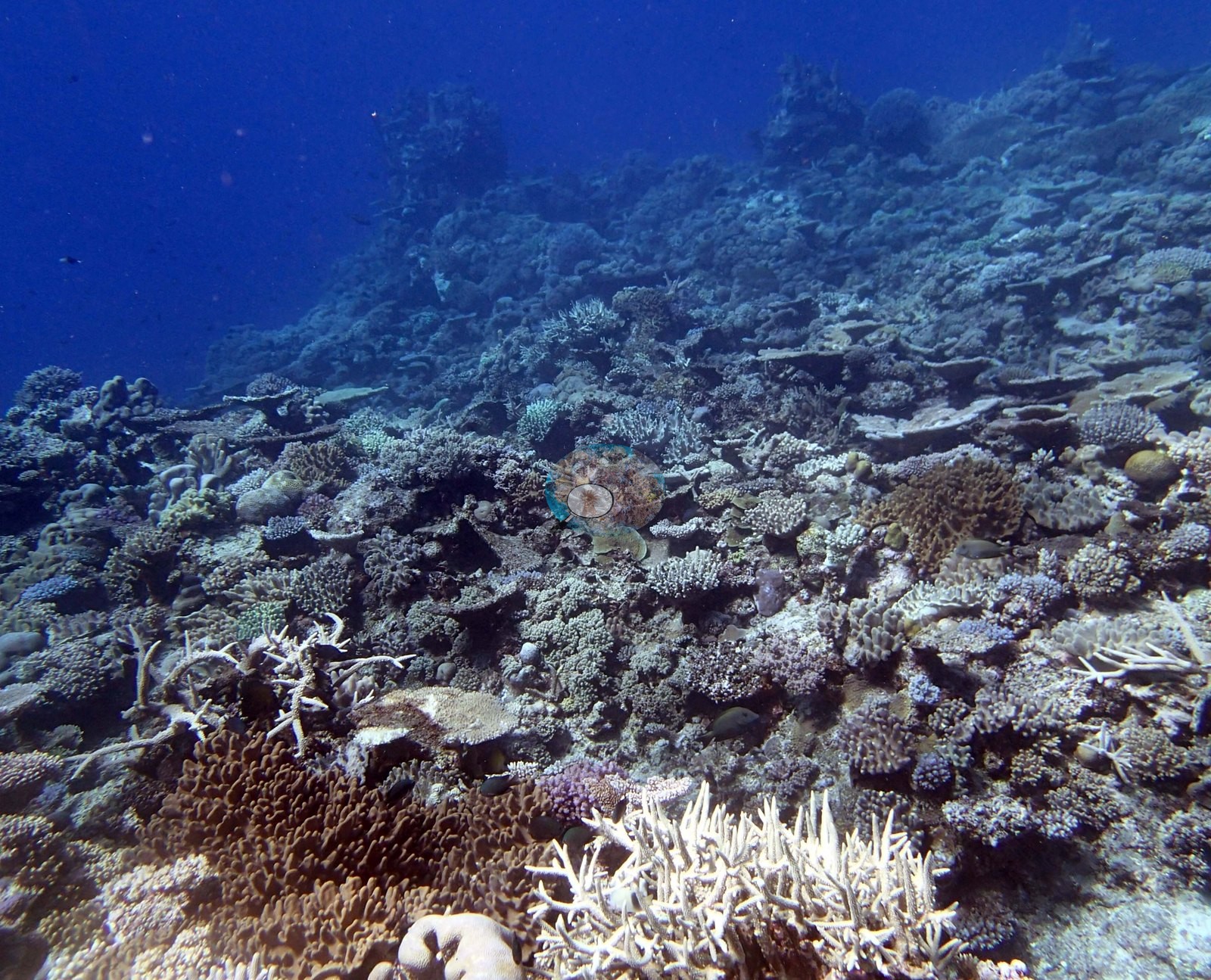
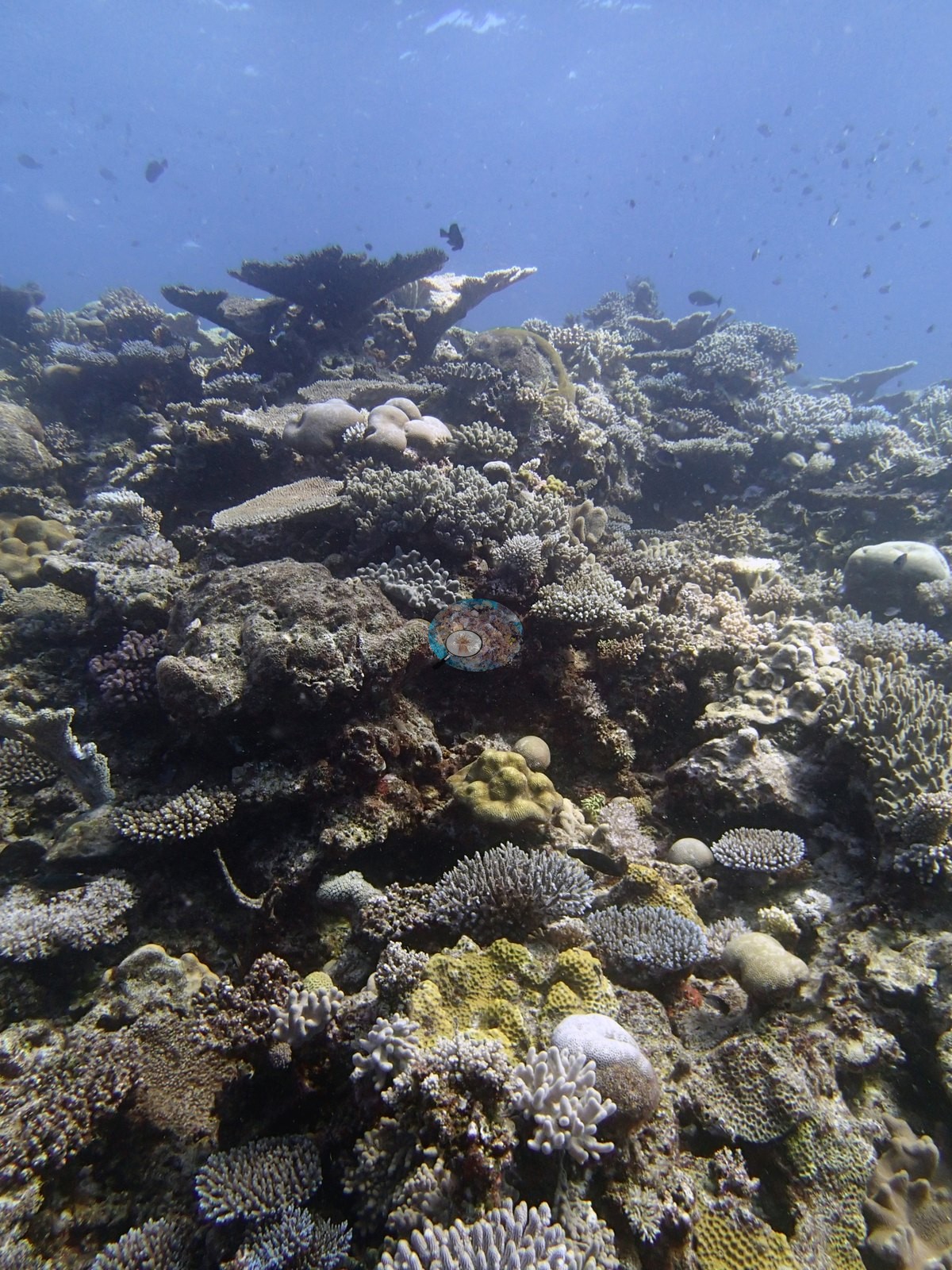
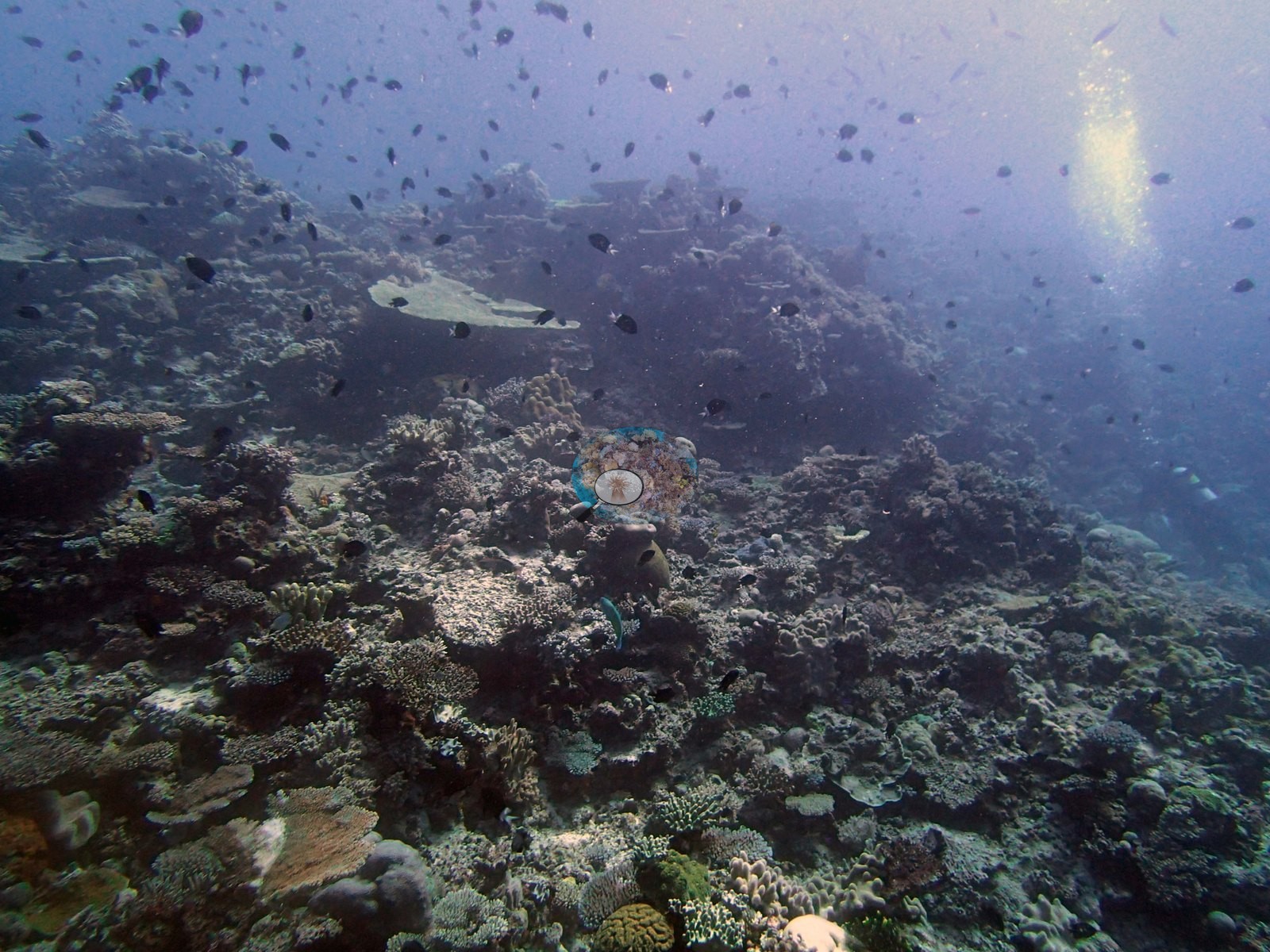
The wall and pillar frames (e.g., Image 6, Image 16, Image 33) show the same community turned vertical, coral to the edge of the drop with blue water beyond.
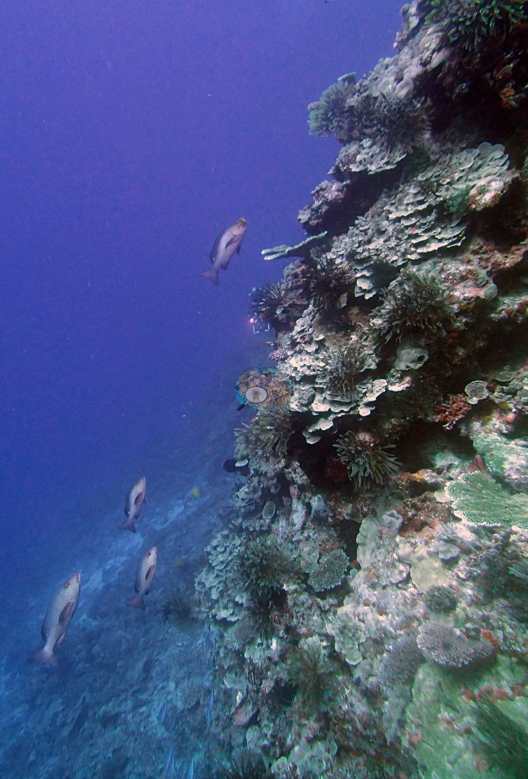
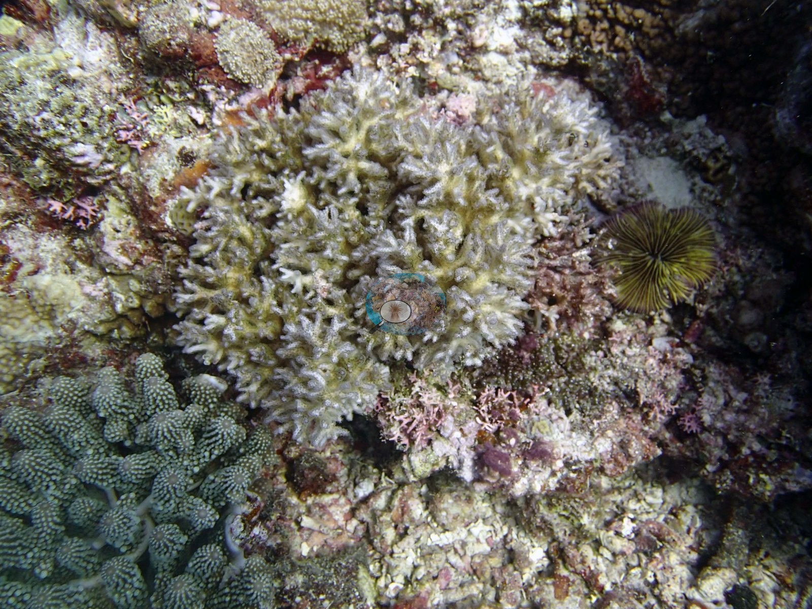
Image 26, a Pocillopora damicornis by Anderson's identification, is one of the close frames: a bushy branching colony with polyps out, a crinoid perched on its edge, and green low-growing colonies and coralline crust below.
Coral cover
- Reference: Khaled bin Sultan Living Oceans Foundation, Global Reef Expedition (in situ transect)
- Coral cover 40%, from LOF-GRE in situ transects (the mean of all of this station's own transects in the expedition's per-transect benthic data) — a diver transect measurement, not a photographic or computer vision estimate.
- Algal cover 46% from the same transect.
- Coral Health Index 2.50/5 (Mayfield et al. 2021, Platax).
- Transect mean 40 ± 2.1% SE (n = 5 transects; Living Oceans Foundation per-transect benthic data); the headline above is this all-transect mean.
Stressors
Stark white colonies appear in Image 2 (a tabulate or branching colony) and Image 37 (a lobed massive colony), and in Image 3 a large table is pale cream.
No percentage is estimated here and none should be inferred — this is a count of frames, not of reef. Nor can these photographs settle the cause. Recent tissue loss is at least as plausible as bleaching: this dive is where the survey recorded high Drupella (corallivorous snail) numbers, white bare branch tips were recorded on a foreground staghorn, and the whole set is blue-shifted enough that color judgment is unsafe. What can be said is that the pattern is not confined to one colony or one frame, and that it sits alongside a great deal of normally pigmented, structurally intact coral in the same frames. No crown-of-thorns seastars are visible anywhere in the set, and no unambiguous disease lesions. The 2013 survey notes for this site record large Drupella snails in high numbers on the reef top at 5-10 m, with large lesions on 5% of the colonies (Khaled bin Sultan Living Oceans Foundation expedition site notes, 2013; regional summary in Bruckner 2014). The Living Oceans Foundation transects here recorded 2.4% cyanobacteria and no recently dead coral.
Fish and other fauna
Image 24 is a fish frame: a tight school of blue planktivores in open water.
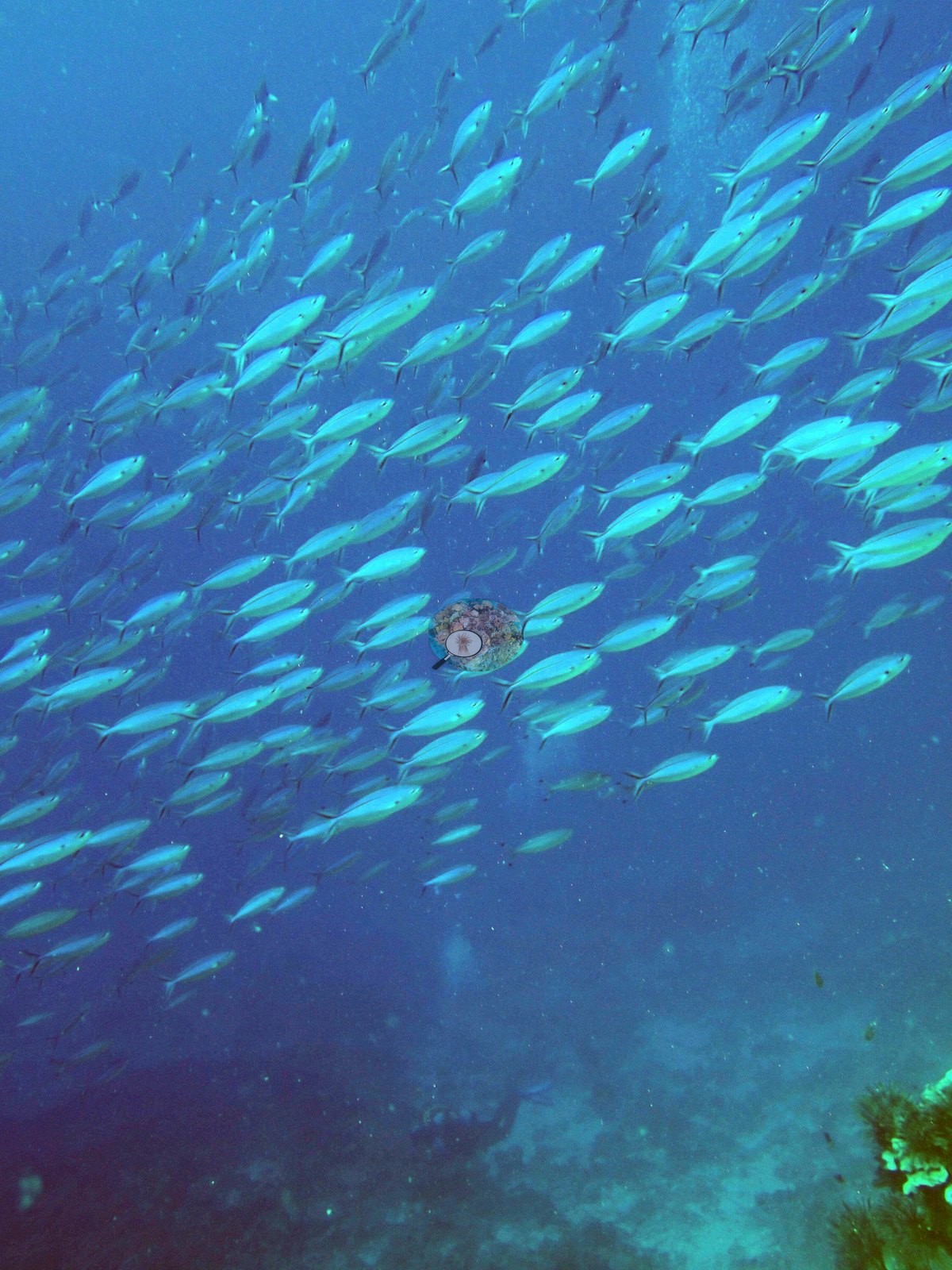
Fish life is conspicuous throughout — Anderson noted abundant, healthy fish life, schooling fish, and fish on the ledge — with dark planktivores hanging in clouds above the coral in Image 38, Image 17, Image 4, Image 15, Image 34, and Image 3, and reef fish working the framework in most others.
Comparisons with other data
Apart from the Living Oceans Foundation survey shown here, we found no other surveys at this site as of 2026. If you would like to share yours, please email us.
Other notes
What the frames cannot settle: depth zonation. Nothing in the set carries a readable gauge, so the platform, wall, and pillar frames cannot be assigned to the depth bands in the survey narrative below.
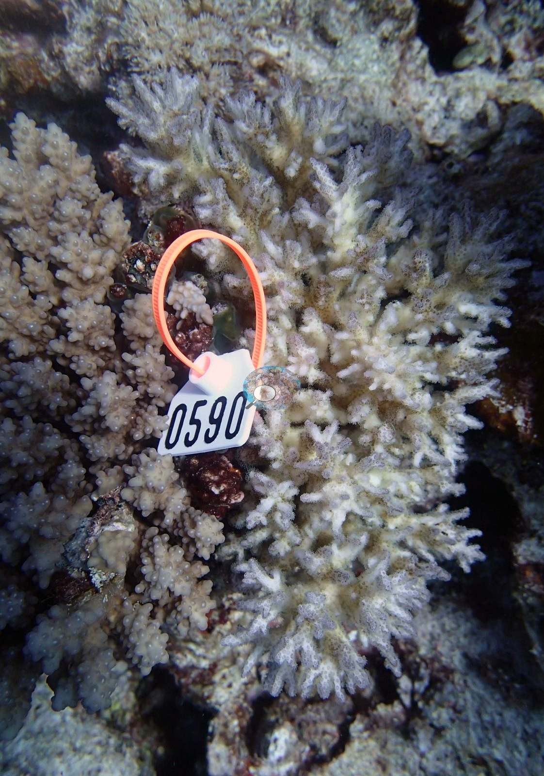
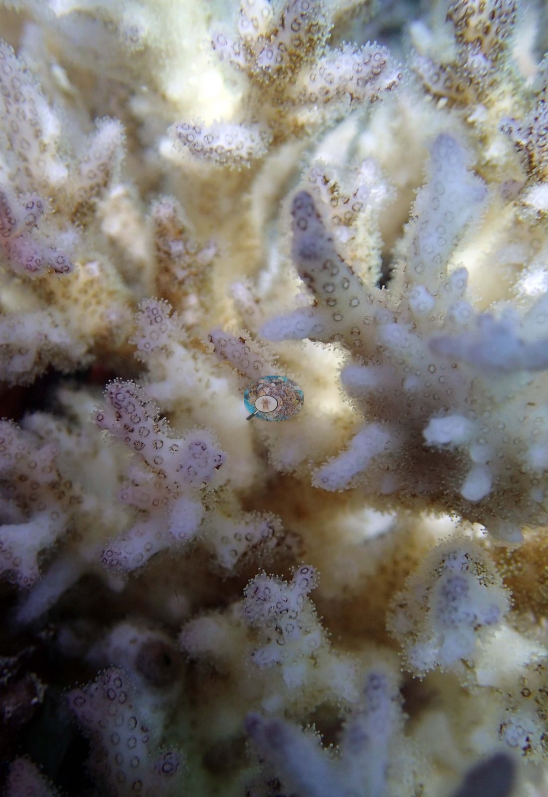
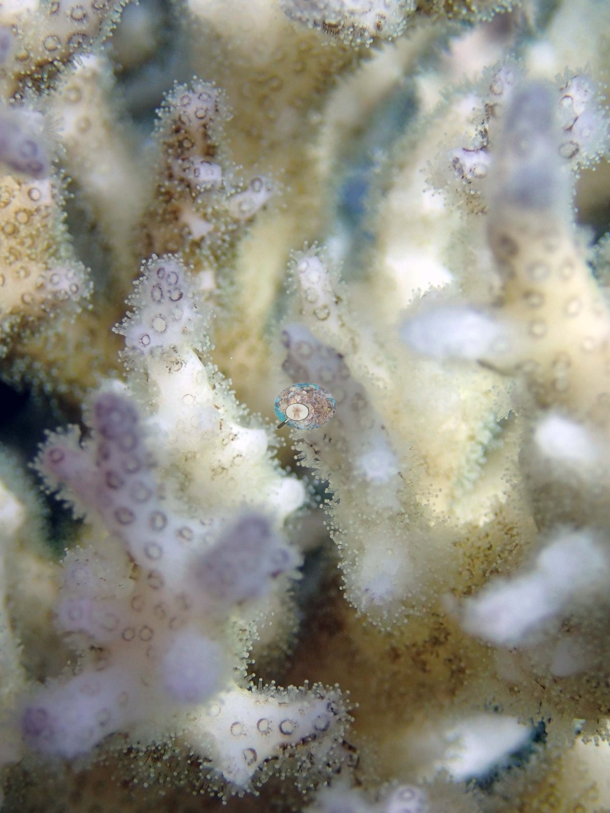
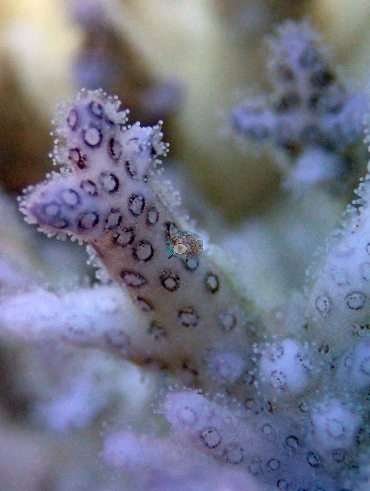
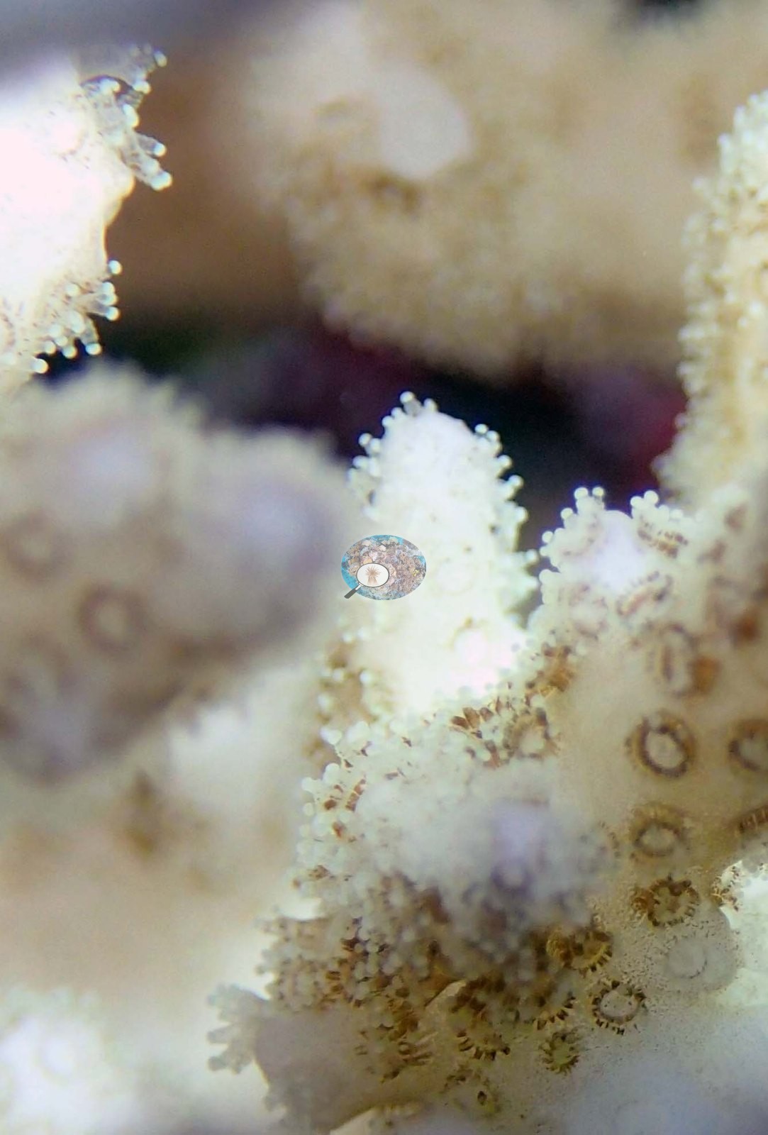
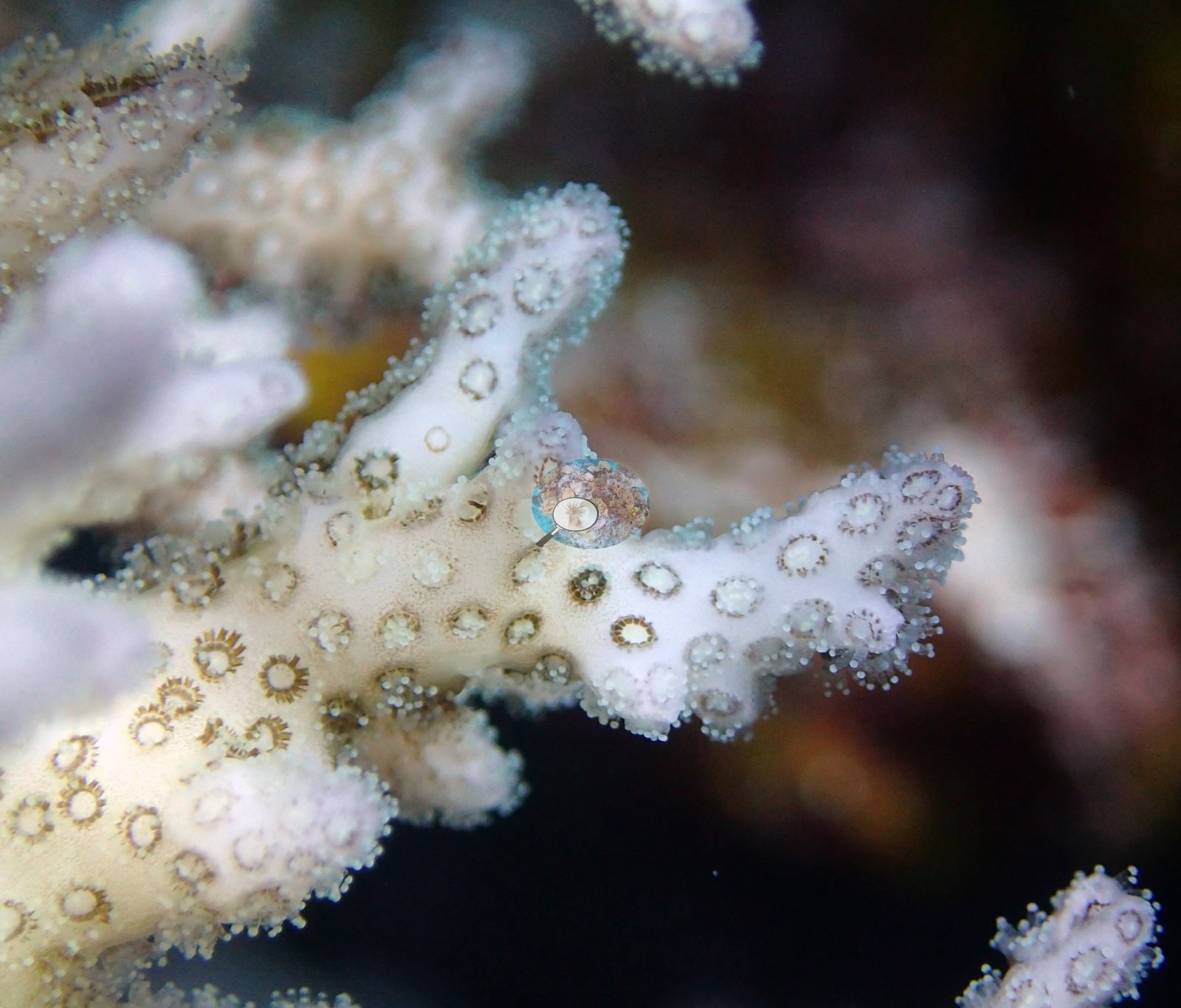
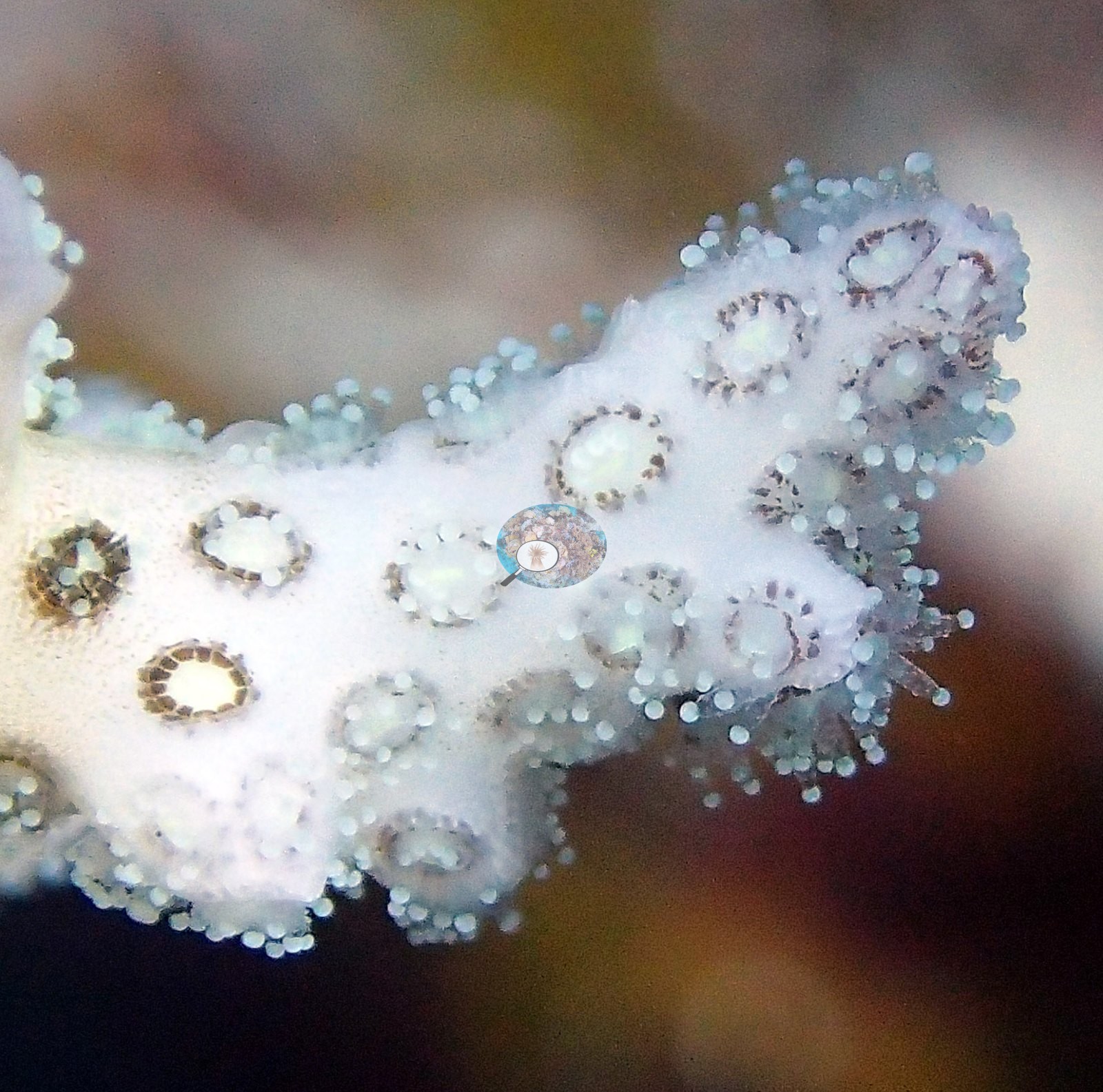
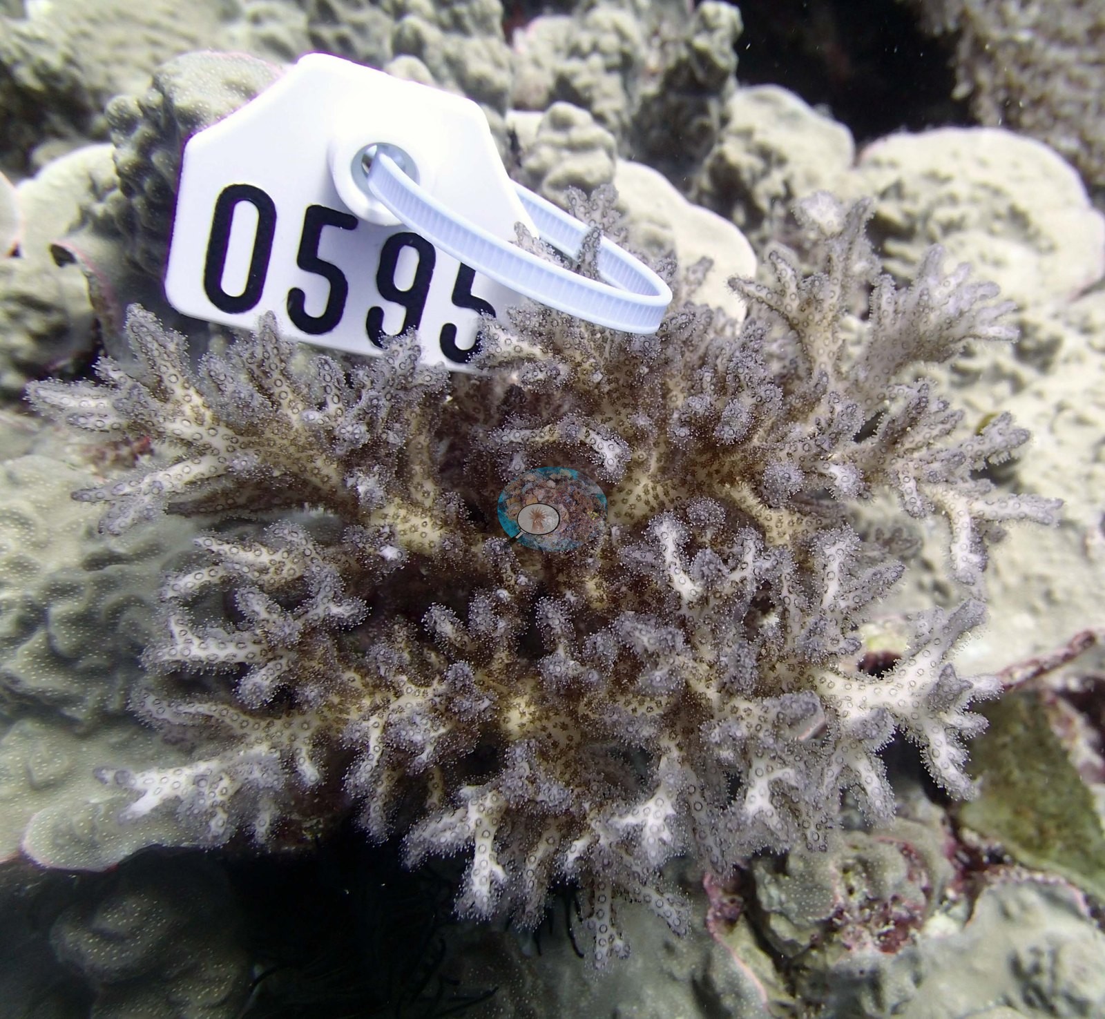
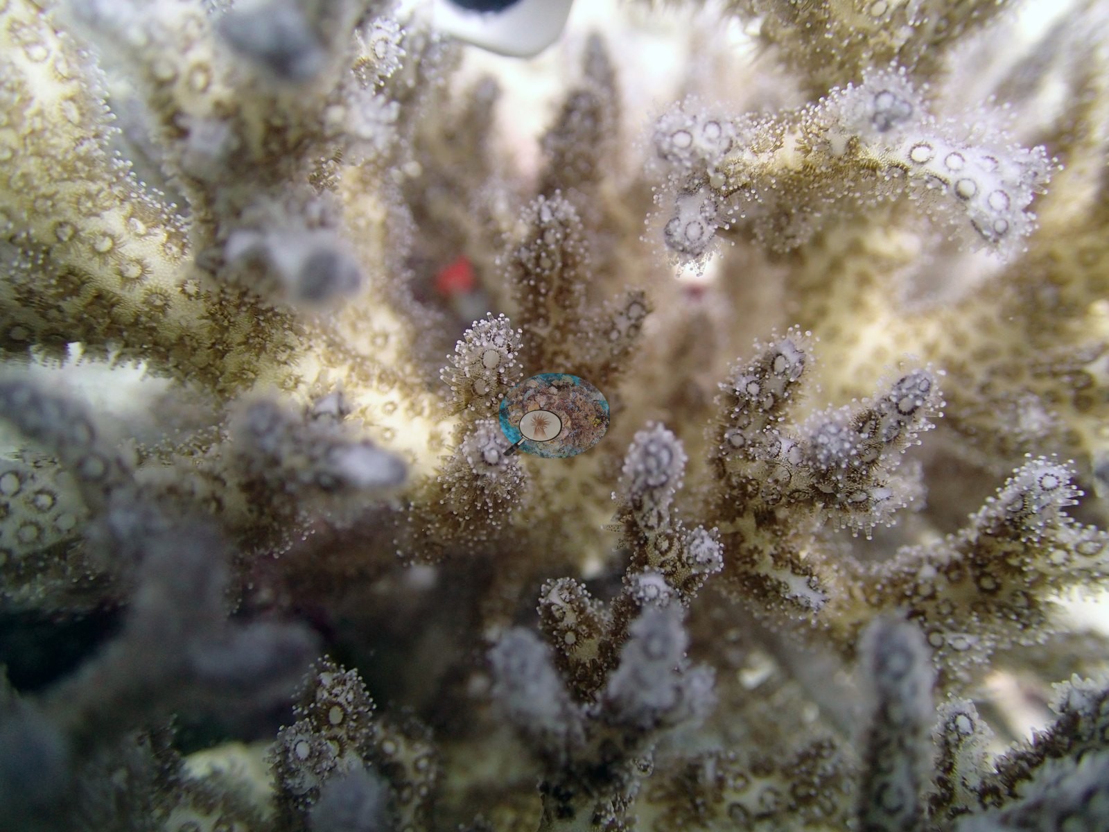
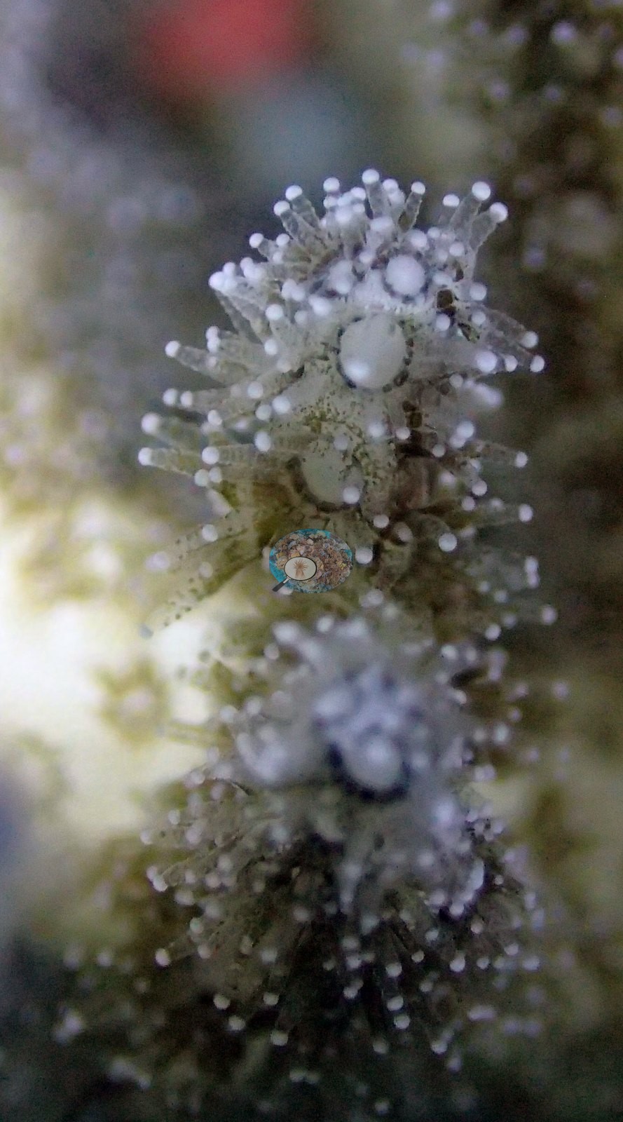
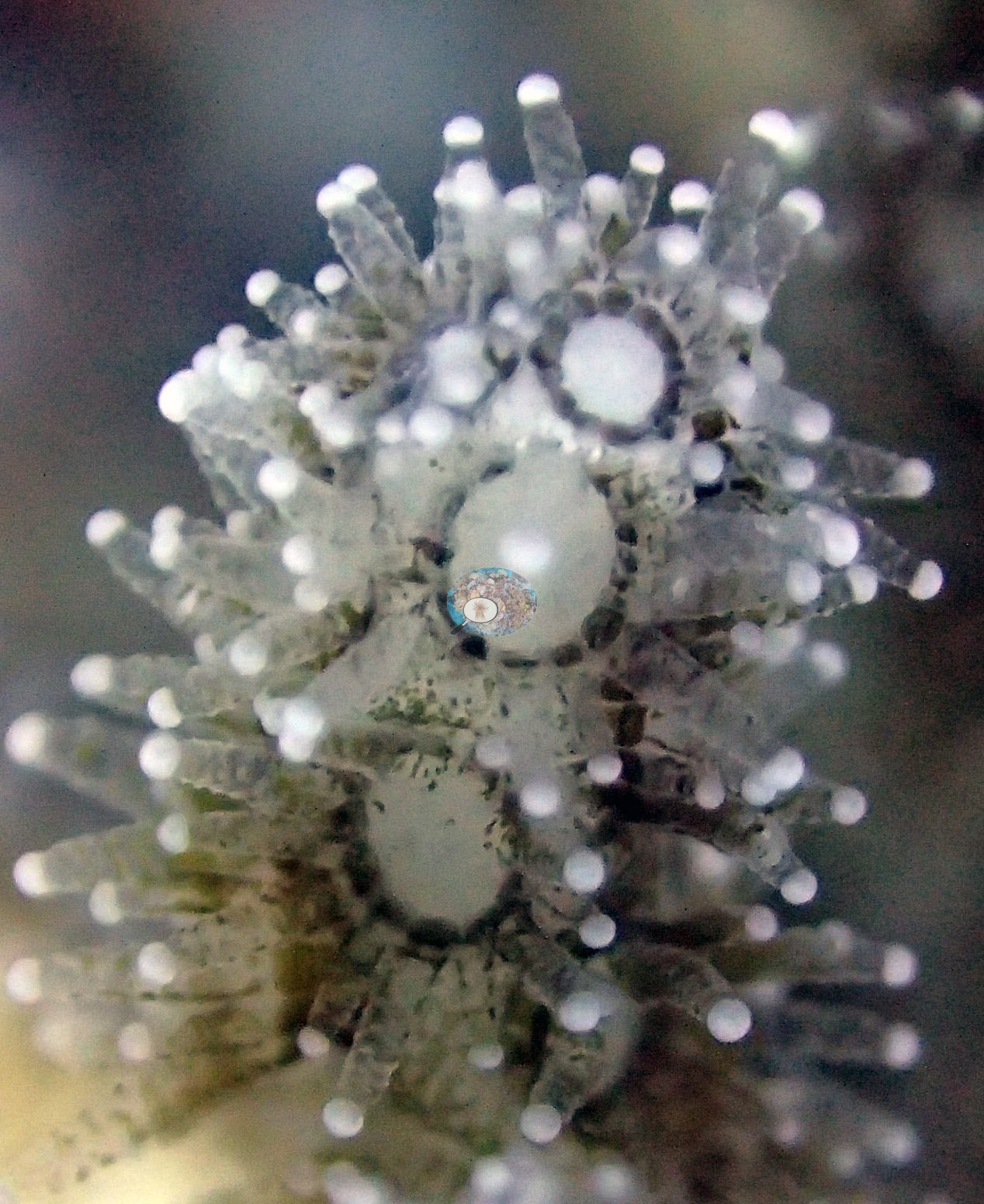
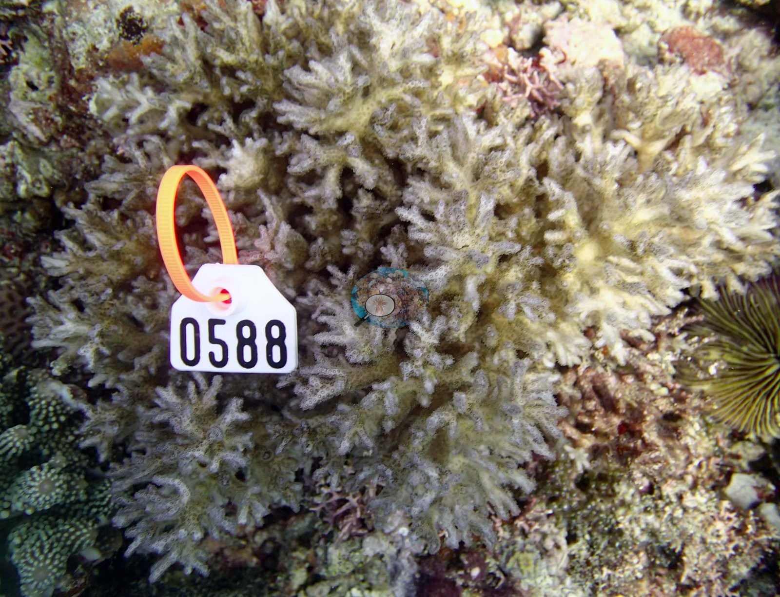
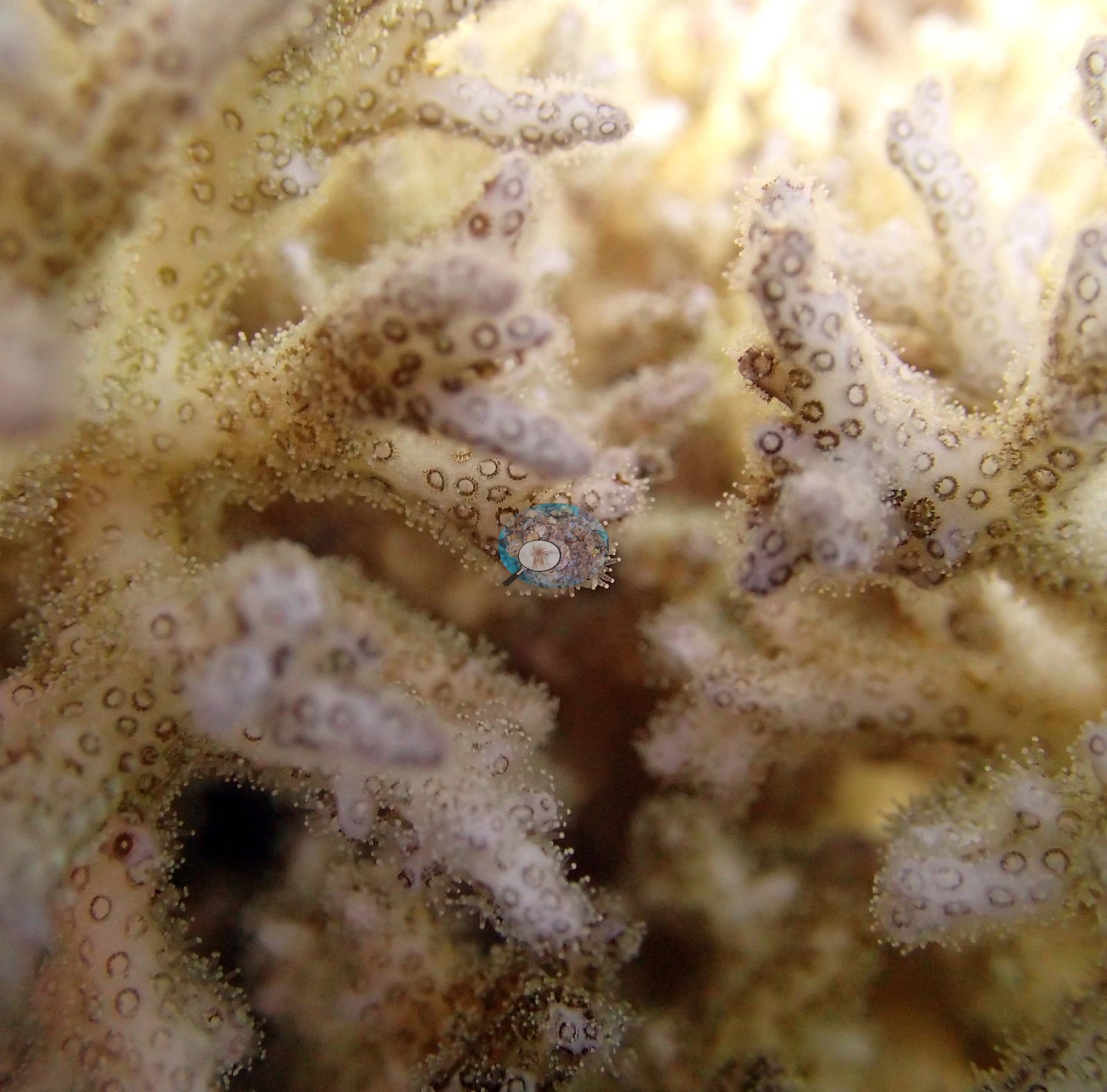
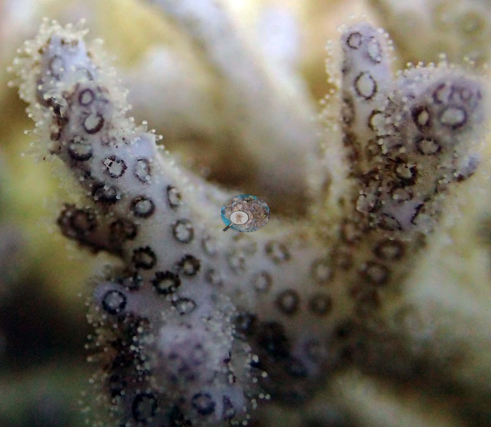
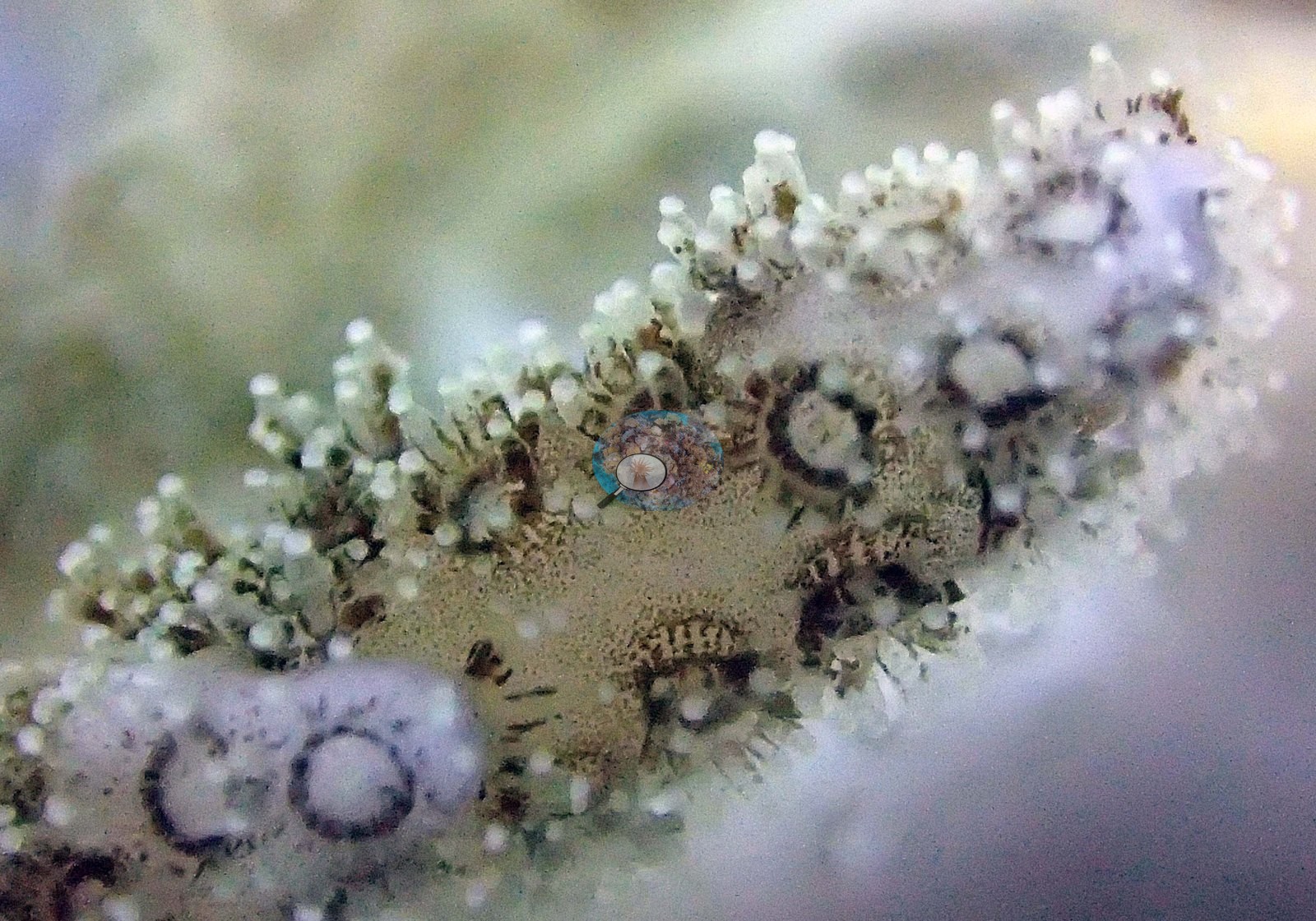
Survey team's site notes: A series of wide, discontinuous spurs then slope very gradually into deeper water. The tabular corals make up 30% of the cover. Very large Porites rus colonies (3-4 m) occur at mid depths (15-20 m) and are intermixed with Lobophora, Echinophyllia, and other species. Cover from 15-25 m is 10-20%, declining to 5.0% at 30 m. Several very large Diploastrea (3 m) occur at 25 m – 30 m. Also numerous larger Montipora plates and crusts cover the bottom. Reef has moderately high CCA in shallow water (30%), which progressively declines with depth. Turf algae is up to 60% cover at 10 m, declining with depth. Soft corals are moderately abundant from 5-10 m (15-20%), declining with depth. There are white lesions on several colonies. Anderson sampled three colonies here (26, 27, and 28, all P. acuta in the photo folders and sheet); the sheet records colony 26 as 20-30% bleached. The colony close-ups are Images 7-13, 19-22, and 28-31.
CV model comparison
More info
The photo row below is a methods comparison only: it is never averaged with the transect and never used as the headline. It compares site means only: the photos come from the same survey dive as the transect but not from the same points.| Method | Hard coral cover (%) | Photos (n) ± SE |
|---|---|---|
| Transect mean (all transects) | 39.7 | |
| CoralSCOP unedited | 25.5 | n = 23 · SE 2.1 |
| Claude (edited photos) | 29.2 | n = 23 · SE 3.0 |
The two computer-vision methods agree within 3.7 pp (25.5–29.2%) and read 10.5–14.2 pp below the in-water transect (39.7%); the headline is the in-water transect because a diver transect is independent of the photographs.
See a more detailed, stand-alone version of this table.
Data & links
- Download benthic survey data (CSV) ↗
- Download all LOF-GRE data (933 sites) ↗
- Photo portfolio ↗
- Coral Reef Diagnostics region page ↗
All images from Coral Reef Diagnostics. Data are from the Khaled bin Sultan Living Oceans Foundation Global Reef Expedition and Coral Reef Diagnostics.
© Coral Reef Diagnostics.
References
- Mayfield AB, Dempsey AC, Chen CS (2021). Assessing coral health in the Kingdom of Tonga with a coral health index. Platax 18:53-78. DOI
- Bruckner AW (2014). Global Reef Expedition: Kingdom of Tonga. Field Report. Khaled bin Sultan Living Oceans Foundation, Landover, MD. 28 pp. PDF