
Part of the regional summaries: Australia (GBR) - Far North: Green stations (no-take reefs) and Australia (GBR)
More info
Far Northern GBR (NOAA CRW virtual station)Anderson called this stop simply gorgeous: broad table Acropora over the slope and walls packed with pink, mauve, mustard, and orange colonies with almost no bare rock between them. Needle corals were common here, new recruits were plentiful, and a 2-3 knot current limited how deep the team could go. Surveyed on the Living Oceans Foundation's Global Reef Expedition.
Eleven other CRD dive sites within 5 km shown in orange — click a marker to open its page.
🗺 Detailed map of the Global Reef Expedition dive sites, unnamed reefs 11-122, 11-096, and 11-091, northern Great Barrier Reef (Khaled bin Sultan Living Oceans Foundation; opens in a new window)
14 of the 15 photographs on this page are shown beside the text that discusses them under "Site description" below.
Site description
Setting
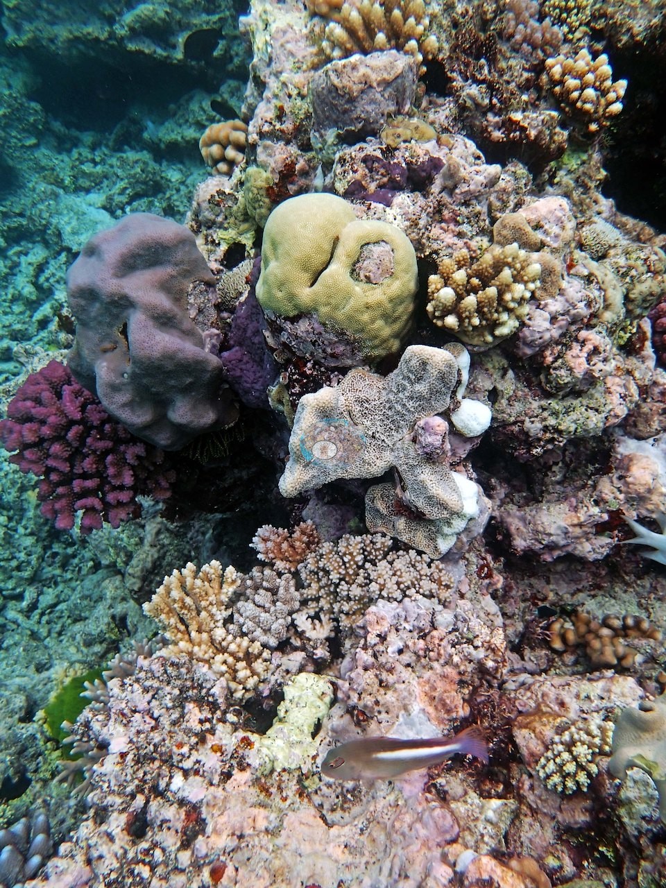
A stiff 2-3 knot current kept the team from pushing past the mid-depths, but what they found there is on show in frame after frame: a broad cream table Acropora spreading beside overlapping gray plates, chartreuse Montipora, and mounding Porites; a close vertical face where a dozen colonies of different genera and colors, pink, mauve, mustard, olive, orange, sit within a hand's span of each other, with almost no bare rock between them (e.g., Image 8).
This reef sits in a no-take Marine National Park (Green) Zone.
General benthic features
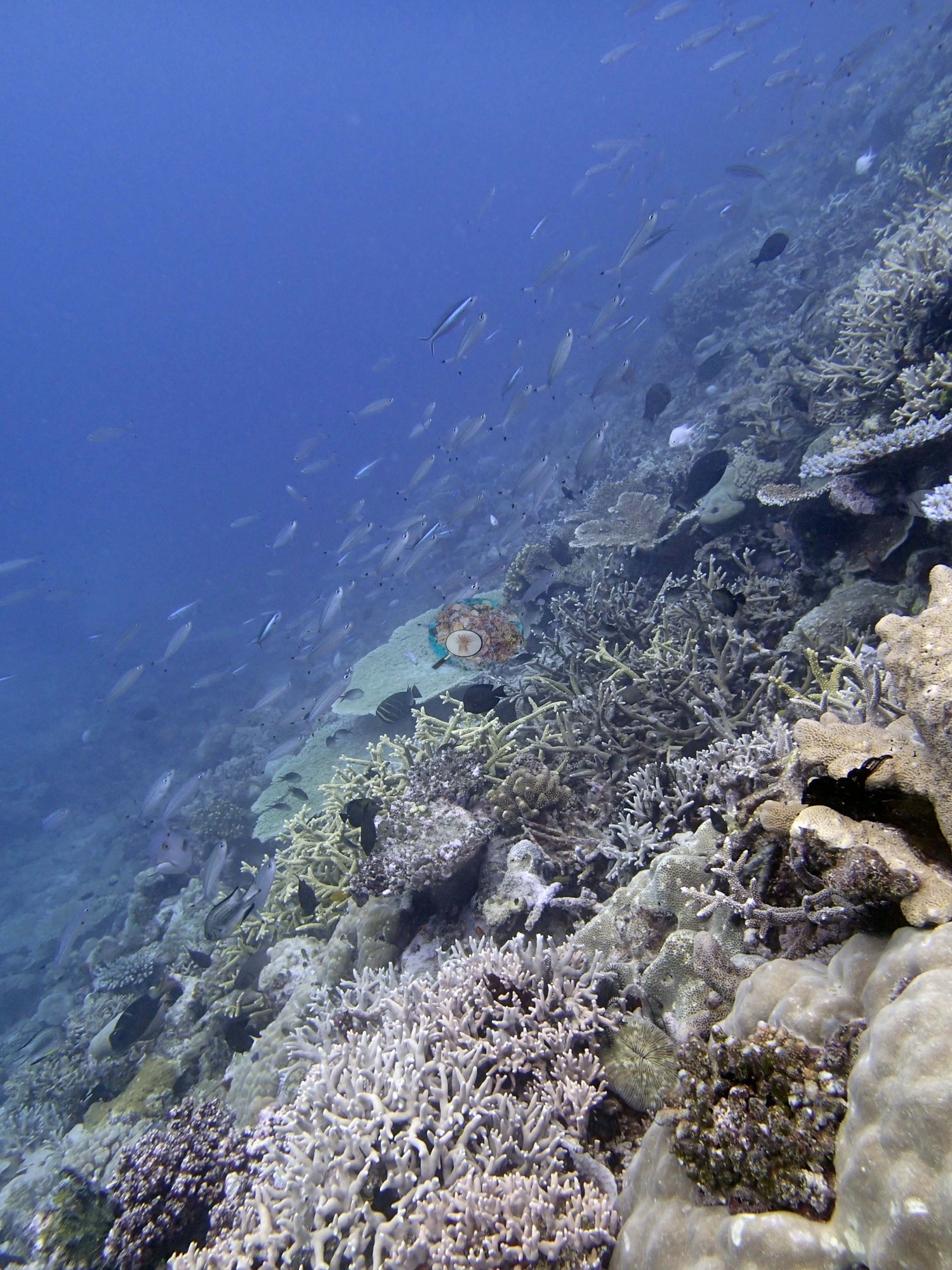
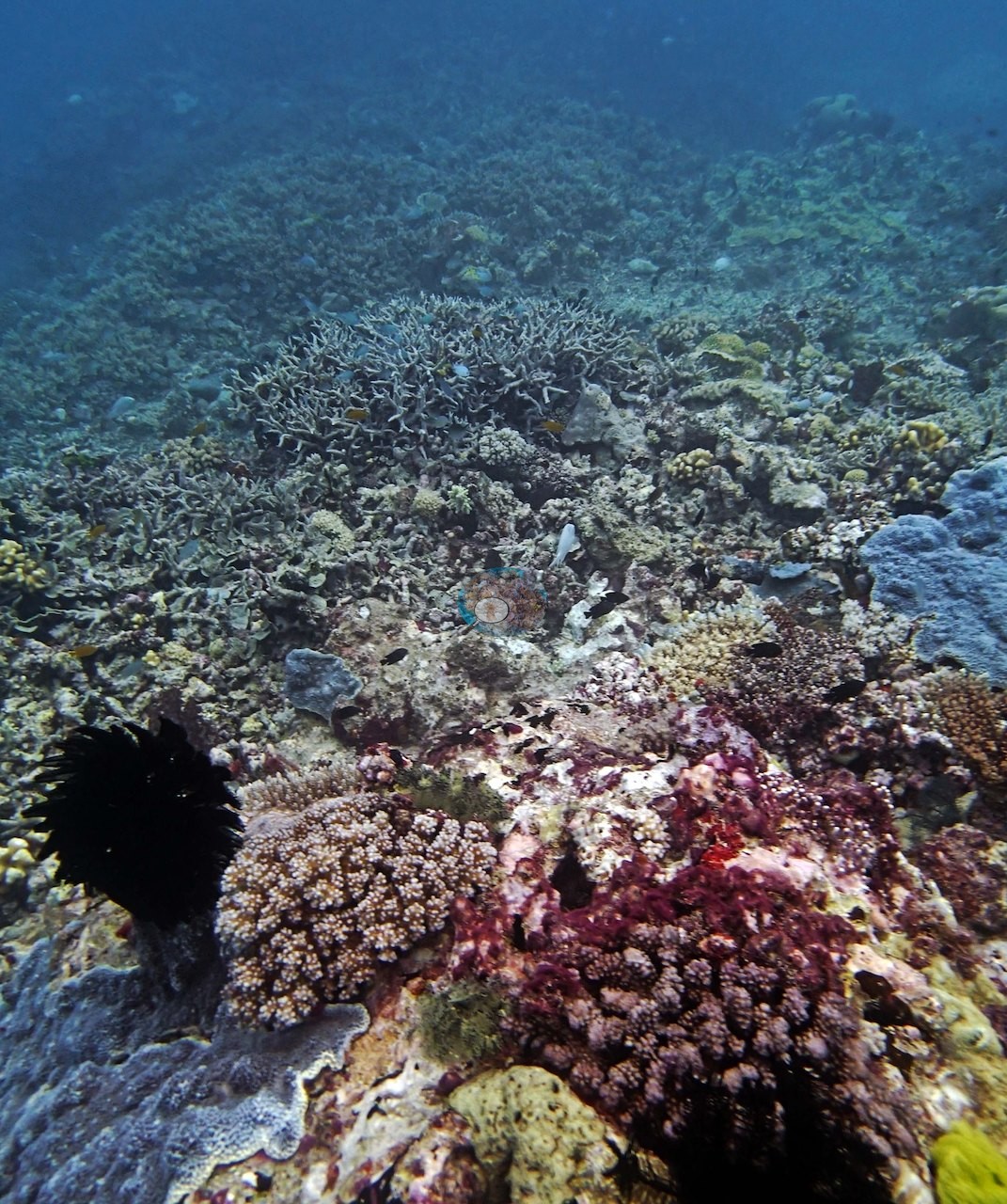
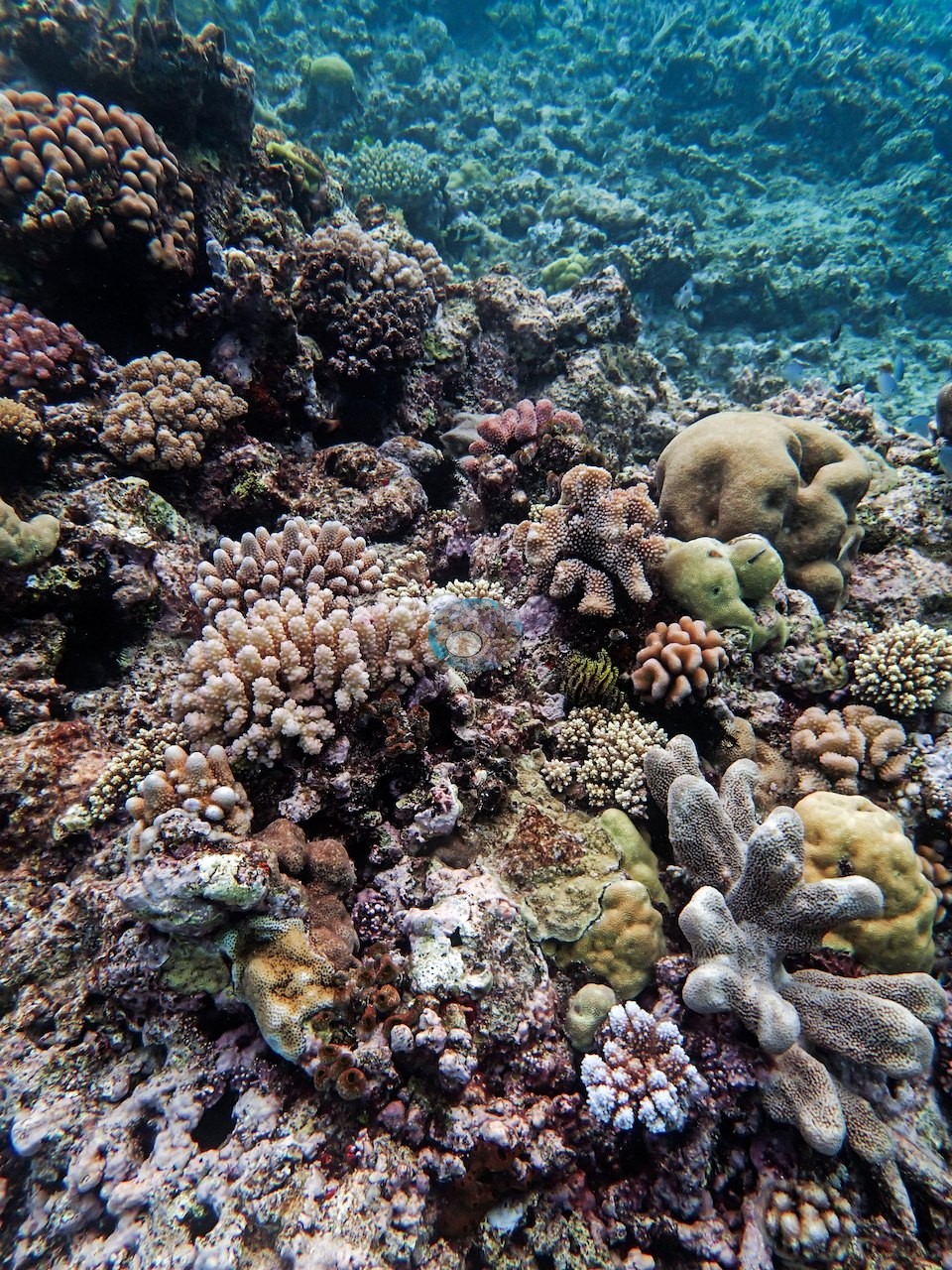
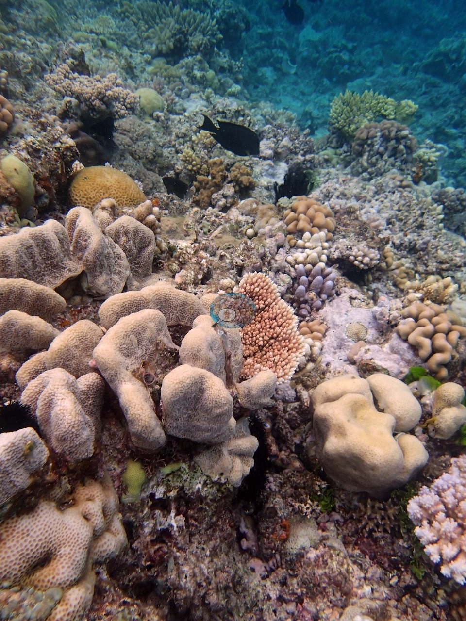
The assemblage Anderson logged, branching Porites, Pocillopora verrucosa and damicornis, Stylophora pistillata, table and branching Acropora, Montipora, and Lobophyllia, is all visible here. Needle coral (Seriatopora hystrix and caliendrum) was common, alongside a strong showing of new recruits. The dominant impression across the wide frames is packing. Image 4, Image 7, Image 9, and Image 15 show framework so densely occupied that bare rock is hard to find: tan and cream digitate and corymbose Acropora, mauve and dusky-rose pocilloporids, brown branching colonies, yellow-green encrusting sheets, green algal tufts, and sponges all interlocking within a hand's span. Image 1 is the shallow reef top with a large cream table Acropora and overlapping gray plates around it; Image 2 is the best structural frame in the set, a big dark-branched table well over a meter across spreading out over the slope into blue water, with a second flat plate below it, lobed brown colonies, and a yellow honeycomb-corallite massive on the pavement beneath. Image 12 looks along that slope dropping away into blue.
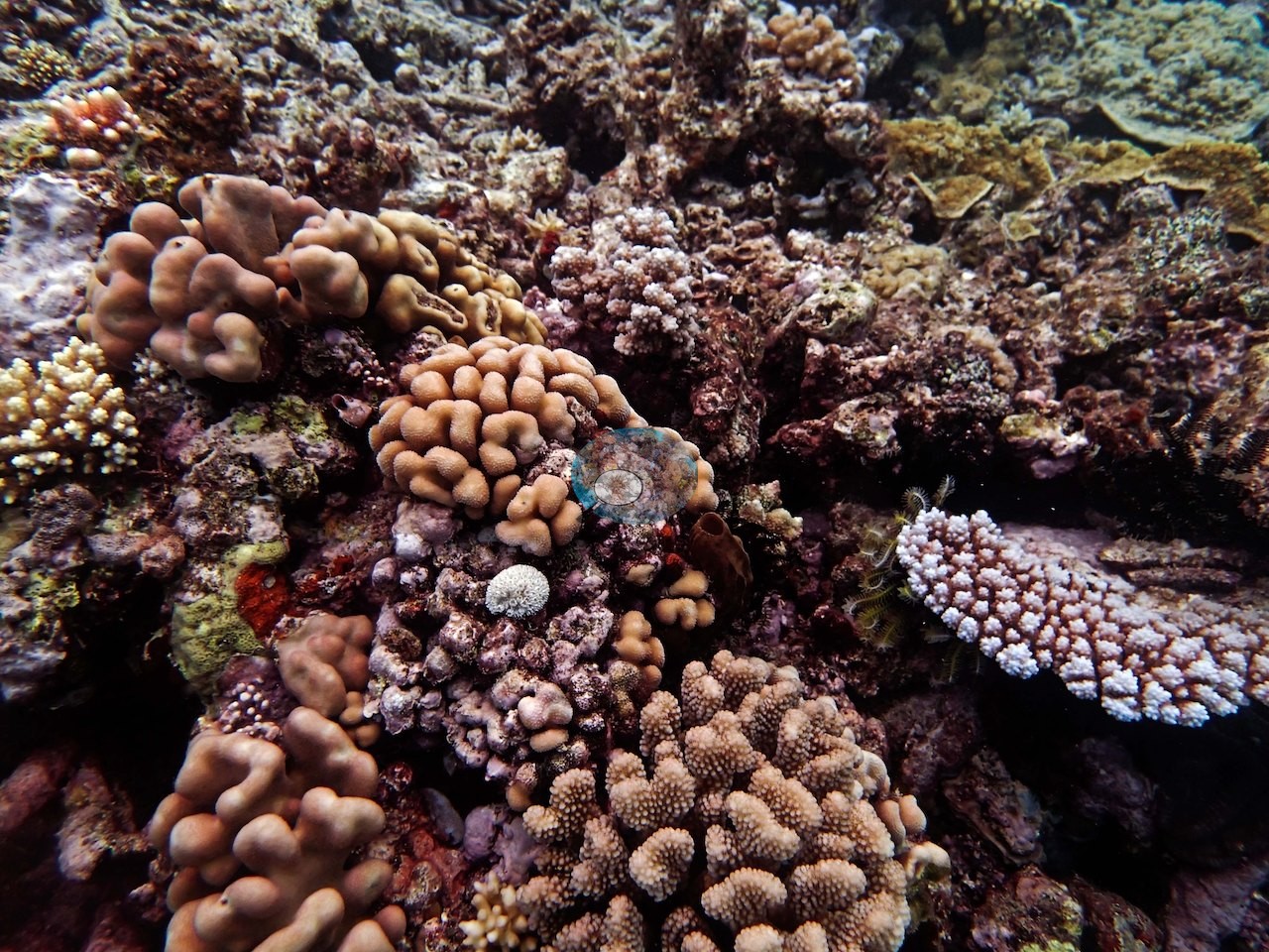
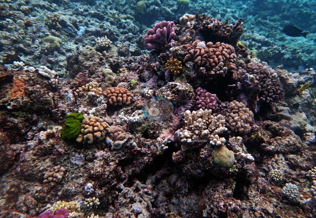
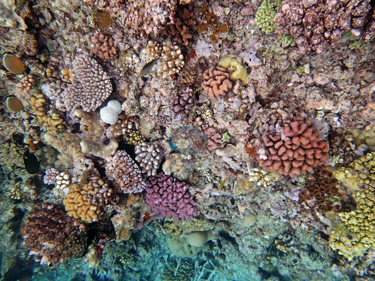
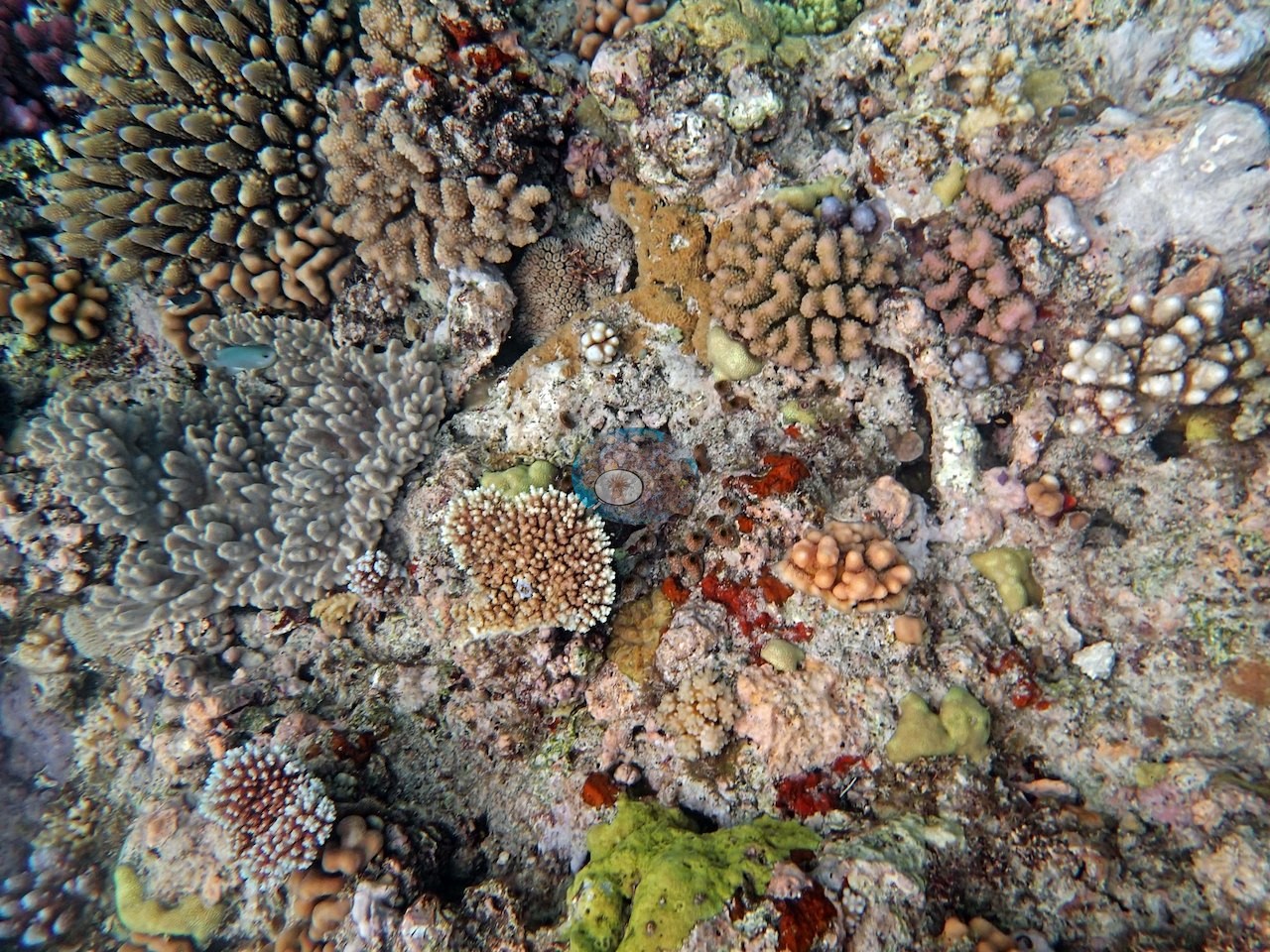
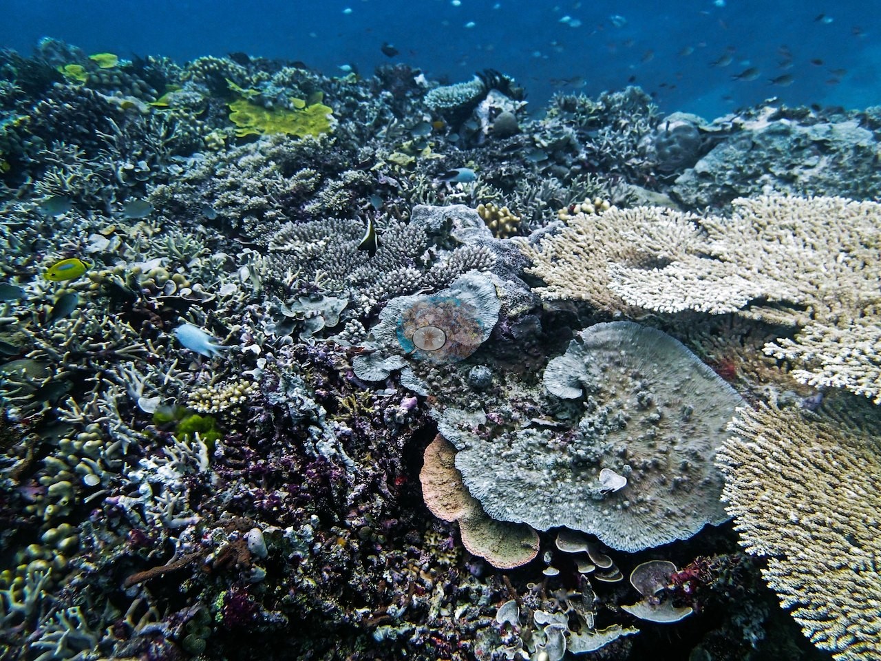
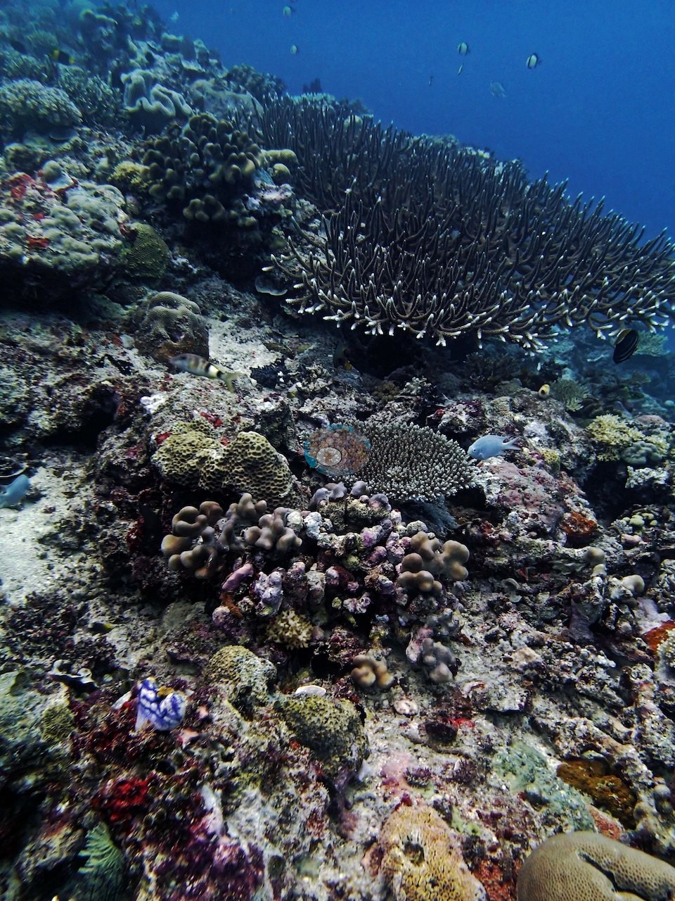
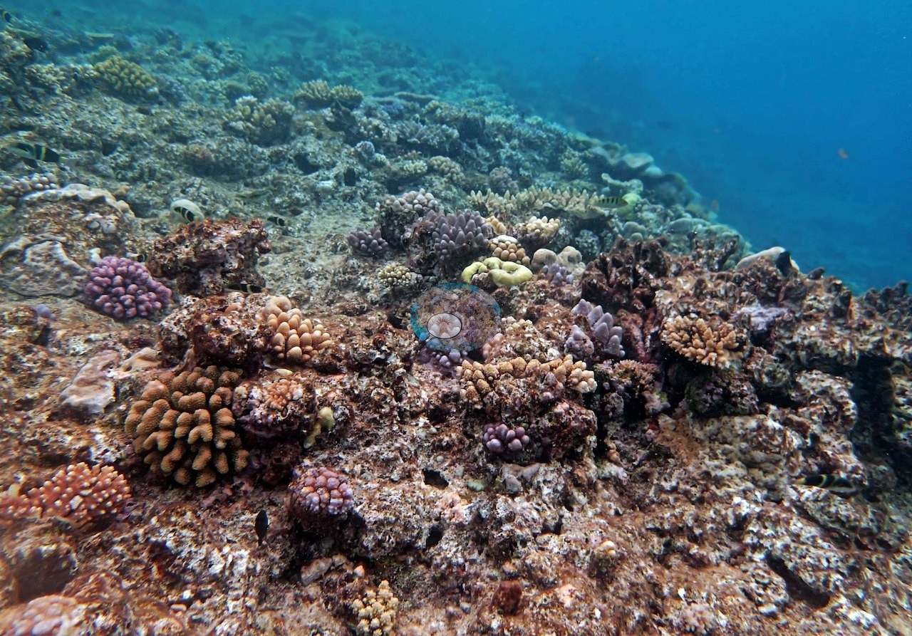
Three frames are single-colony portraits. Image 10 is a deep maroon-purple colony of thick knobby branches filling the frame; Image 13 is a bright pink pocilloporid, its clustered branch tips rounded and crowded, set on pale pavement; and Image 11 is a peach-cream knobby pocilloporid wedged between a tan massive colony and a lilac encrusting colony, with a tuft of green filamentous algae behind it.
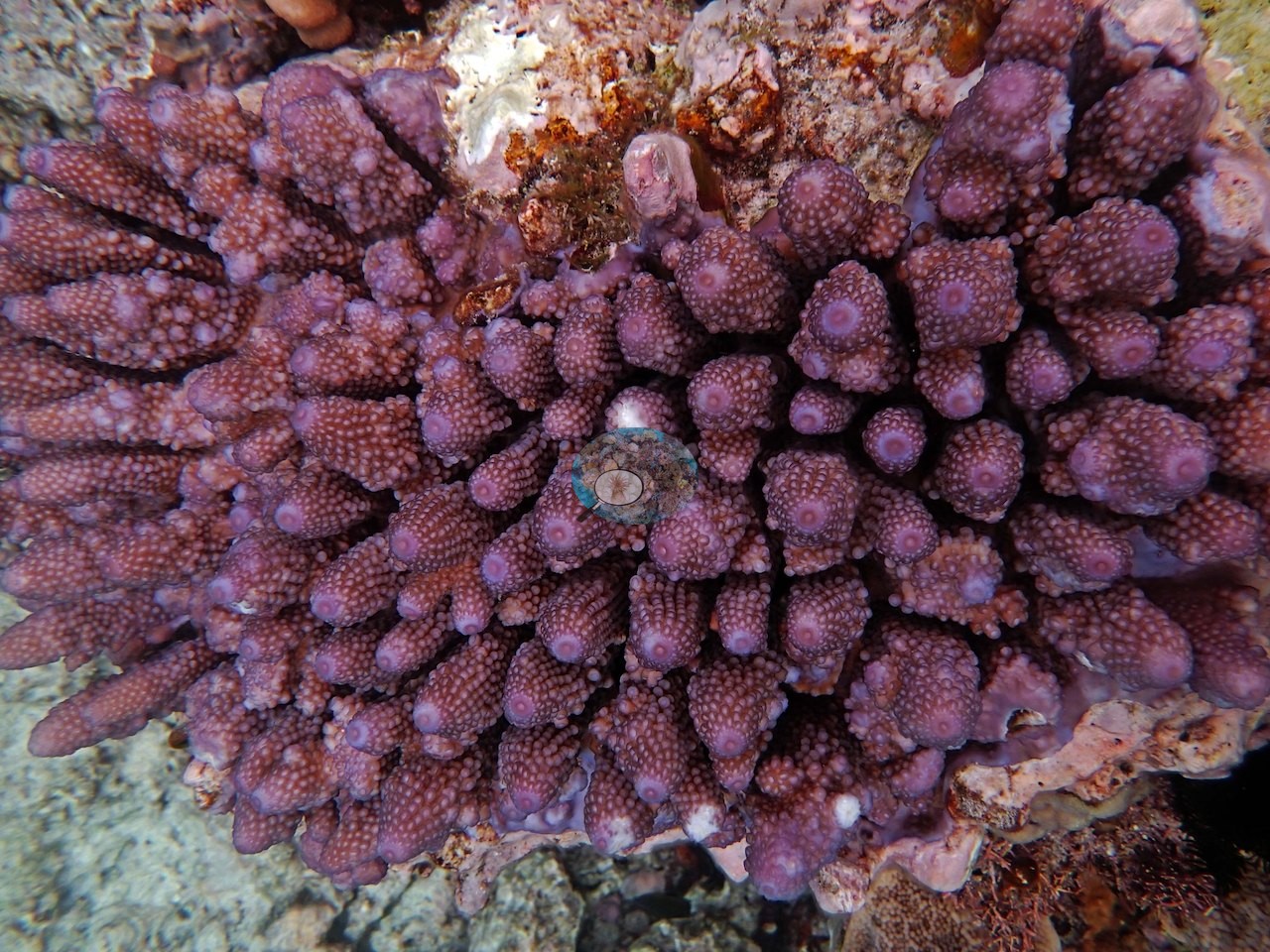
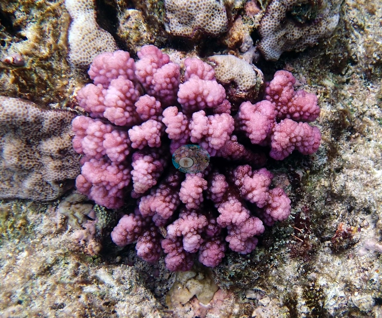
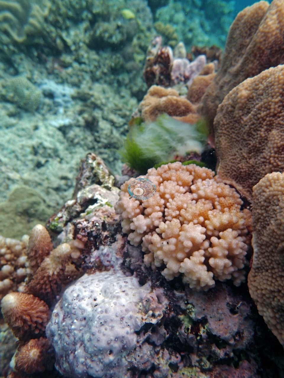
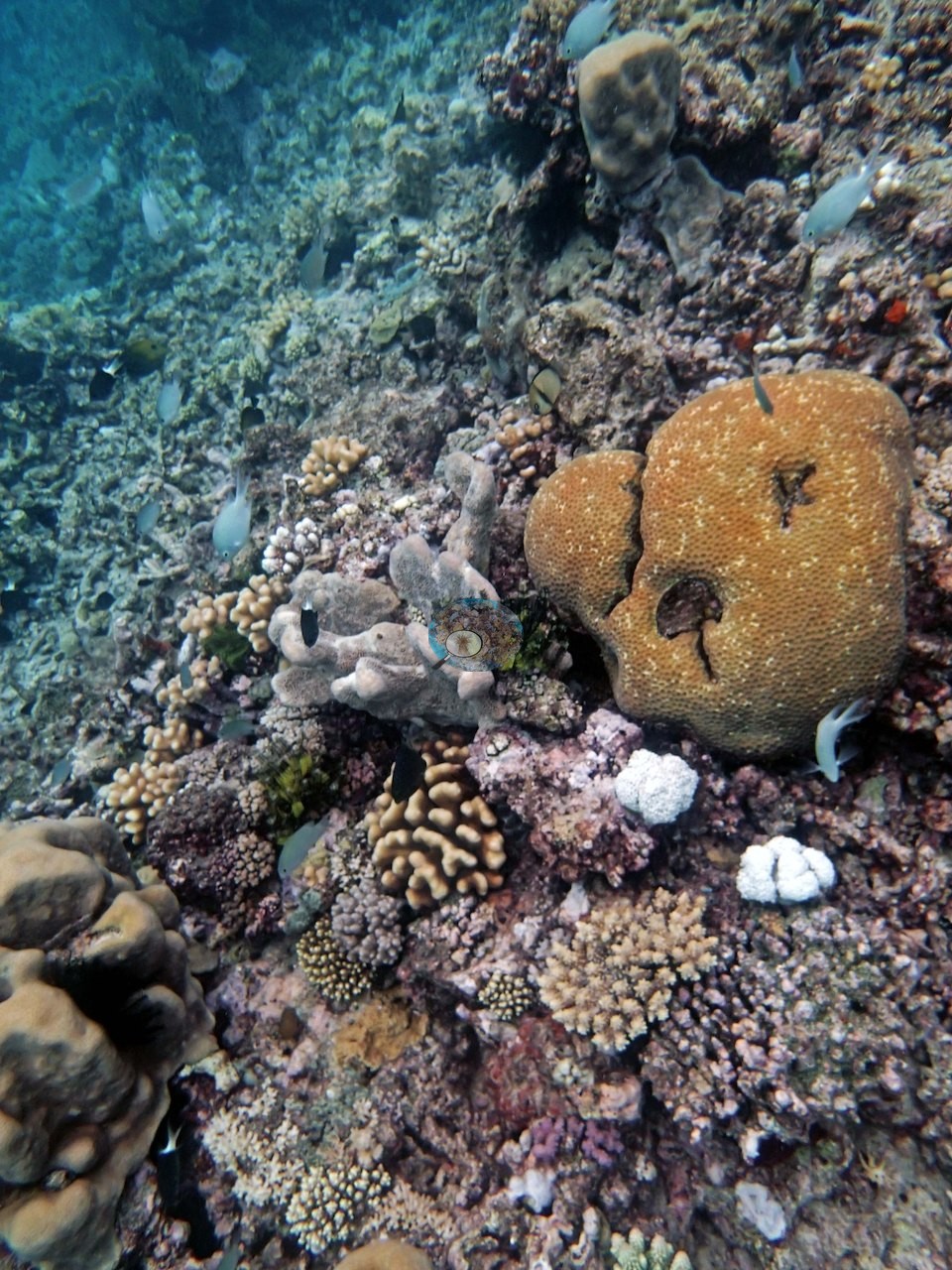
Two of them, the knobby-branched colonies in Image 10 and Image 13, match the Pocillopora verrucosa Anderson logged here and are reported on that basis rather than from the photograph alone. Image 5, seen close, centers on a large orange-tan massive with fine even corallites carrying two dark irregular cavities in its surface, with gray-tan lobed soft corals of leather-coral form beside it, a black-and-green algal tuft, and a cloud of small blue-gray damselfish over the whole scene.
Those fleshy lobed soft corals recur in Image 6 and Image 14 and are a real component of this reef.
Coral cover
- Reference: Khaled bin Sultan Living Oceans Foundation, Global Reef Expedition (in situ transect)
- Point-count cover measured 36%, with one transect at 15.7 m running as high as 50-60%.
- Transect mean 36 ± 22% SE (n = 2 transects; Living Oceans Foundation per-transect benthic data); the headline above is the expedition's station value.
Stressors
Two small knobby colonies on the pavement in the foreground of Image 5 are bleached: stark white with their tissue intact, beside tan-to-pink knobby colonies of the same growth form. These are two bleached colonies in one frame, so no bleaching extent is estimated. Elsewhere in the 15 frames there is no visible disease lesion, crown-of-thorns feeding scar, or field of drained-white branches.
Comparisons with other data
No prior survey data from this site. If you would like to share yours, please email us.
Other notes
Anderson called this stop simply "gorgeous," and the photographs justify it. The station's 15 frames from that dive are all reef imagery — no quadrats, no topside, no divers.
CV model comparison
More info
The photo row below is a methods comparison only: it is never averaged with the transect and never used as the headline. It compares site means only: the photos come from the same survey dive as the transect but not from the same points.| Method | Hard-coral cover (%) | Photos (n) ± SE |
|---|---|---|
| Transect (published) | 35.5 | |
| CoralSCOP edited | 36.4* | n = 12 · SE 2.5 |
| Claude (edited photos) | 29.1* | n = 12 · SE 4.4 |
The two computer-vision methods differ by 7.3 pp (29–36%) and read within 6.4 pp of the in-water transect (36%); the headline is the in-water transect because a diver transect is independent of the photographs.
*Based on only 12 reef-habitat photos; this estimate is less certain.
Data & links
- Download benthic survey data (CSV) ↗
- Download all LOF-GRE data (933 sites) ↗
- Photo portfolio ↗
- Coral Reef Diagnostics country page ↗
All images from Coral Reef Diagnostics. Data are from the Khaled bin Sultan Living Oceans Foundation Global Reef Expedition and Coral Reef Diagnostics.
 © Coral Reef Diagnostics.