
Part of the regional summaries: Australia (GBR) - Far North: Green stations (no-take reefs) and Australia (GBR)
More info
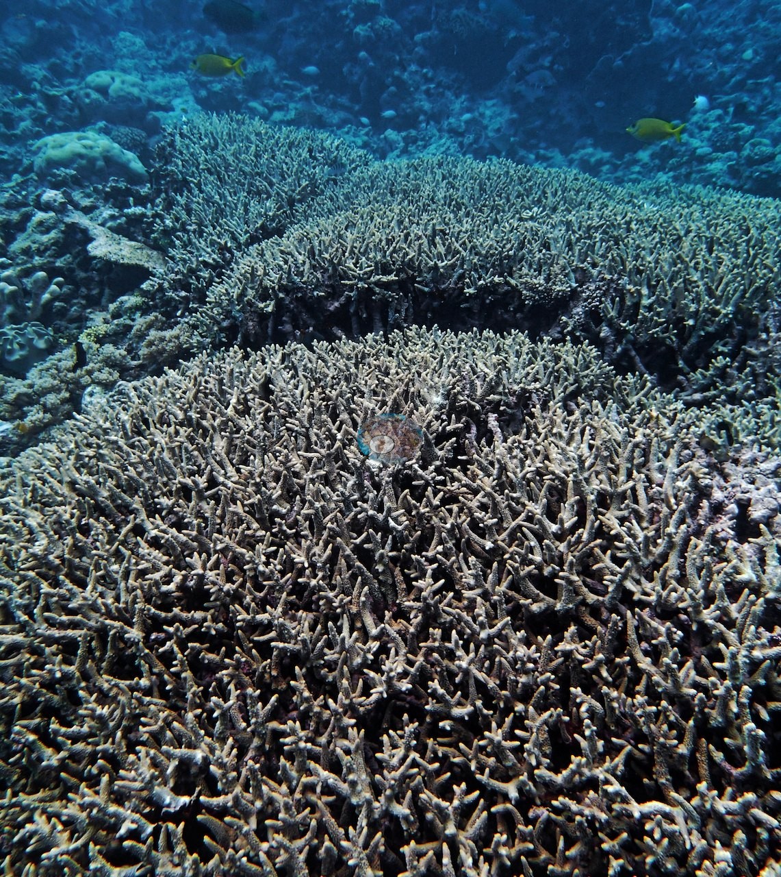
Far Northern GBR (NOAA CRW virtual station)Anderson called this stop strikingly beautiful and estimated 80-90% live coral cover by eye along his own 10 m transect (the expedition's point-count transect scored 36%), with dense Isopora, Pocillopora, and staghorn thickets running unbroken to the limit of visibility, strong recruitment, and tons of small fish. The one blemish was dying or sickly needle coral at 20 m. Surveyed on the Living Oceans Foundation's Global Reef Expedition.
Eleven other CRD dive sites within 5 km shown in orange — click a marker to open its page.
🗺 Detailed map of the Global Reef Expedition dive sites, unnamed reefs 11-122, 11-096, and 11-091, northern Great Barrier Reef (Khaled bin Sultan Living Oceans Foundation; opens in a new window)
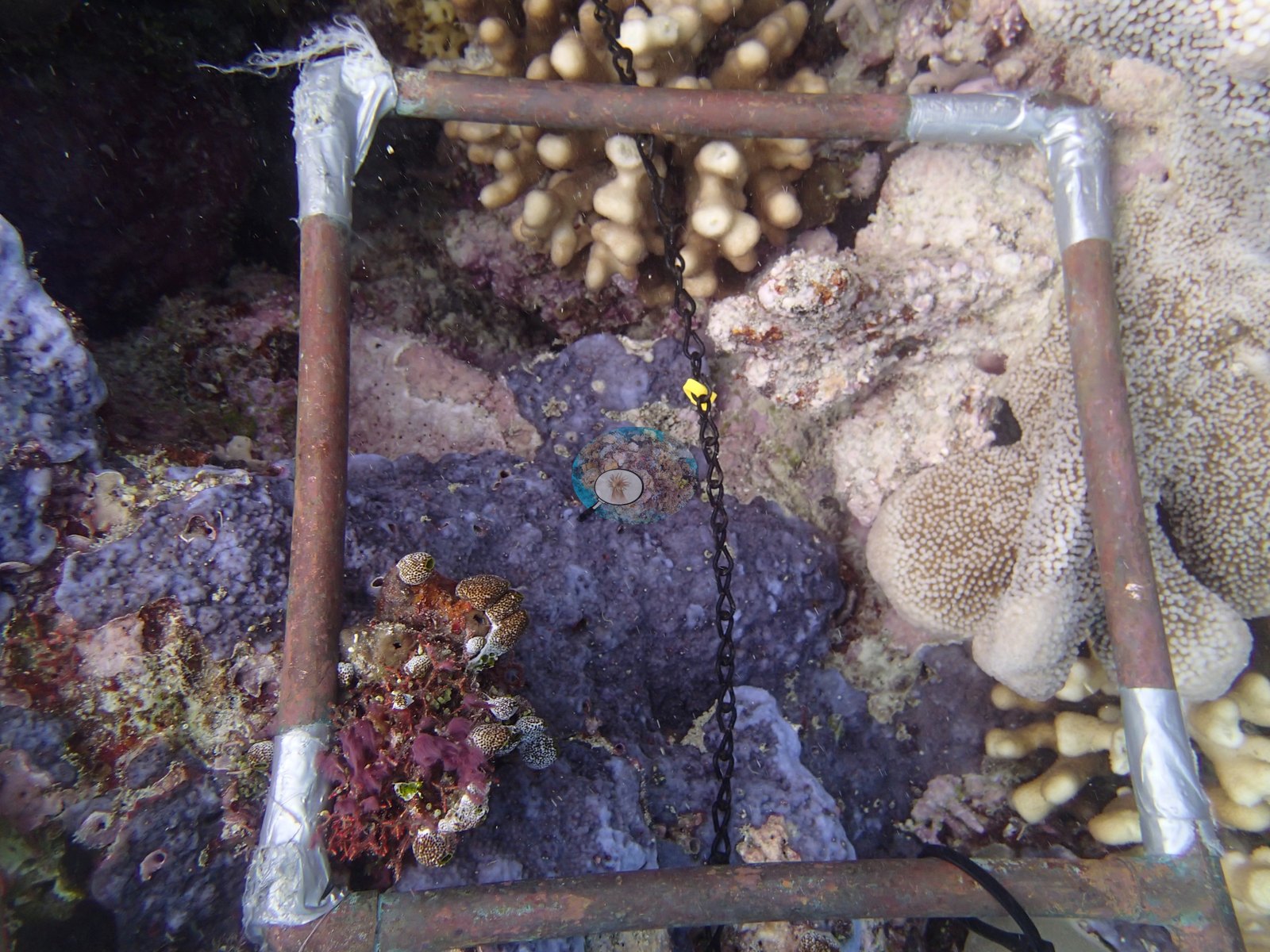
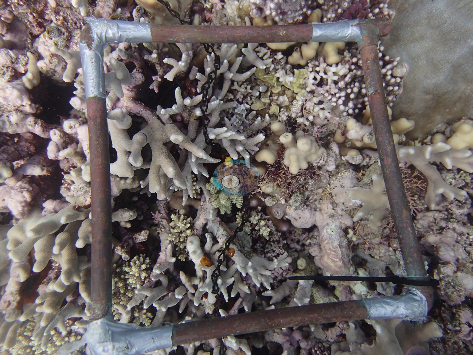
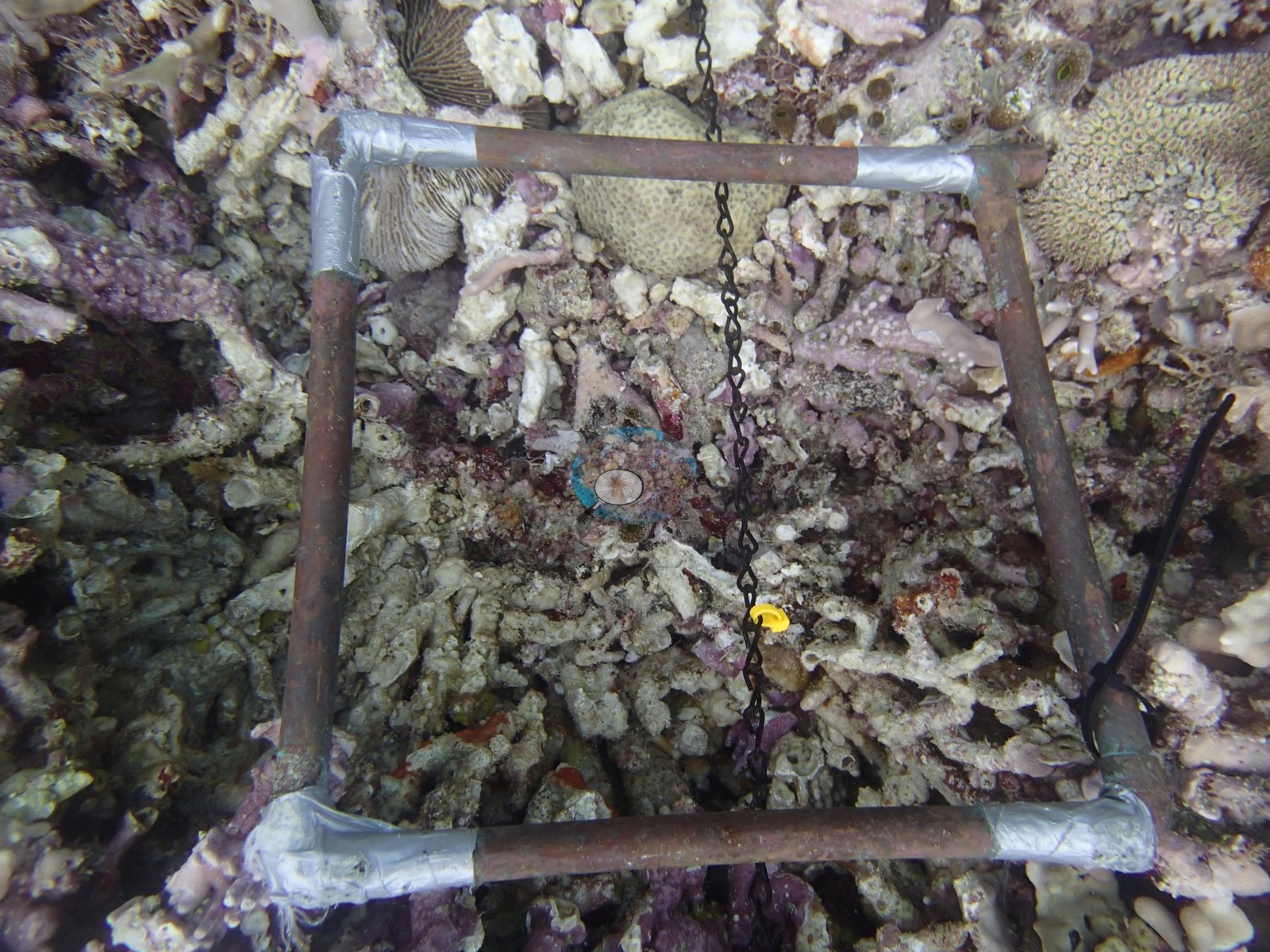
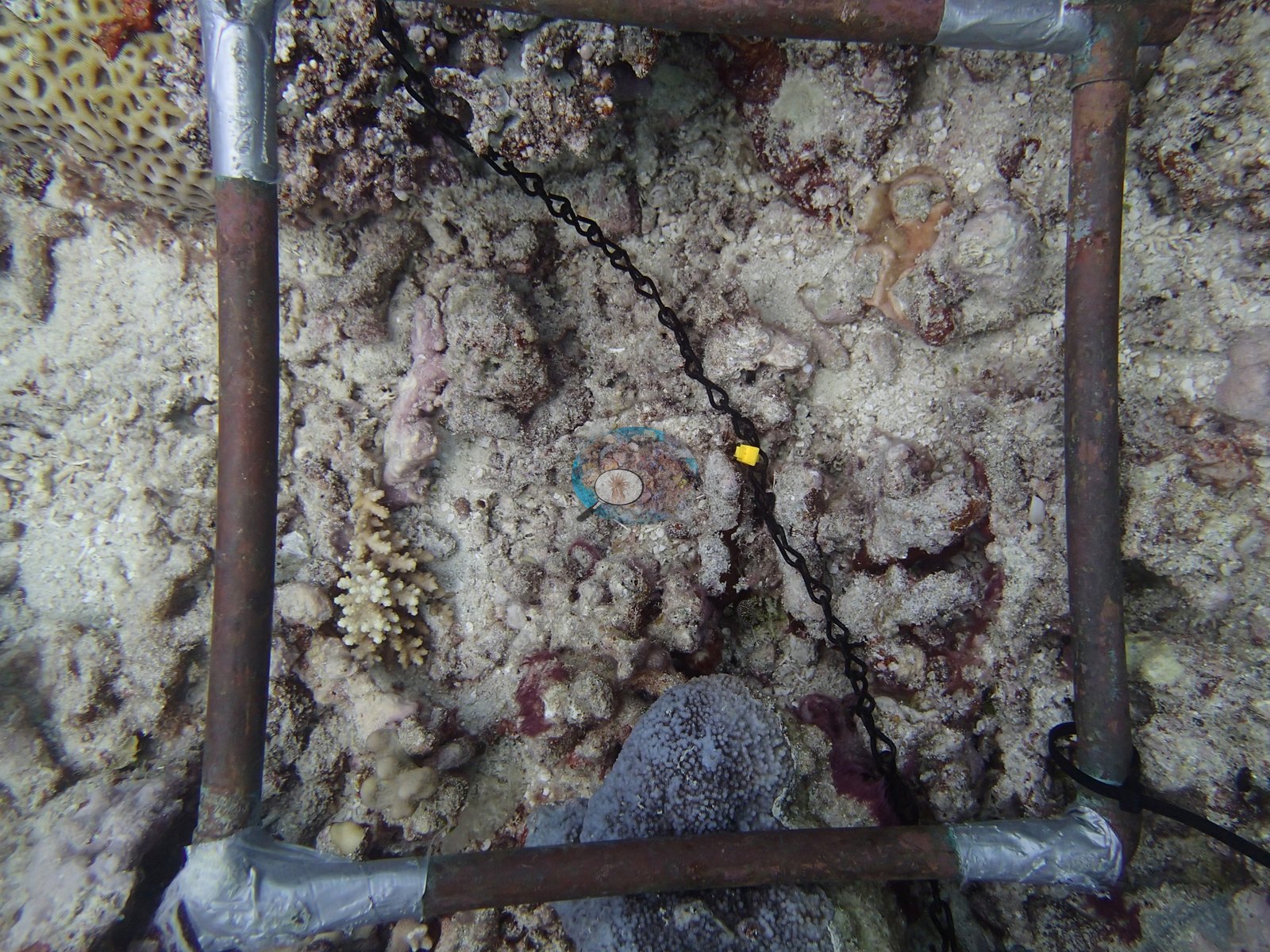
22 of the 38 photographs on this page are shown beside the text that discusses them under "Site description" below.
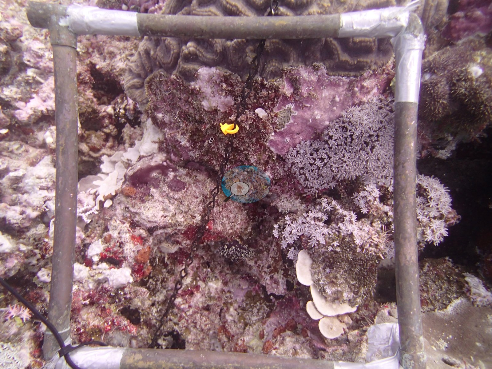
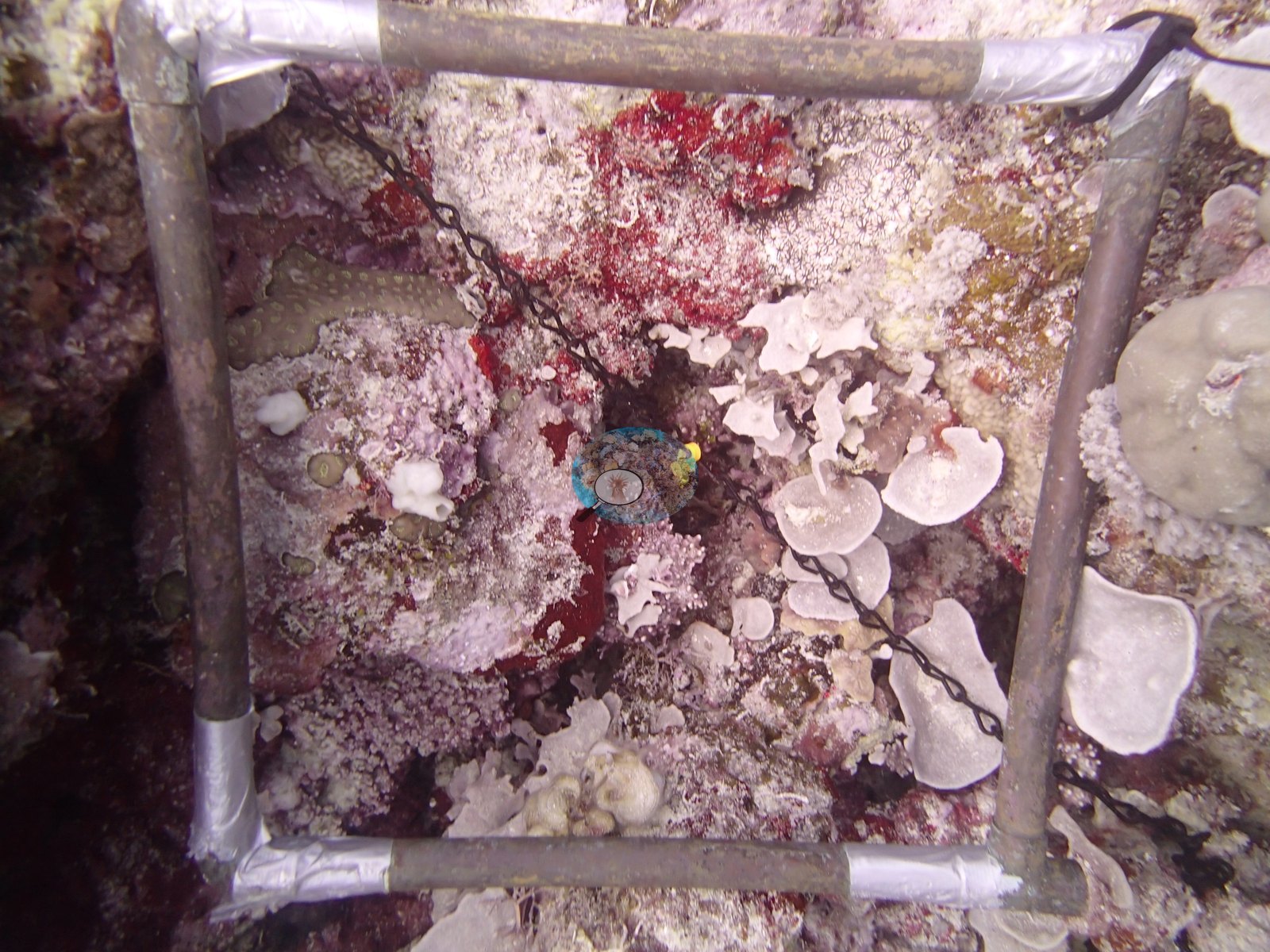
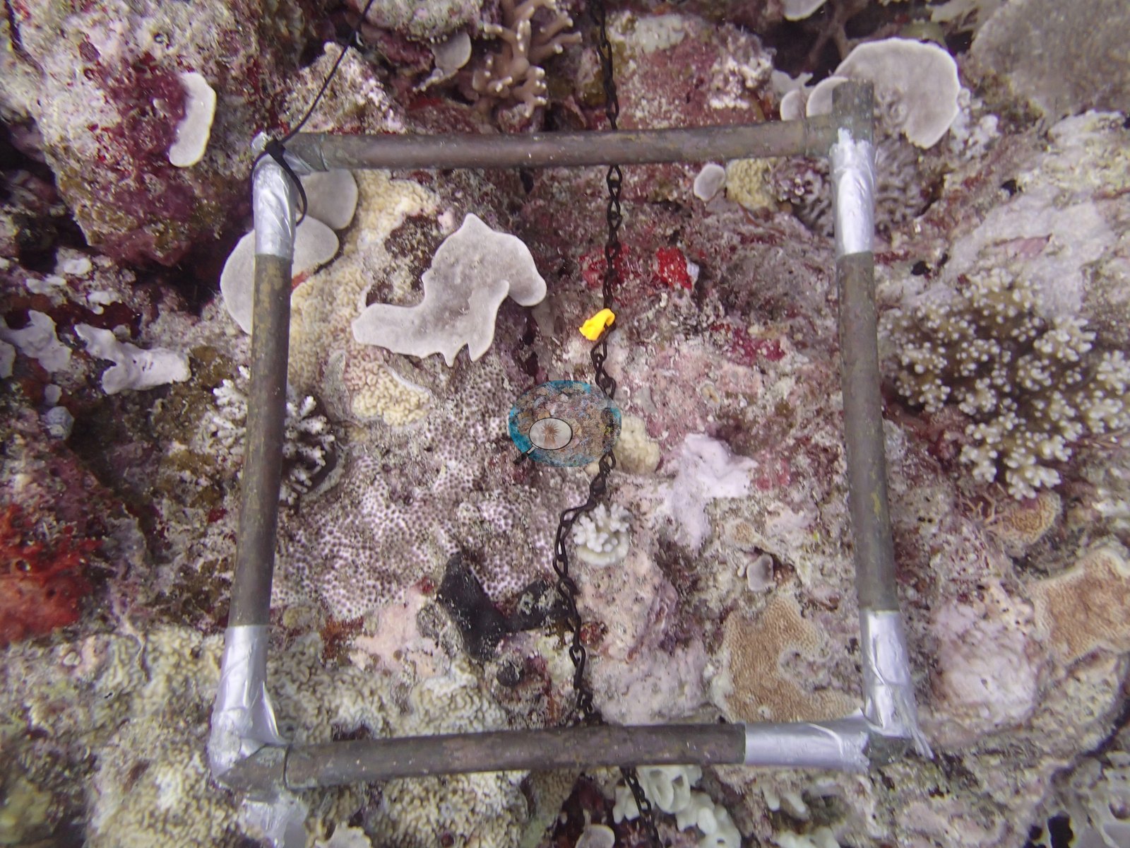
Site description
Setting
As a Marine National Park (Green) Zone, this reef is open to divers and snorkelers on a no-take basis.
General benthic features
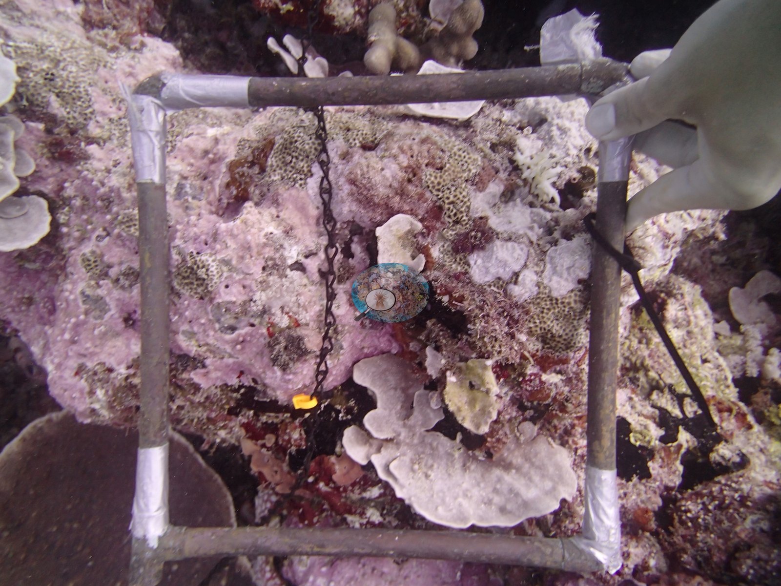
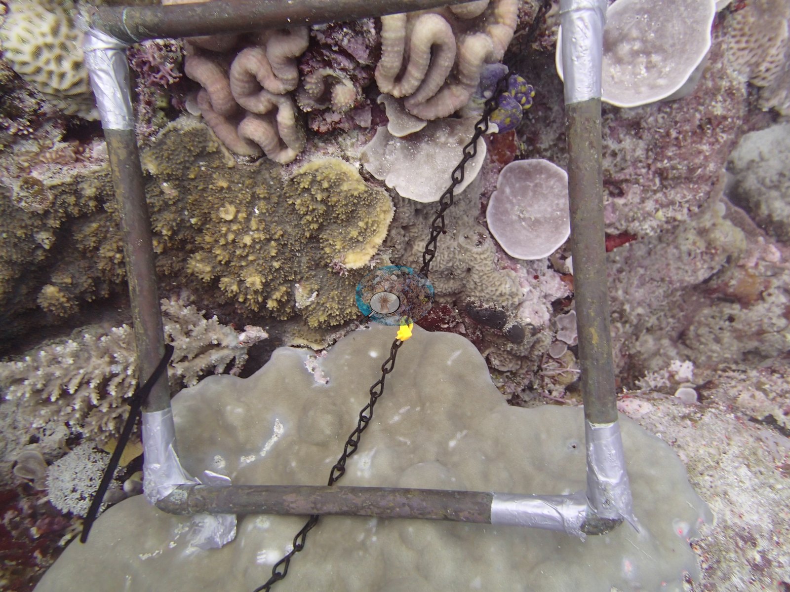
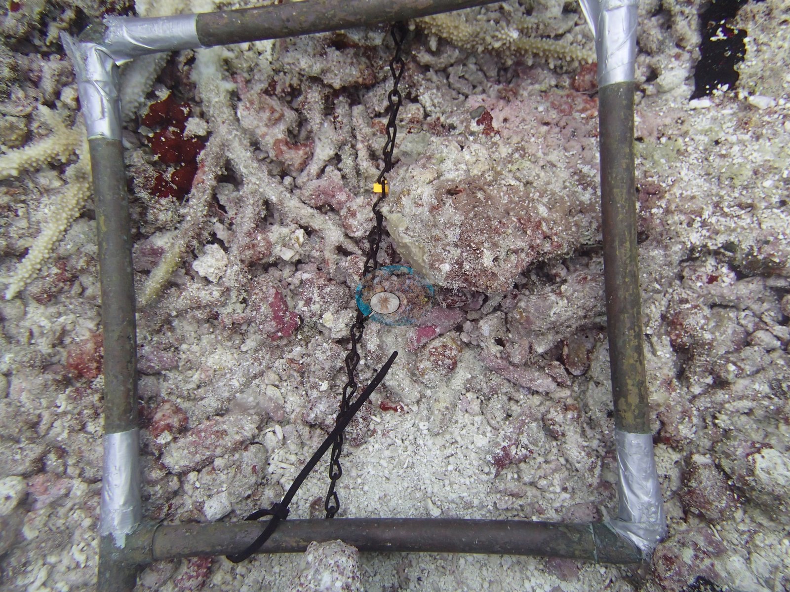
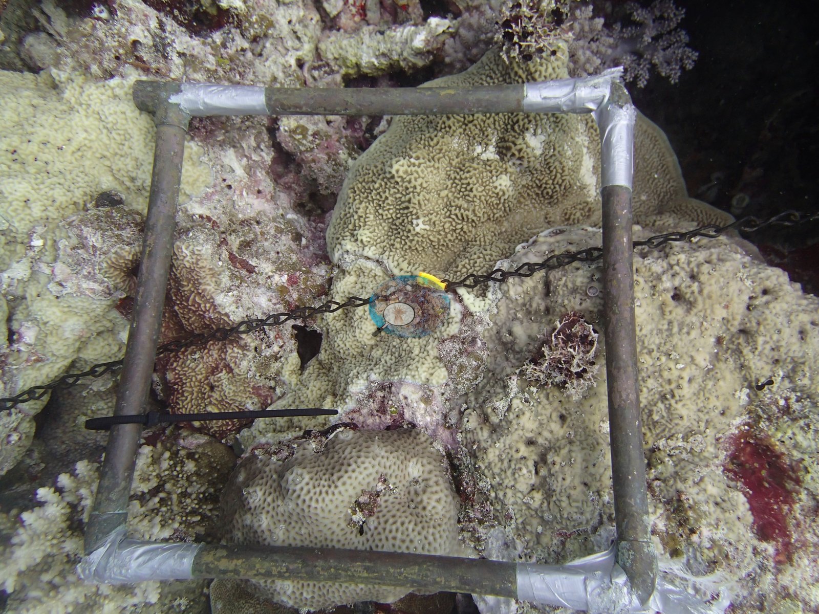
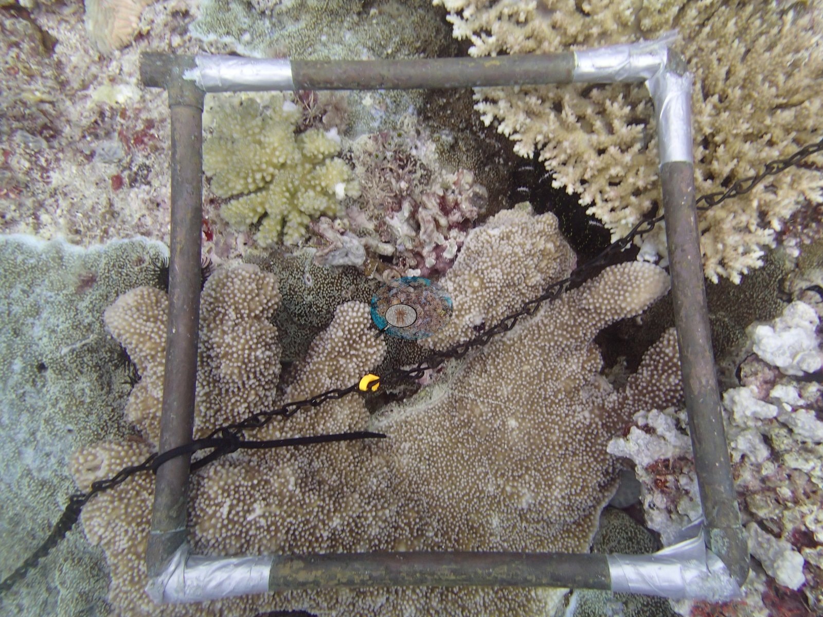
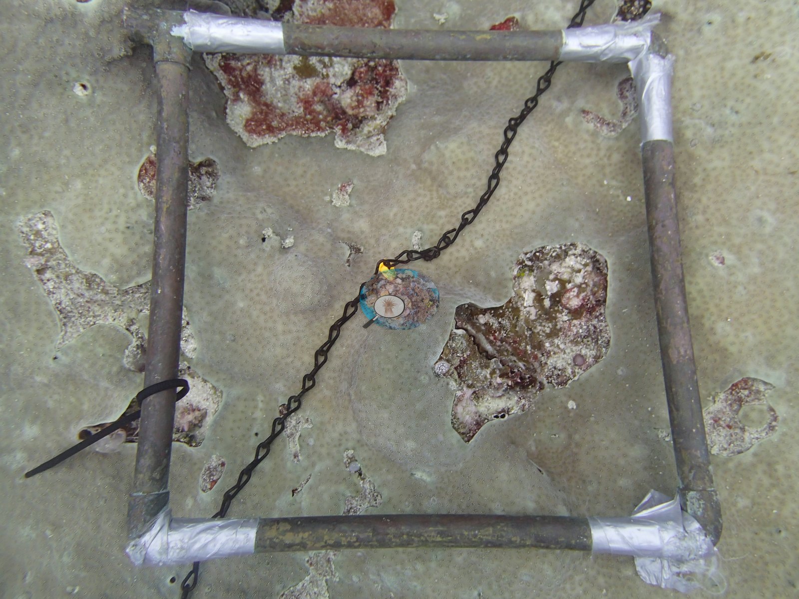
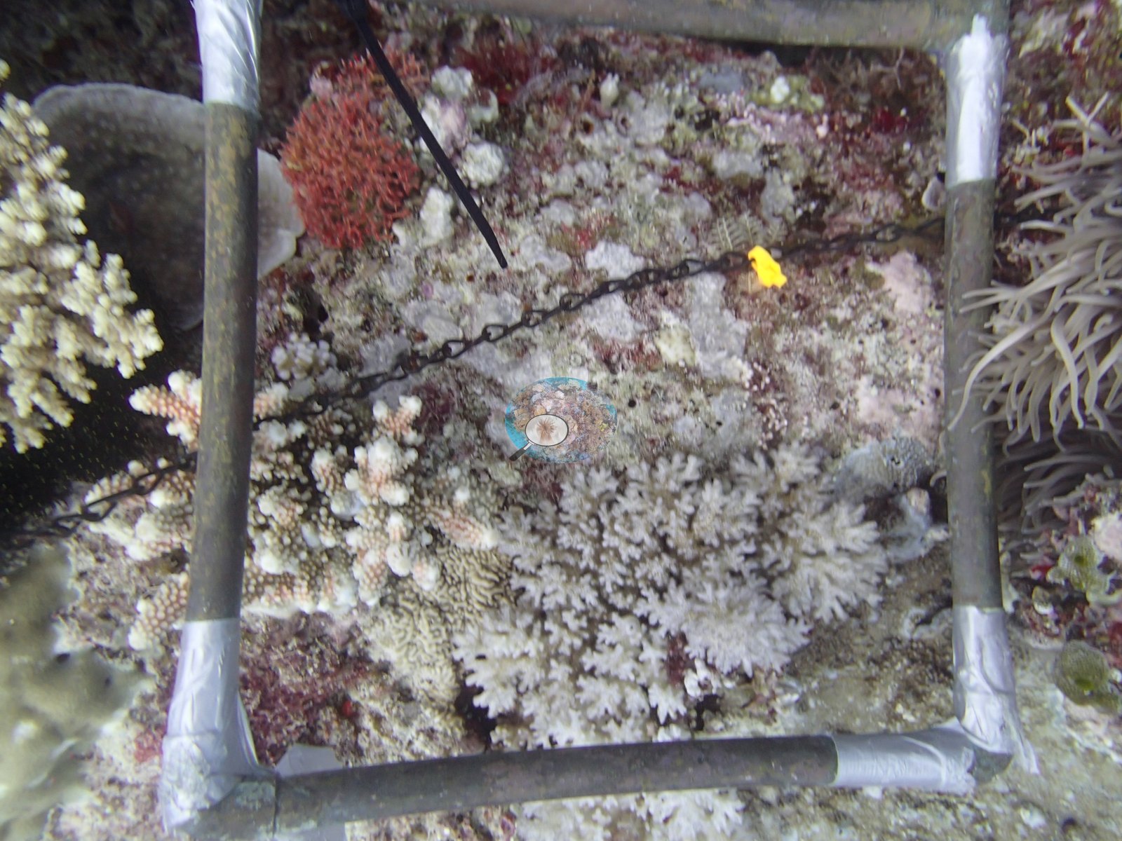
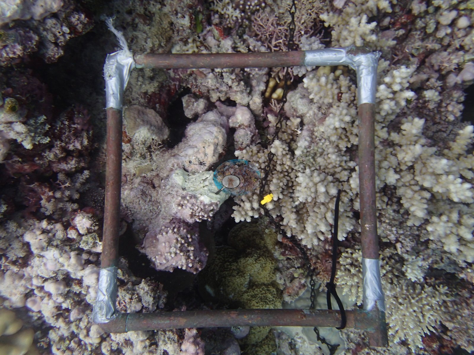
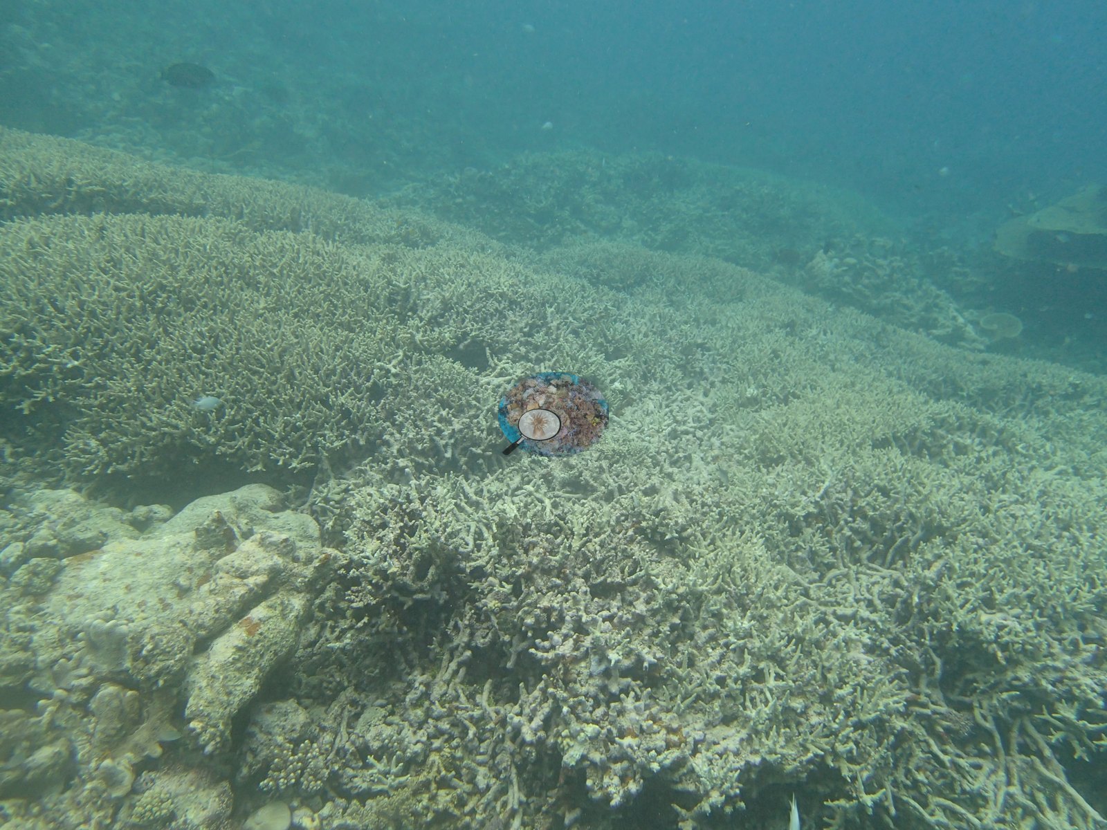
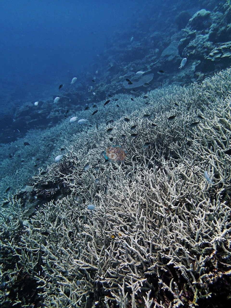
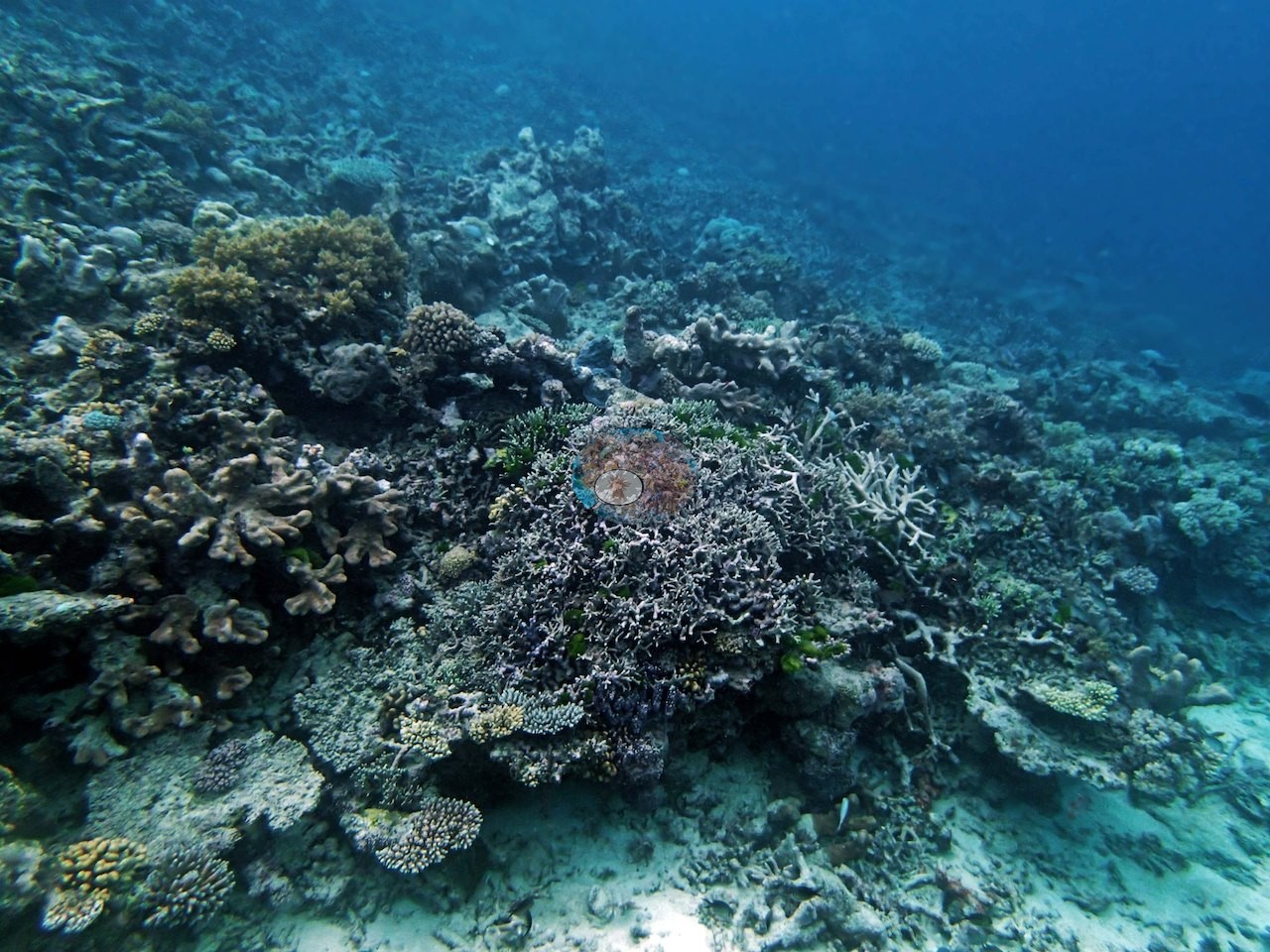
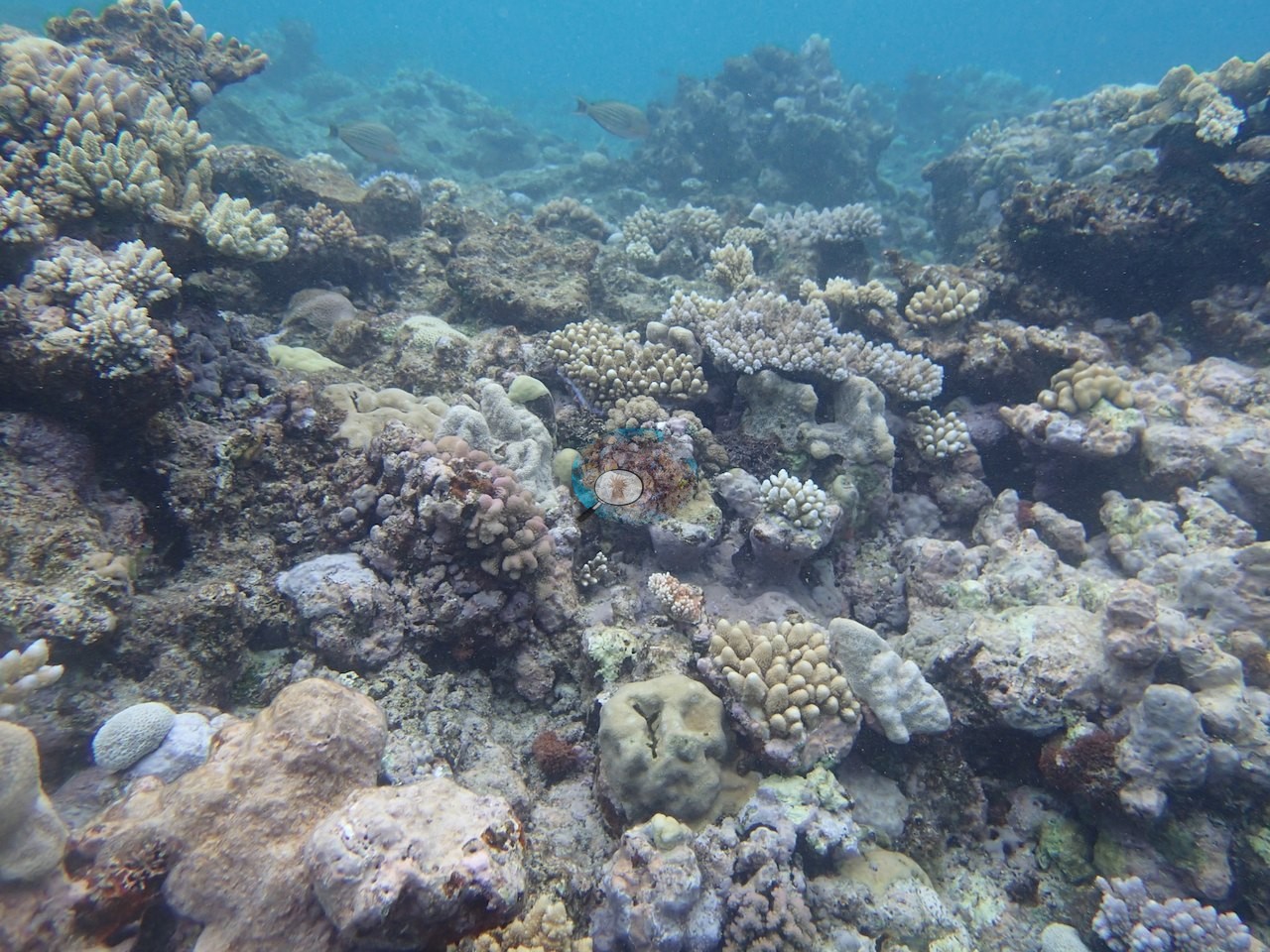
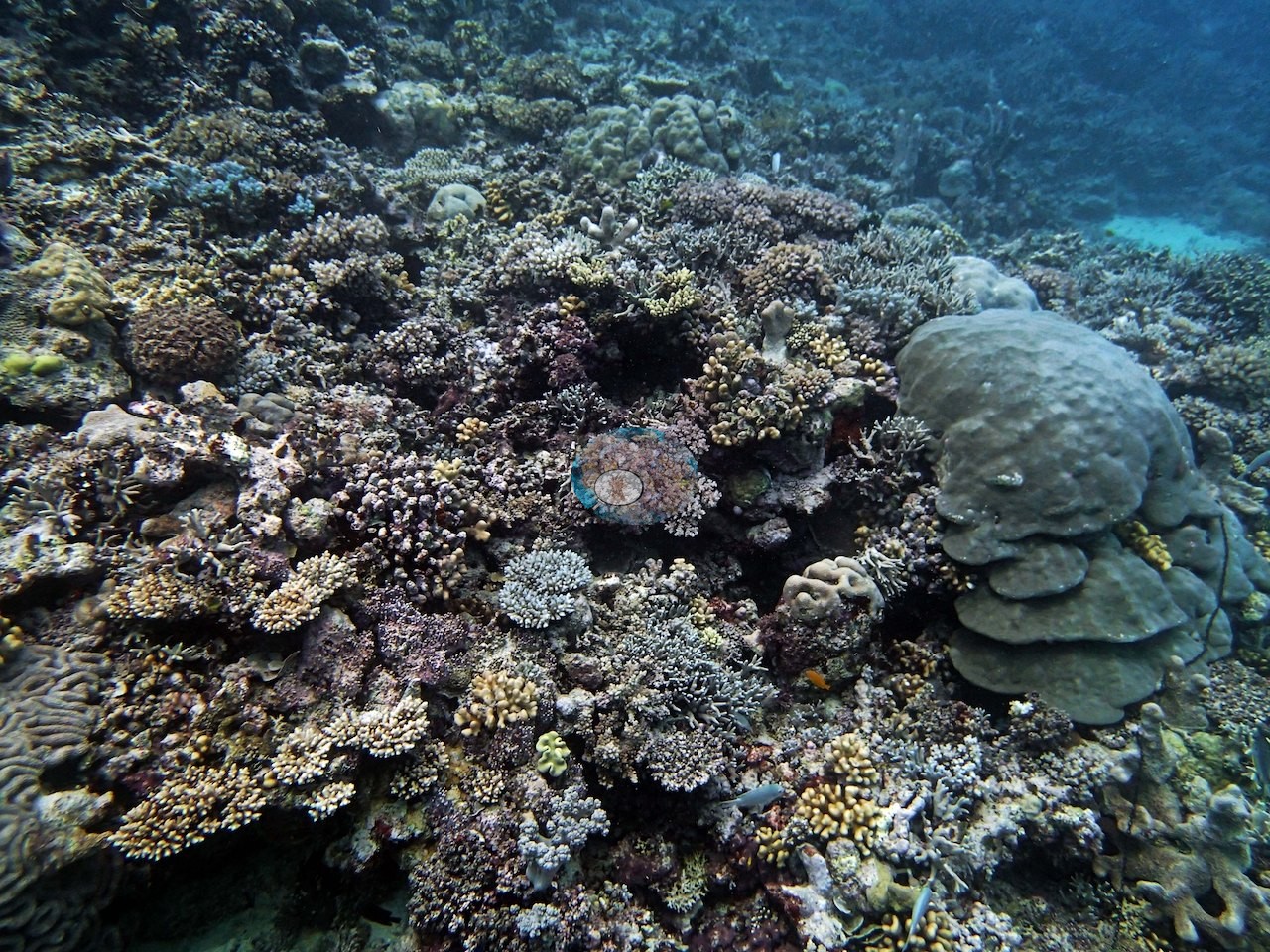
The reef built from a dense mix of Isopora, Pocillopora damicornis, P. verrucosa, and P. acuta, Stylophora pistillata, and lush Acropora thickets, with recruitment running strong throughout (e.g., Image 3, Image 4, Image 5).
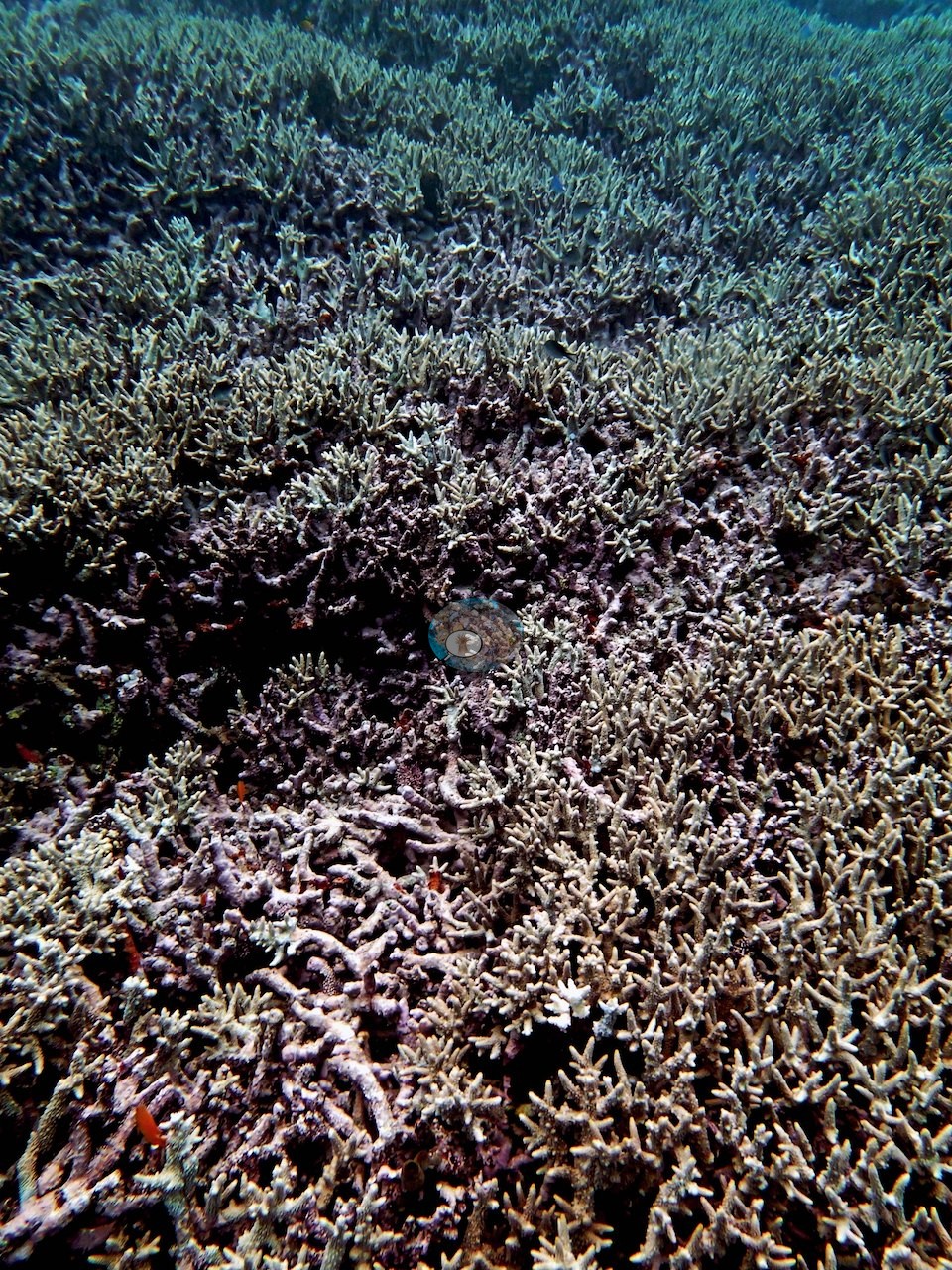
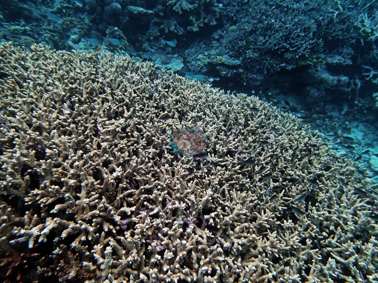
The thickets vary in texture: a blunt-branched, cream-tipped stand grades into finer gray branching in one frame (e.g., Image 9), and a pale gray staghorn stand spreads down the slope in another (e.g., Image 10).
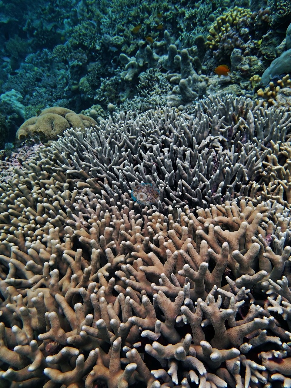
The mixed pavement is just as dense close in: pink-tipped green branching colonies, knobby cream colonies, blue-tipped corymbose colonies, and small massives packed around a bright green algal clump (e.g., Image 12), bright orange-red meandroid brain colonies wrapped around a cream branching colony (e.g., Image 17), and a round orange-red corymbose colony beside a purple branching colony and small tables (e.g., Image 19).
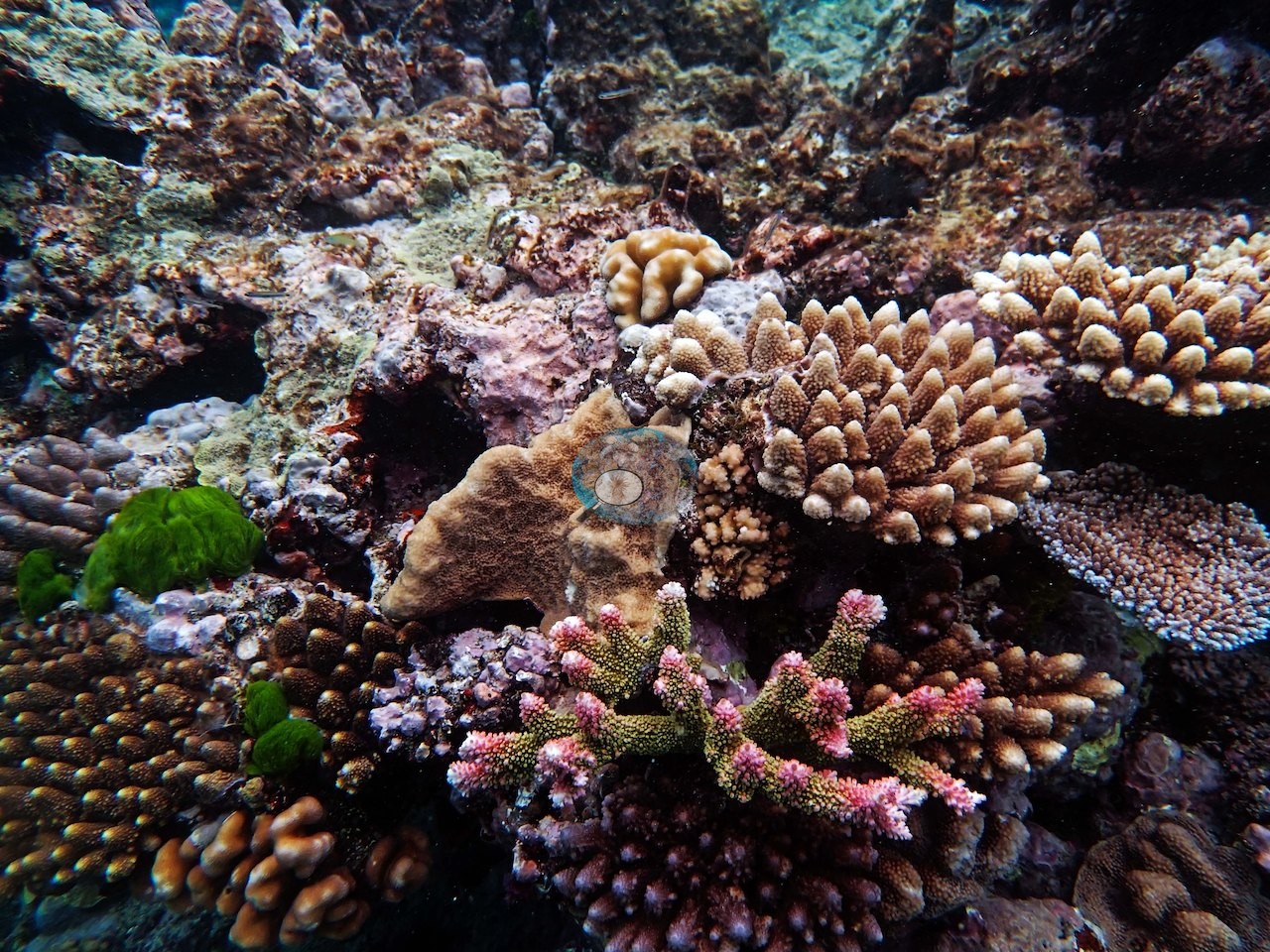
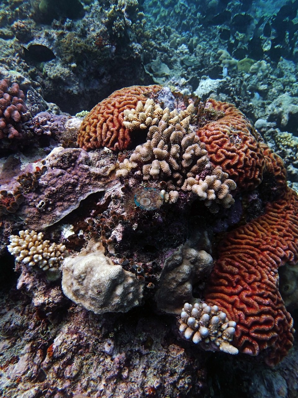
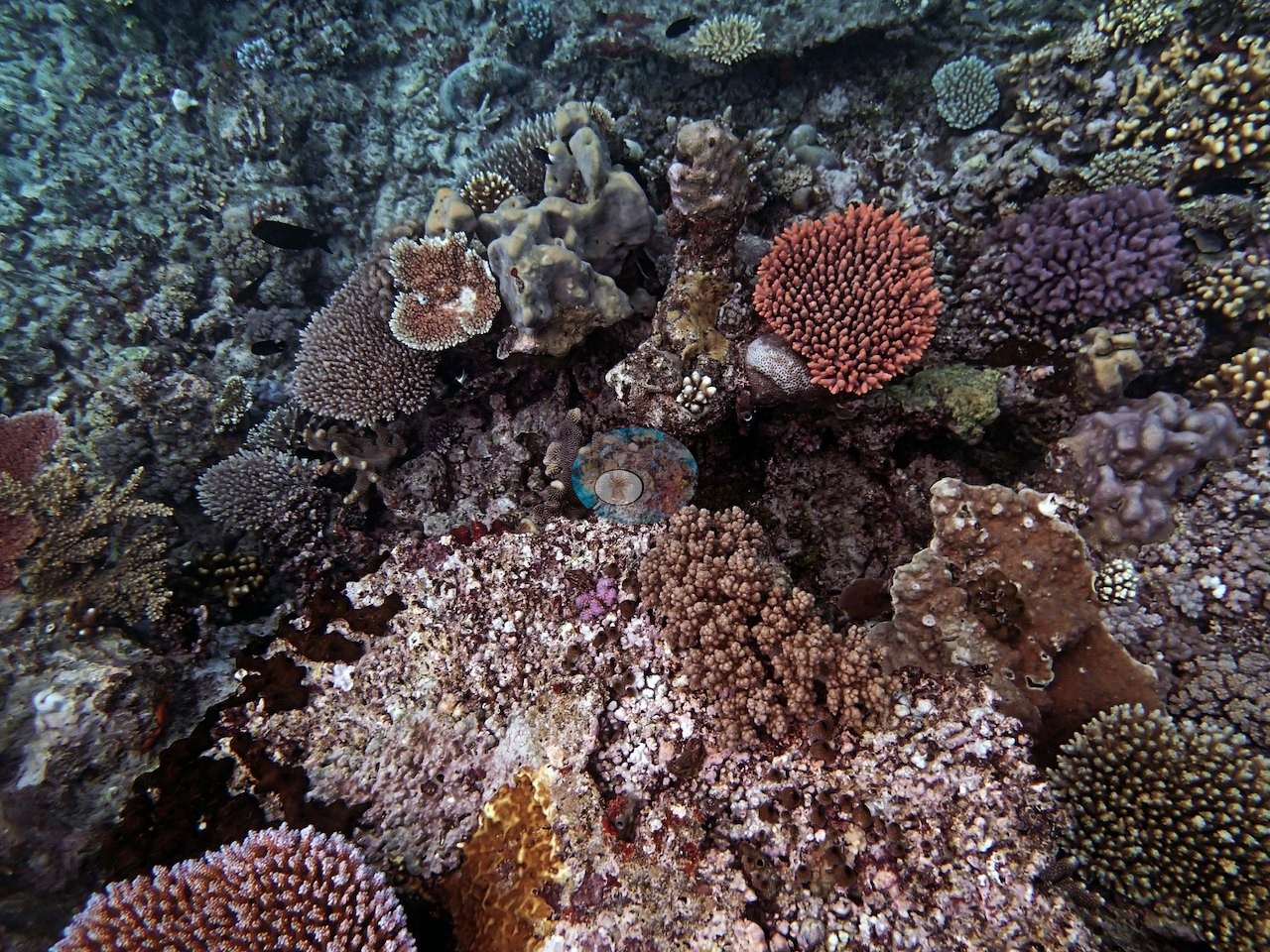
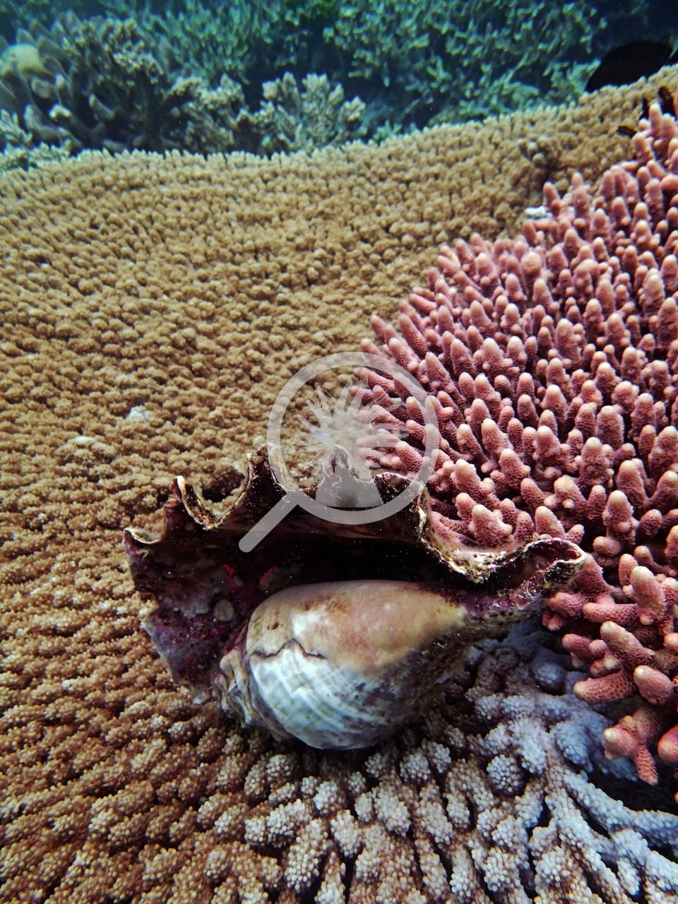
A large gastropod shell lay wedged where a tan tabular colony met pink and blue-tipped branching colonies (e.g., Image 7).
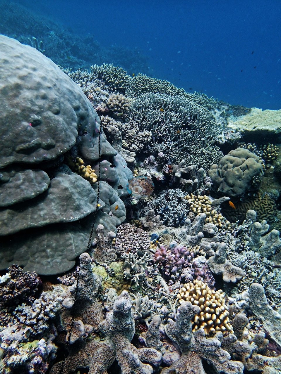
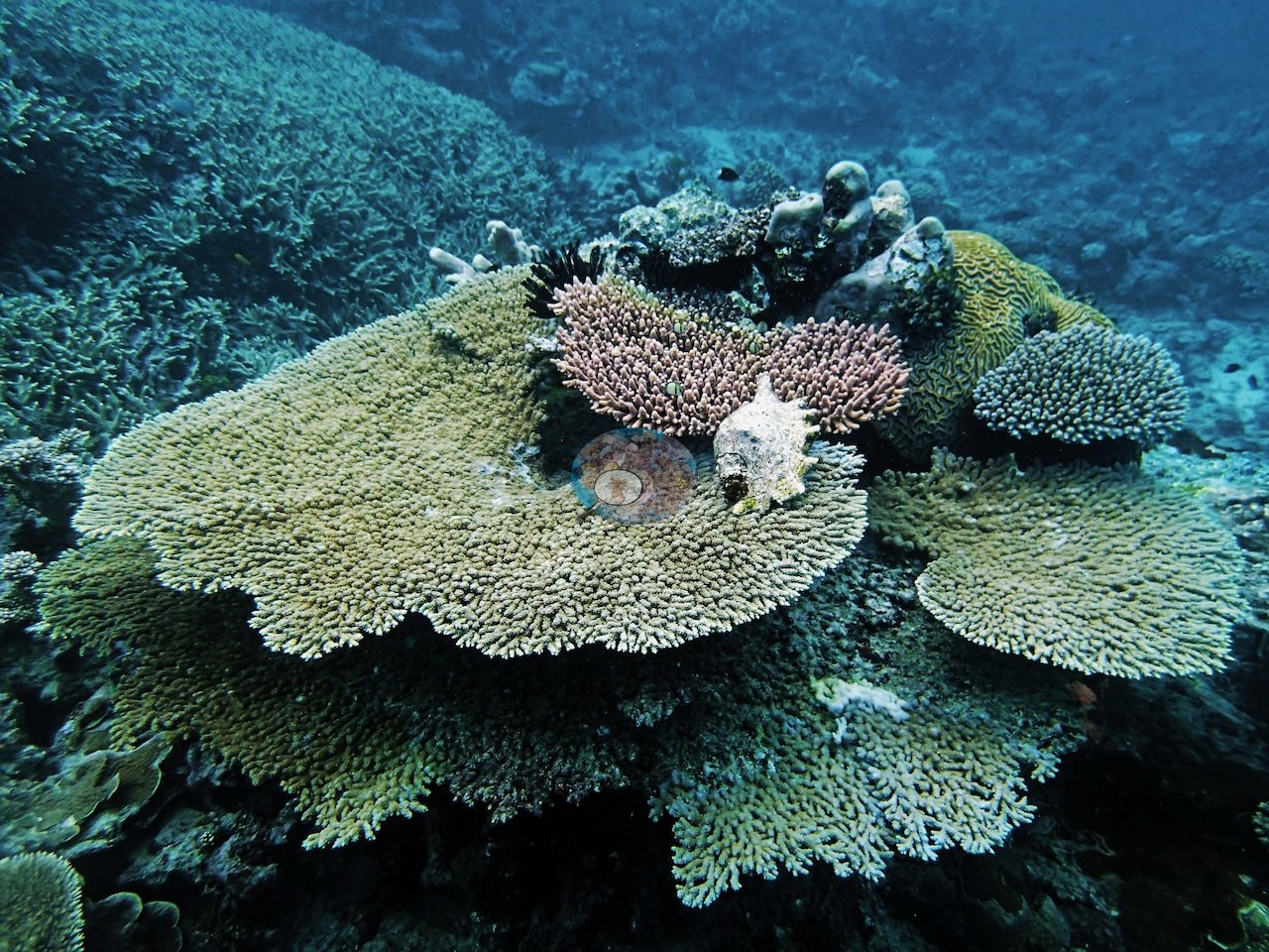
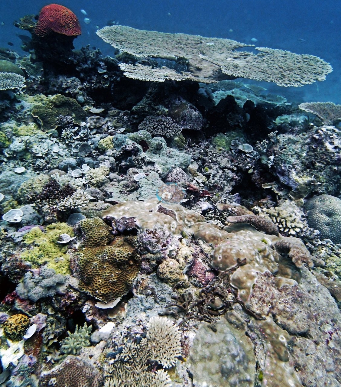
Toward the reef edge, a large tiered massive colony stood beside a staghorn field and upright lobed colonies (e.g., Image 13), and broad horizontal Acropora tables stacked in tiers, a pink bushy colony and a large spiny gastropod shell resting on top (e.g., Image 14).
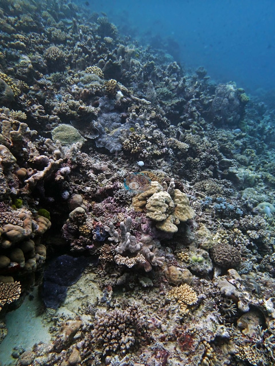
Down the slope the same mosaic of massive, corymbose, branching, and tabular colonies continued into deep water (e.g., Image 18, Image 20, Image 26, Image 27), with a reddish-purple table colony standing out among gray-green plating colonies (e.g., Image 24).
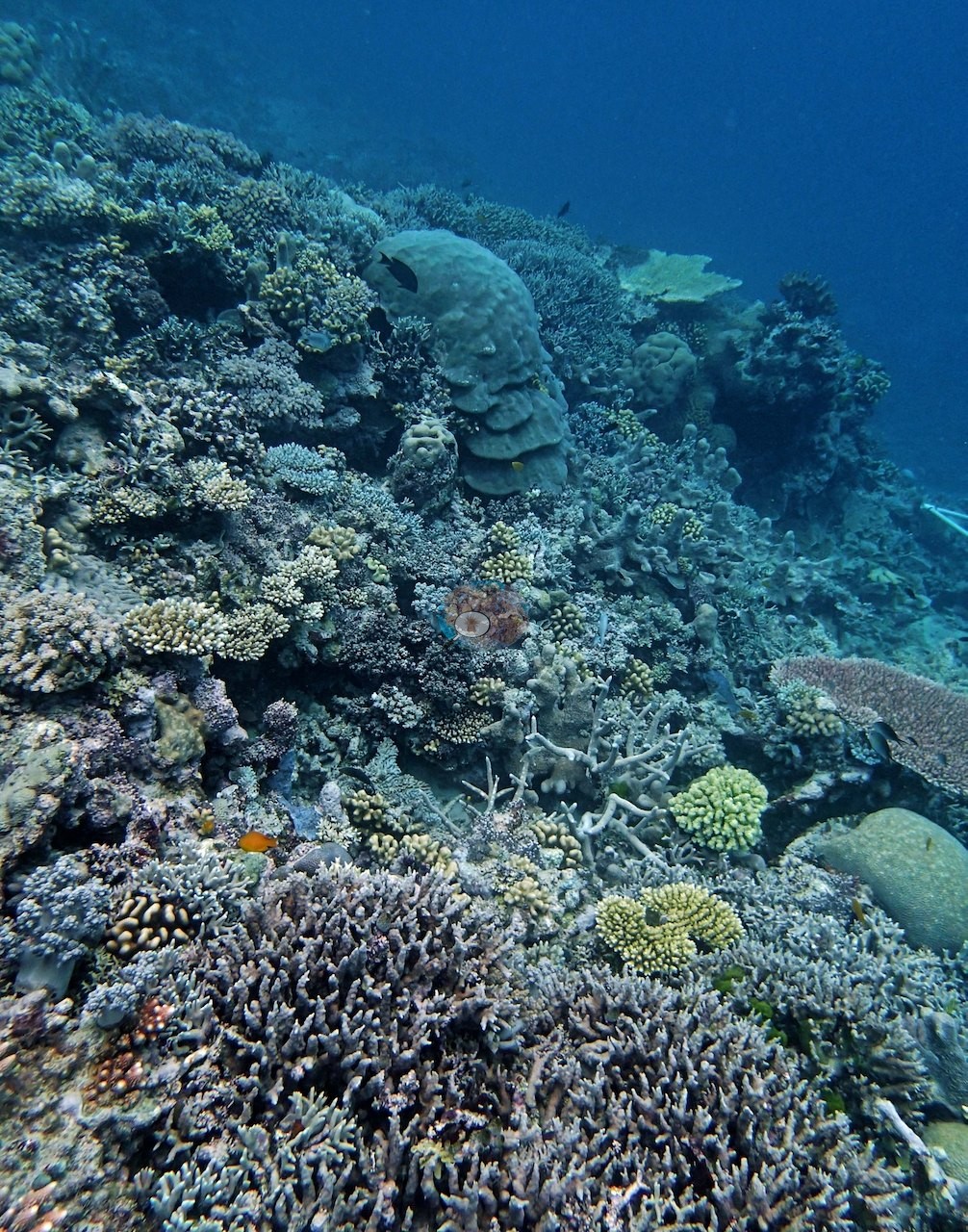
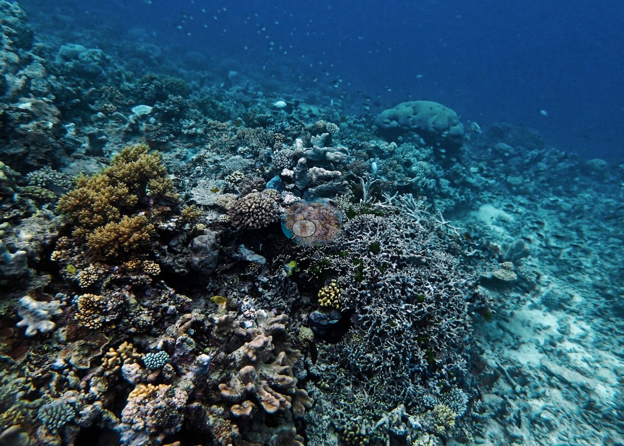
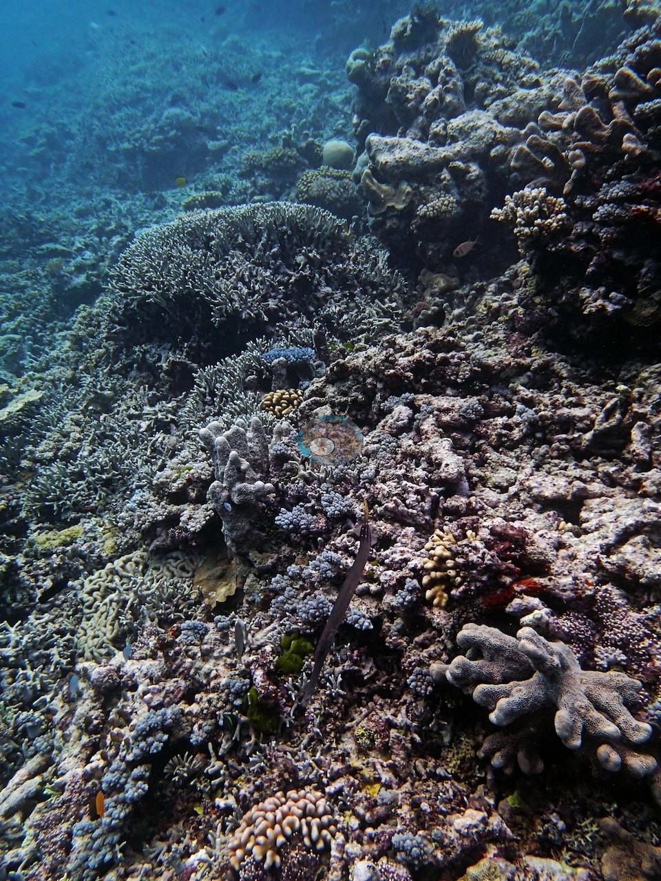
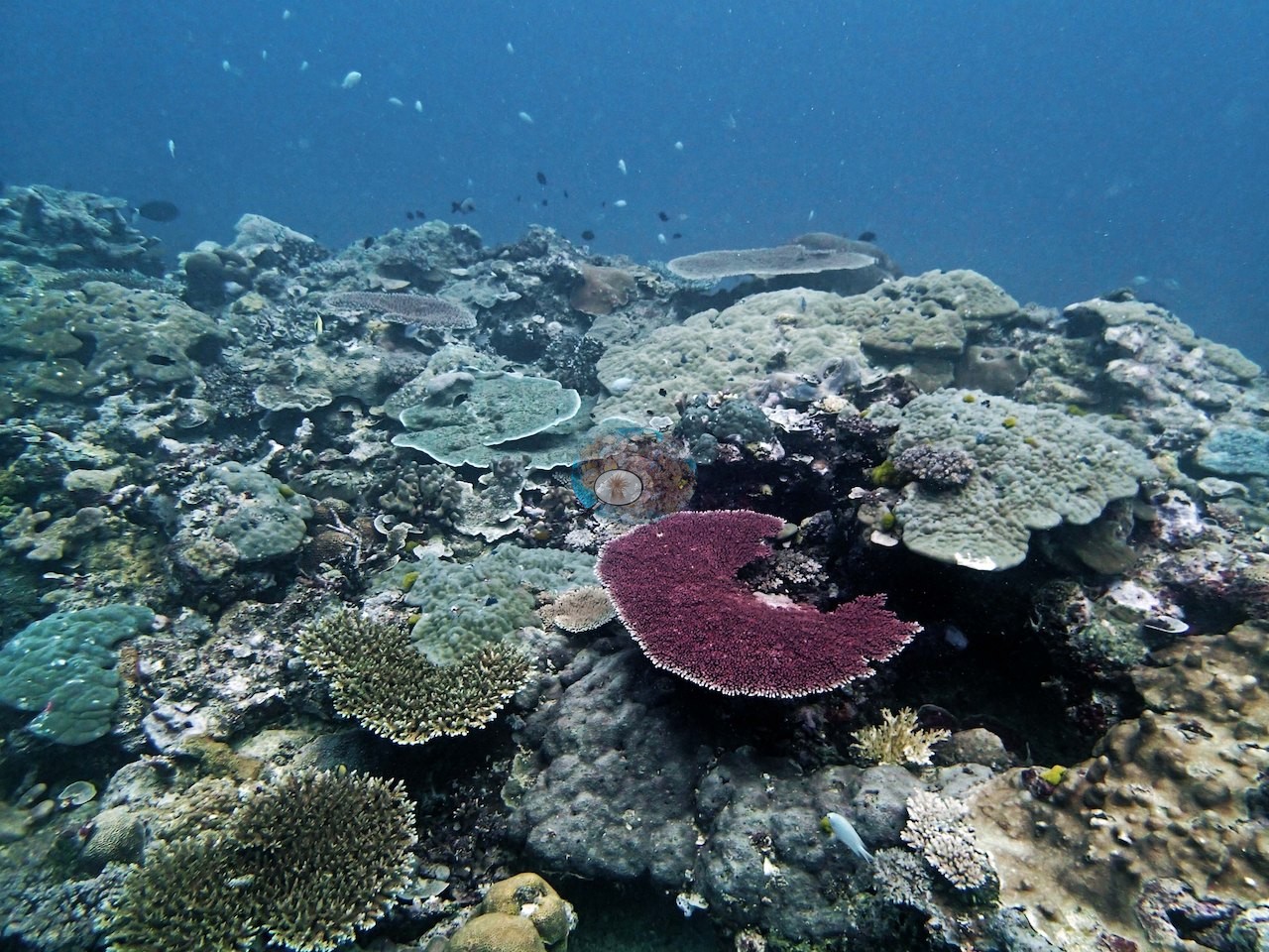
At the base, branching patch heads rose from sand (e.g., Image 21), and in hazier shallow water the cover shifted to knobby corymbose colonies and small massives on turf-covered rock (e.g., Image 22).
Coral cover
- Reference: Khaled bin Sultan Living Oceans Foundation, Global Reef Expedition (in situ transect)
- What the data say. Overall cover across the site's point-count survey stood at 36%.
- One transect was scored at this station (36%; n = 1, so no SE; Living Oceans Foundation per-transect benthic data); it is the expedition's station value printed above.
- What Anderson saw. His field notes call this stop at Unnamed Reef 11-096 a "strikingly beautiful site with high coral cover and diversity" and estimate live coral cover by eye along his own three transects at 40% (19.5 m), 60% (19 m), and 80-90% (10 m) (e.g., Image 1, Image 2).
Stressors
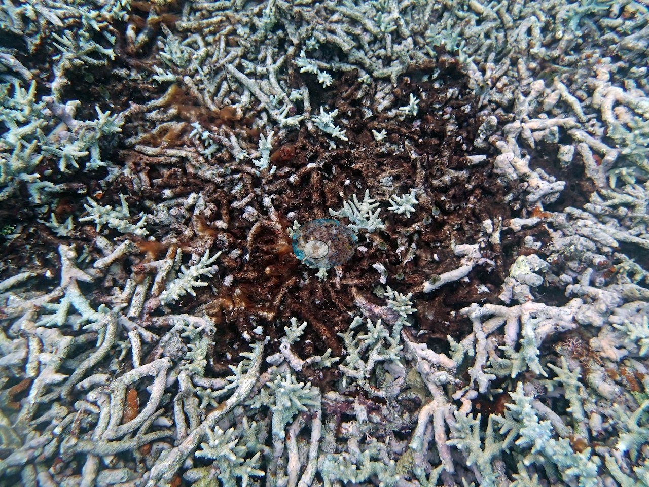
The one blemish on an otherwise thriving picture: at 20 m, needle coral (Seriatopora hystrix) was flagged as dying or sickly. One close frame shows a patch of staghorn skeleton overgrown with brown turf in the middle of a pale thicket, a small patch of mortality the photograph cannot date or assign a cause to Image 16.
Fish and other fauna
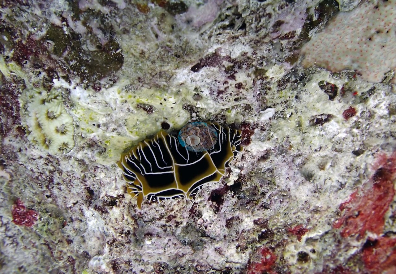
Fish life was abundant too, described plainly as "tons of small fish." A black-and-yellow polyclad flatworm crept over coralline-crusted rock (e.g., Image 23).
Comparisons with other data
No prior survey data from this site. If you would like to share yours, please email us.
CV model comparison
More info
The photo row below is a methods comparison only: it is never averaged with the transect and never used as the headline. It compares site means only: the photos come from the same survey dive as the transect but not from the same points.| Method | Hard-coral cover (%) | Photos (n) ± SE |
|---|---|---|
| Transect (published) | 36.0 | |
| CoralSCOP edited | 39.1* | n = 18 · SE 5.3 |
| Claude (edited photos) | 46.9* | n = 18 · SE 4.7 |
The two computer-vision methods differ by 7.8 pp (39–47%) and read 3.1–10.9 pp above the in-water transect (36%); the headline is the in-water transect because a diver transect is independent of the photographs.
*Based on only 18 reef-habitat photos; this estimate is less certain.
Data & links
- Download benthic survey data (CSV) ↗
- Download all LOF-GRE data (933 sites) ↗
- Photo portfolio ↗
- Coral Reef Diagnostics country page ↗
All images from Coral Reef Diagnostics. Data are from the Khaled bin Sultan Living Oceans Foundation Global Reef Expedition and Coral Reef Diagnostics.
 © Coral Reef Diagnostics.