
Part of the regional summaries: Fiji - Vanua Balavu, Fiji - Northern Lau (Vanua Balavu / Cicia / Nayau), and Fiji - Lau
More info
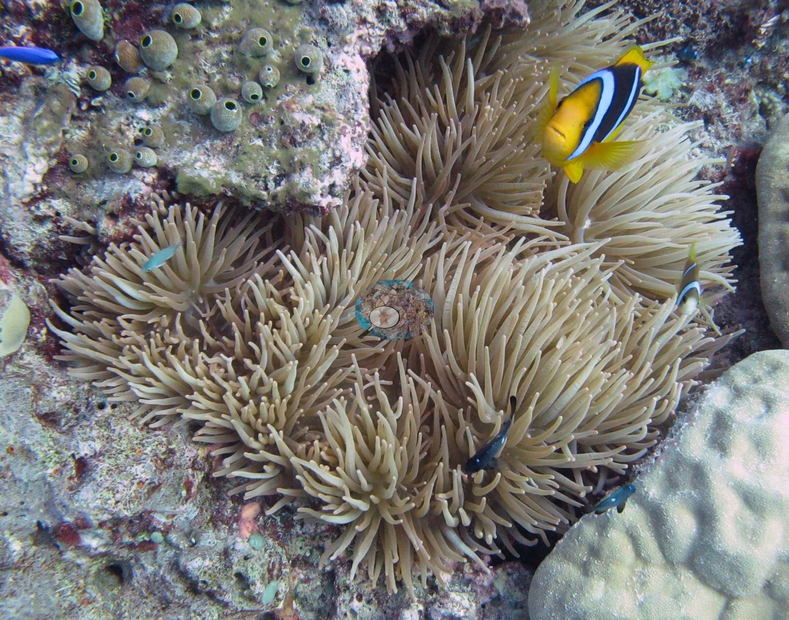
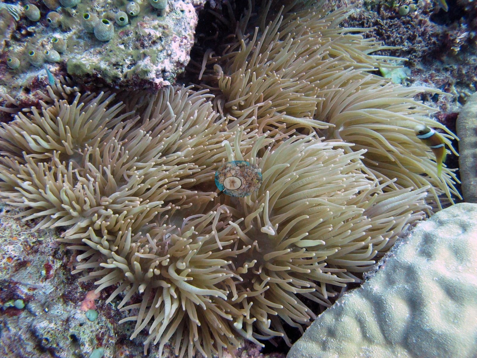
Fiji (NOAA CRW virtual station)A Vanua Balavu site photographed only in close-up, featuring a Porites colony studded with colorful Christmas tree worms, a host anemone with its anemonefish, and the find of the dive, a large triton's trumpet shell. The one colony Anderson sampled here was flagged as a physiological stress outlier in a 2017 study, and cover was 22%. Surveyed on the Living Oceans Foundation's Global Reef Expedition.
E dua na vanua e Vanua Balavu a taba voleka ga, e vakaraitaki kina e dua na lase Porites e vakaiukuuku ena ulo (Christmas tree worms) e roka vakaidina, e dua na anemone kei na nona ika (anemonefish), kei na ka e kunei vinaka duadua ena nunu, e dua na davui levu (triton's trumpet shell). Na dua ga na lase a taura kina na ivakaraitaki ko Anderson eke a vakaraitaki me duatani ena leqa vakayago (physiological stress outlier) ena dua na vakadidike ni 2017, ka 22% na ubi ni lase (coral cover).
AI translation of this summary; the English is authoritative. (low confidence)
CTD-cast midpoint; the cast read 26.8°C throughout
🗺 Detailed map of the Global Reef Expedition dive sites, Vanua Balavu, Fiji (Khaled bin Sultan Living Oceans Foundation; opens in a new window)
Site description
General benthic features
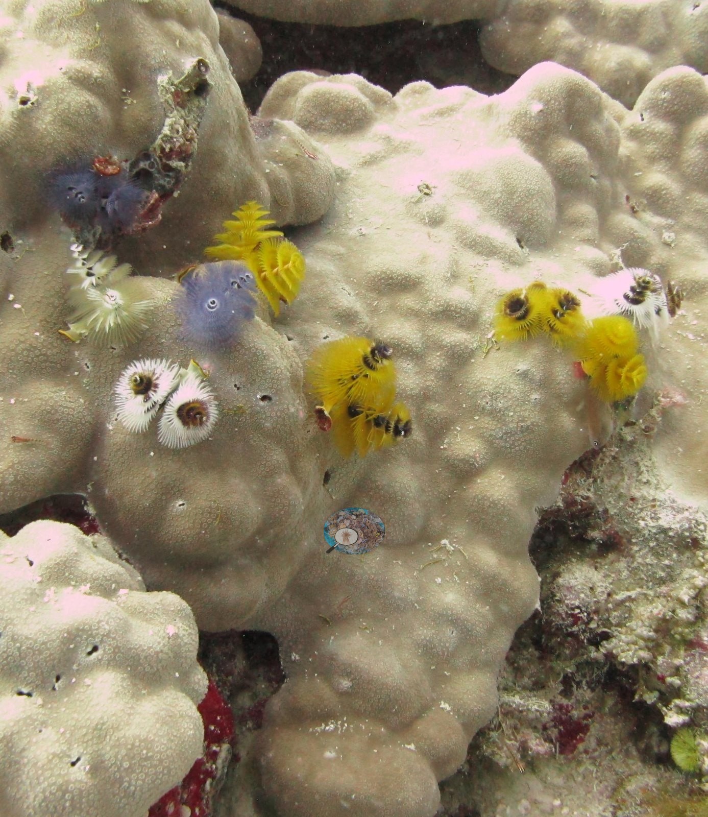
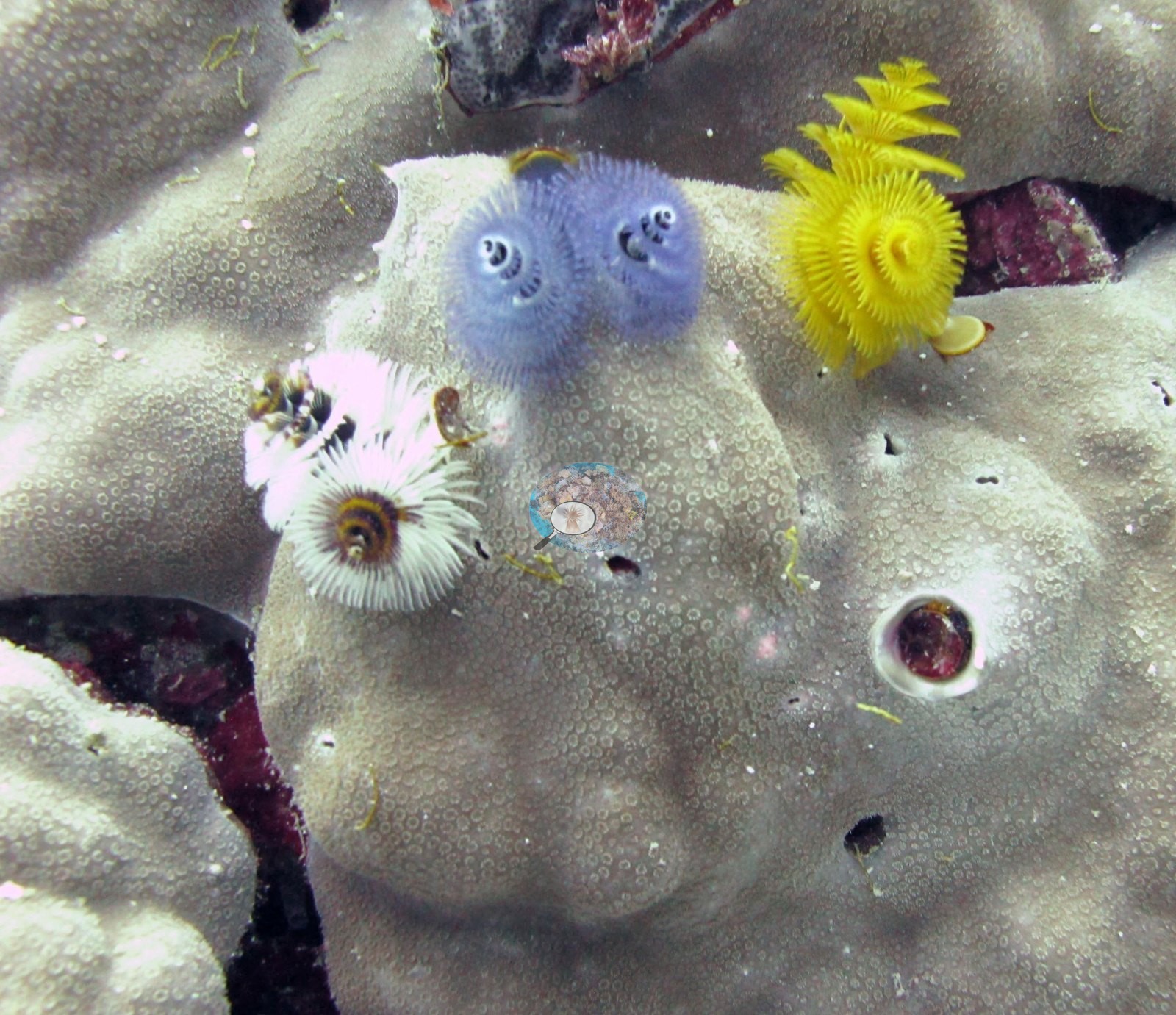
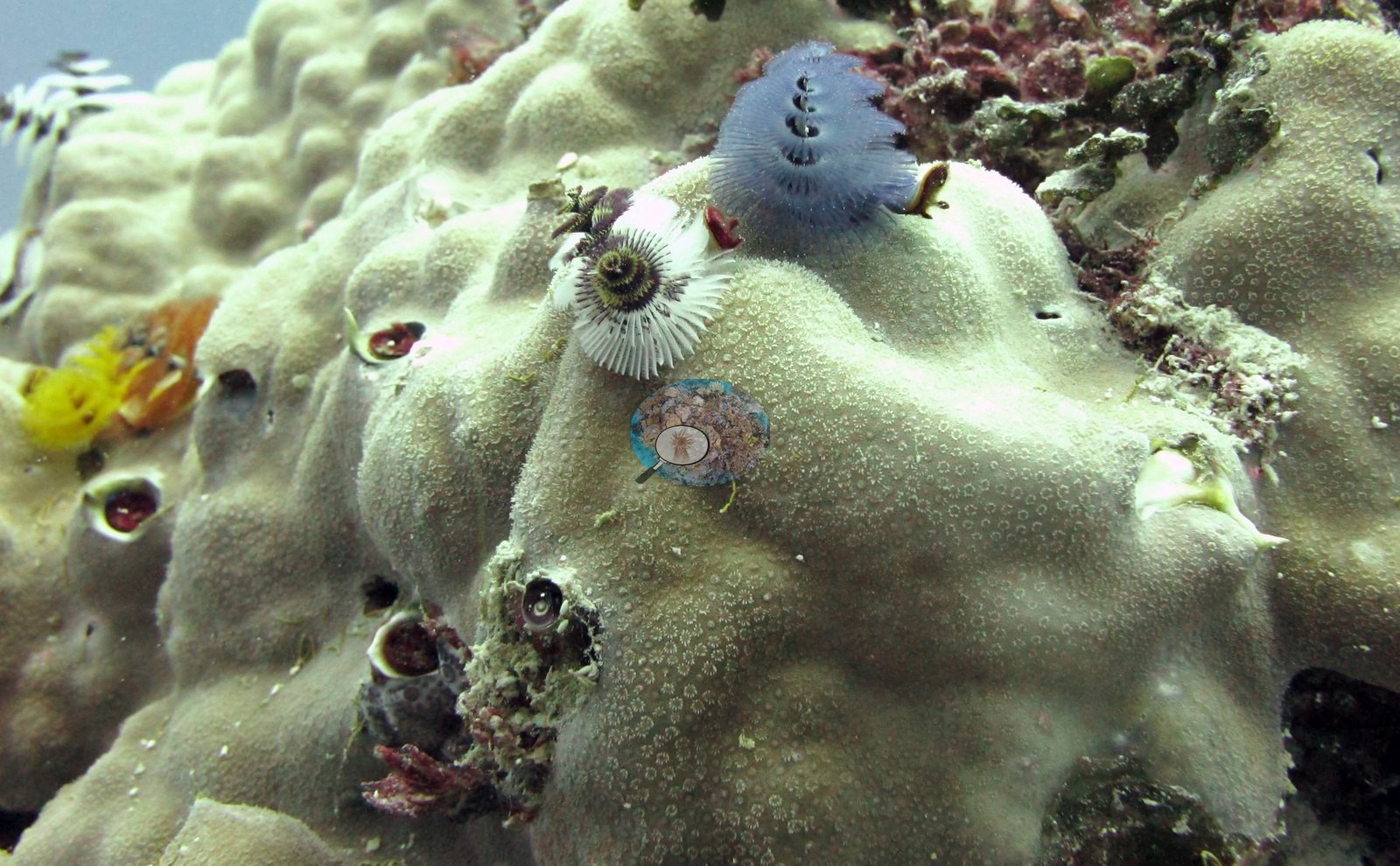
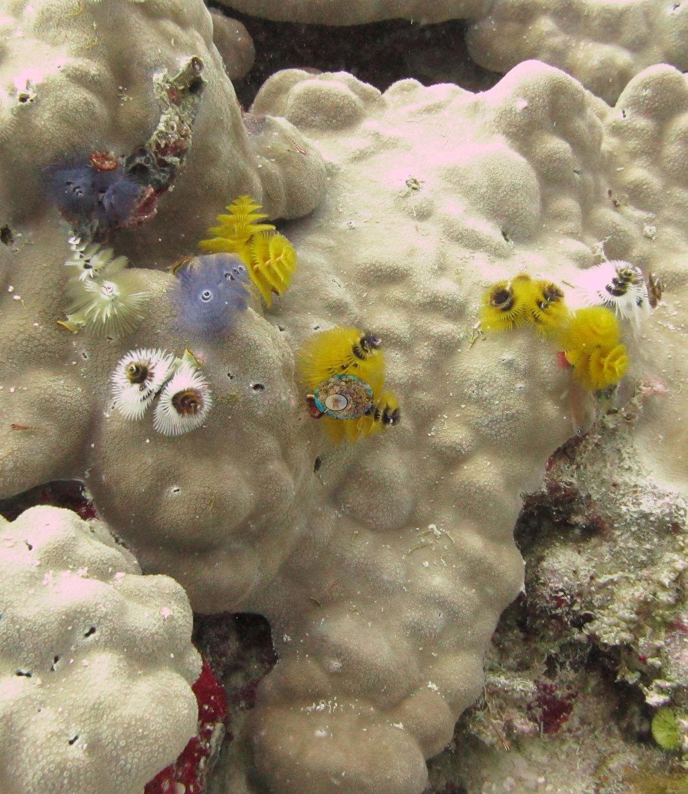
Three frames are given to Christmas tree worms (Spirobranchus) on the cream-green lobed surface of massive Porites: a tight macro of slate-blue, yellow, white, and pale pink crowns among the fine porous polyps, with the gaping shells of embedded bivalves beside them (e.g., Image 4); a wider view of the same colony surface showing worms scattered across the lobes and dark bivalve openings sunk into the skeleton (e.g., Image 5); and a third on knobbier Porites framework with blue and yellow crowns and pink coralline crust along the margins (e.g., Image 3).
Two frames cover the host anemone: one from above with the tan, pale-tipped tentacle field spread over the rock and an anemonefish with a white head bar in the tentacles (e.g., Image 9), and one from the side showing the tentacle mass draped between boulders with a smooth Porites dome below and small damselfishes at the edge (e.g., Image 10).
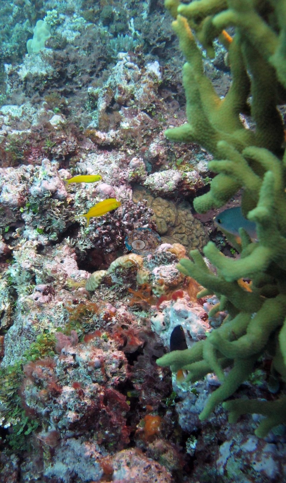
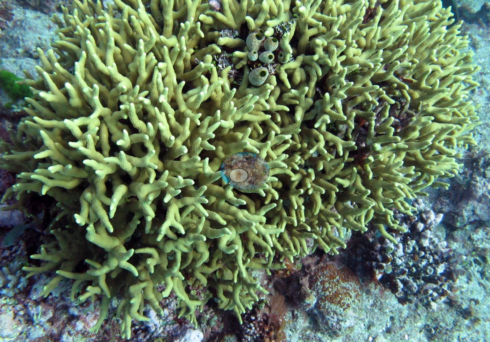
Two frames add further subjects: a green branching Porites-type colony with fine, tightly packed branchlets and small fish among them, which Anderson identified as Fiji fang blennies (e.g., Image 11), and a second, larger green colony of the same branching form with thicker, more irregular contorted branches, identified as an unusual Porites (e.g., Image 12).
One frame shows a colleague's tagged pale pink-and-cream branching colony with a numbered tag fixed beside it on a cable tie, the antler branches threading up through olive turf and red algal crust (e.g., Image 6).
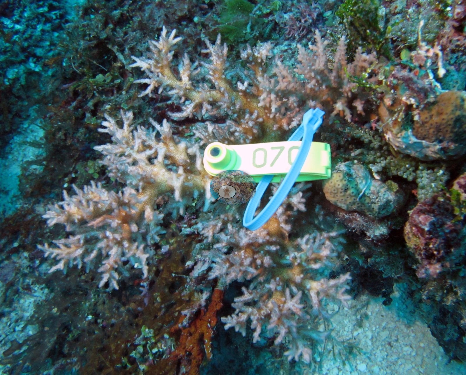
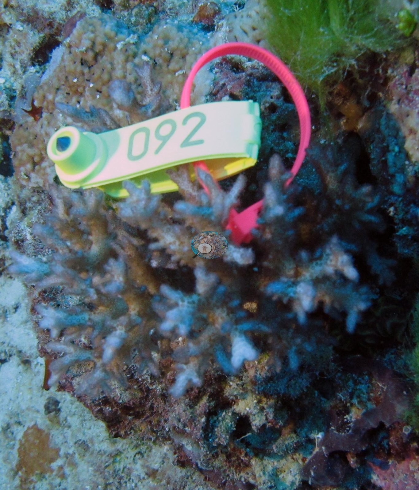
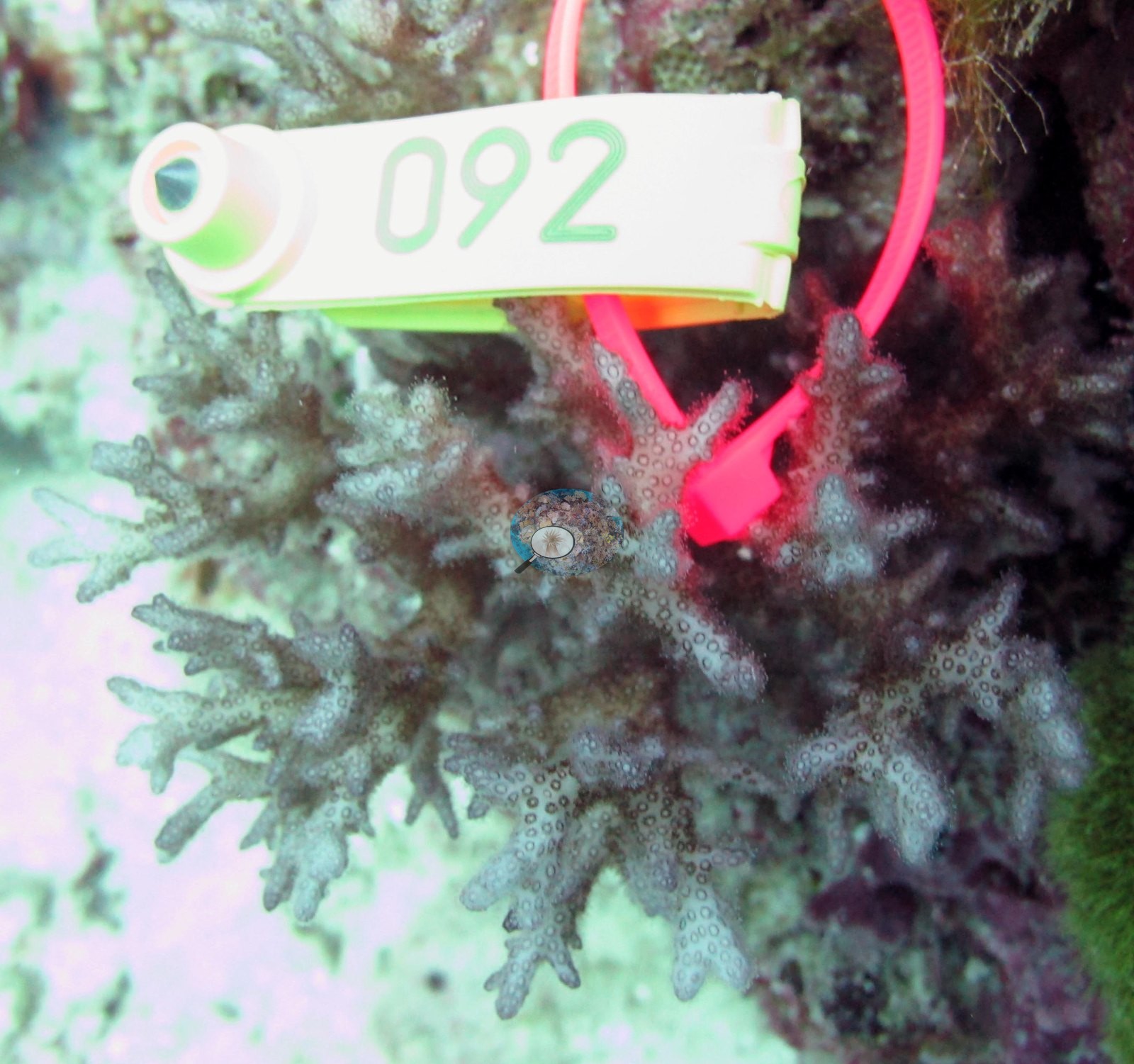
The colony Anderson sampled here, F153 (P. acuta on the sample sheet), is the tagged branching colony in Image 1 and Image 2.
Coral cover
- Reference: Khaled bin Sultan Living Oceans Foundation, Global Reef Expedition (in situ transect)
- Coral cover: 22%. Live hard coral measured on in situ benthic transects during the Khaled bin Sultan Living Oceans Foundation Global Reef Expedition. Transect data are this site's authoritative cover value; no computer vision or image-derived product overrides it.
- Transect mean 22 ± 4.2% SE (n = 4 transects; Living Oceans Foundation per-transect benthic data); the headline above is this all-transect mean.
Stressors
Apart from the sampled colony F153 (below), no colony in view is bleached or shows a disease lesion or crown-of-thorns scar, but with twelve close-ups of hand-picked subjects that is not a statement about the station. Anderson's sample sheet records the colony sampled here (F153) as 10% bleached; the bleached portion was sampled. The one coral colony from this site with an outlier score was flagged as a physiological outlier (Mayfield et al. 2017, PLoS ONE). The Living Oceans Foundation transects here recorded 6.8% cyanobacteria and no recently dead coral.
Fish and other fauna
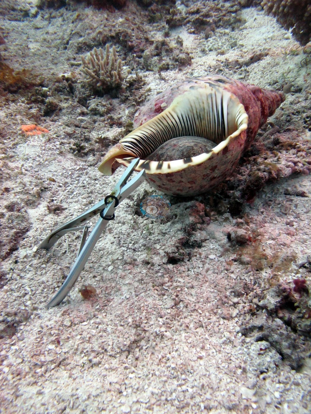
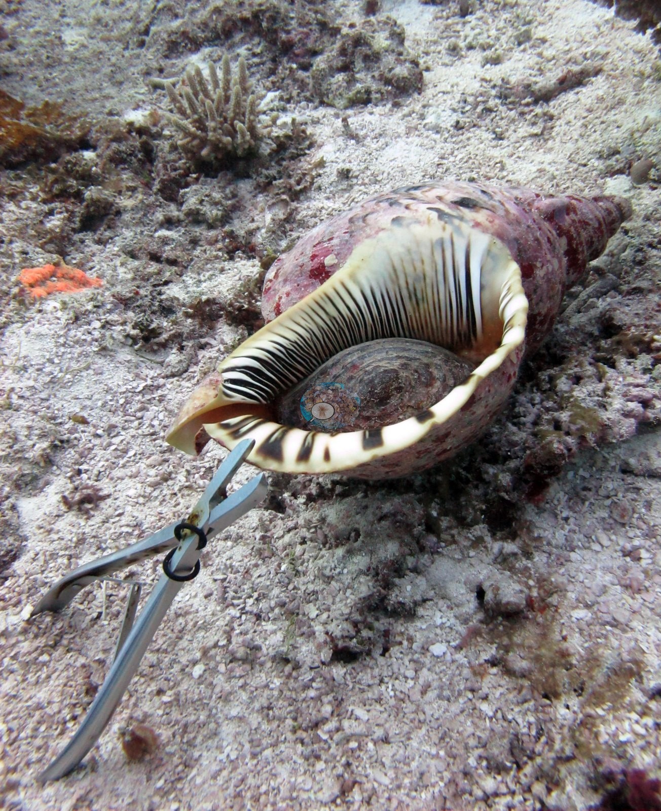
The find of the dive (Anderson's log: 'Saw huge trumpet triton') is a triton's trumpet (Charonia tritonis), photographed twice, from two angles on coarse pale rubble, its cream aperture ridged with dark brown and its outer lip barred black and white, with a pair of bone cutters propped alongside for scale in both (e.g., Image 8, Image 7).
Comparisons with other data
Apart from the Living Oceans Foundation survey shown here, we found no other surveys at this site as of 2026. If you would like to share yours, please email us.
Other notes
The complete photo set for this station contains no reef-scale photography at all — every frame is a close-range subject portrait. What these frames cannot settle: there is not a single wide or medium-range frame in this photo set, so nothing in it speaks to the reef's structure, relief, spatial variation, or the algal dominance recorded in the survey — the site's 22% cover figure and the surrounding benthos are known from the transect, not from this imagery.
Data & links
- Download benthic survey data (CSV) ↗
- Download all LOF-GRE data (933 sites) ↗
- Photo portfolio ↗
- Coral Reef Diagnostics page for this area ↗
All images from Coral Reef Diagnostics. Data are from the Khaled bin Sultan Living Oceans Foundation Global Reef Expedition and Coral Reef Diagnostics.
 © Coral Reef Diagnostics.