
Part of the regional summaries: Fiji - Vanua Balavu, Fiji - Northern Lau (Vanua Balavu / Cicia / Nayau), and Fiji - Lau
2013-06-24
More info
Fiji (NOAA CRW virtual station)A Vanua Balavu site dived twice, with milky, silty water over sand and rubble patched with algae-crusted framework, and a reef shark cruising low. The close-ups are the highlight: a fully pigmented massive coral seen polyp by polyp and a large mauve anemone (Anderson's identification) ringed with pale tentacles, at 30% cover. Surveyed on the Living Oceans Foundation's Global Reef Expedition.
E dua na vanua e Vanua Balavu a nunu kina vakarua, e vulavula vakasusu ka veidrukai na wai ena dela ni nuku kei na tikitiki ni lase mate (rubble) e veitawasei ena ivakatudei e ubi ena lumi, kei na dua na qio (reef shark) e qalo voli e ra. Na itaba taba voleka e ka vinaka duadua: e dua na lase levu (massive) e roka vinaka vakadua (fully pigmented) e raici vakapolyp, kei na dua na anemone levu e roka (mauve) (e vakatakilai ko Anderson) e vakavolivoliti ena tentacle vulavula, ka 30% na ubi ni lase (coral cover).
AI translation of this summary; the English is authoritative. (low confidence)
CTD-cast midpoint of 26.9–27.0°C; 23 Jun 2013 dive
One other CRD dive site with its own page within 5 km shown in orange — click a marker to open its page.
🗺 Detailed map of the Global Reef Expedition dive sites, Vanua Balavu, Fiji (Khaled bin Sultan Living Oceans Foundation; opens in a new window)
16 of the 24 photographs on this page are shown beside the text that discusses them under "Site description" below.
Site description
Setting
FJVB62 sits on the Vanua Balavu lagoon and was worked over two days, 23-24 June 2013, during the Khaled bin Sultan Living Oceans Foundation Global Reef Expedition; the dive reached 18.3 m. Visibility was clearly poor, which is probably why the camera went to macro.
General benthic features
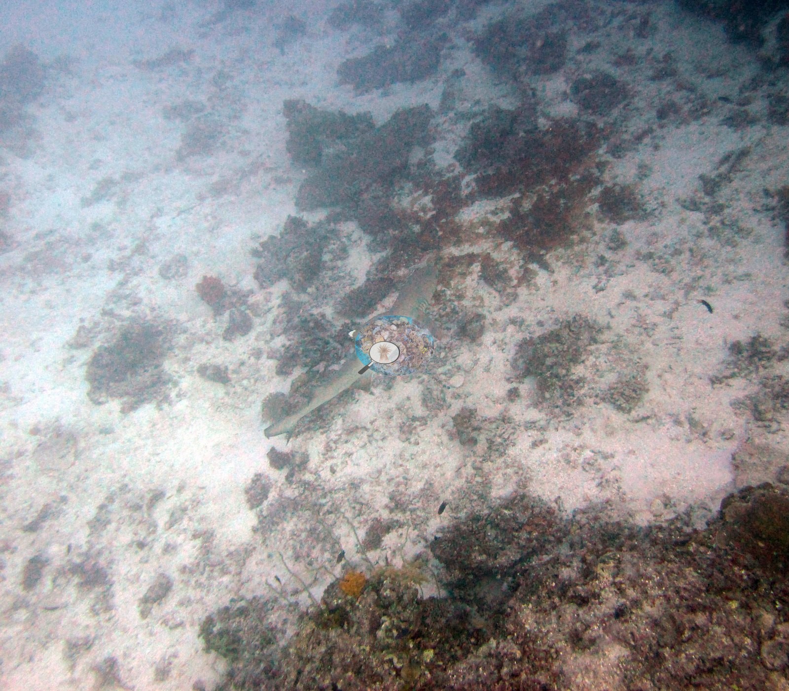
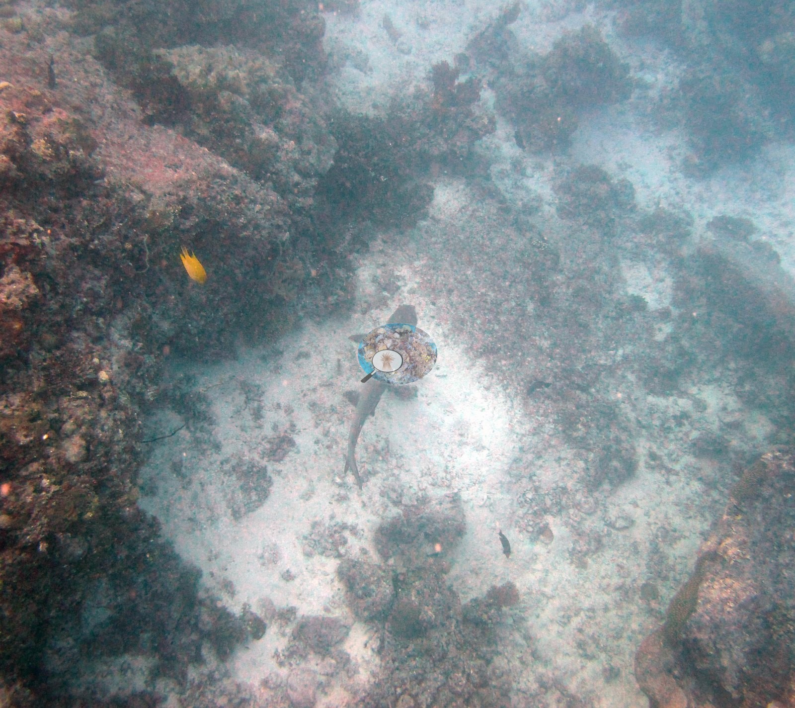
The only two frames with any spatial reach look down through a milky, sediment-laden water column onto pale sand and rubble patched with dark algal-crusted framework, a reef shark cruising low across both and a yellow damselfish holding near the reef edge (e.g., Image 15, Image 21); they are too hazy to settle relief, zonation, or how the coral is distributed here.
The close-ups record what was present rather than how much of it. Image 22 fills entirely with the surface of a massive coral head, hundreds of round, evenly spaced, fully pigmented corallites in orange and olive-green, each with a pale central mouth; Anderson identified the colony as Diploastrea (a field identification).
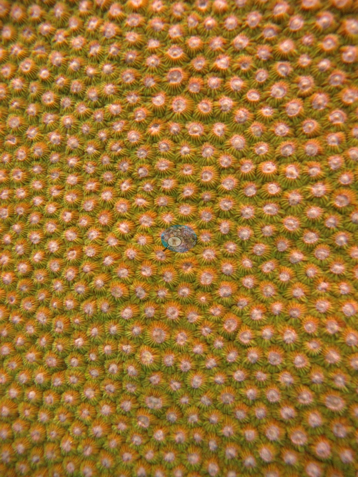
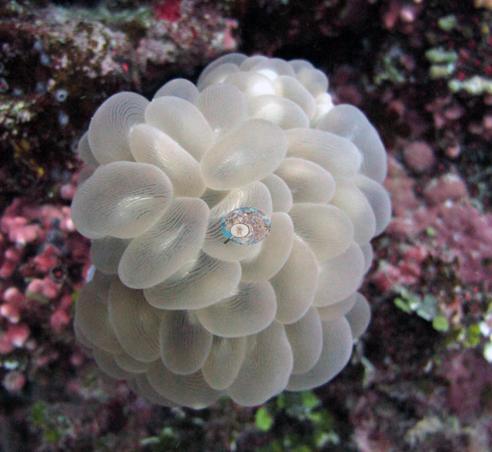
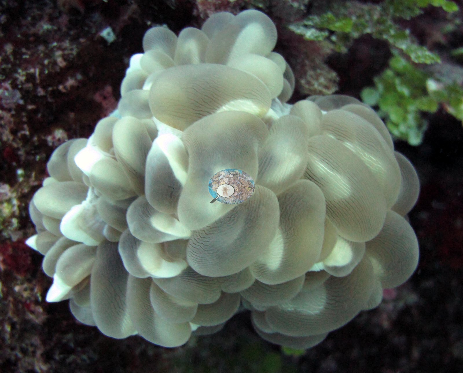
Image 25 and Image 23, identified as bubble coral, show its vesicles inflated over framework crusted with pink coralline algae, green segmented calcareous algae, and green algal tufts.
Coral cover
- Reference: Khaled bin Sultan Living Oceans Foundation, Global Reef Expedition (in situ transect)
- The in situ point-count transect recorded 30% live coral cover against 43% algal cover, and those figures — not the photographs — are the quantitative account of the site.
- Transect mean 30 ± 4.5% SE (n = 4 transects; Living Oceans Foundation per-transect benthic data); the headline above is this all-transect mean.
Stressors
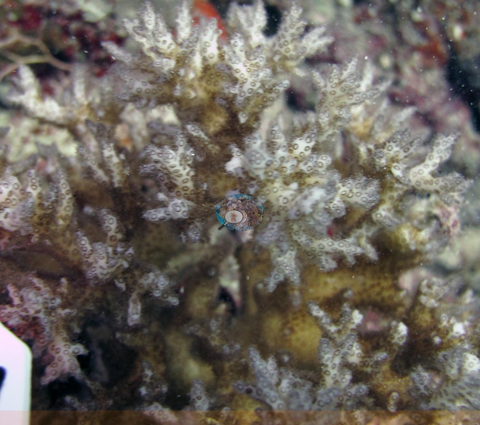
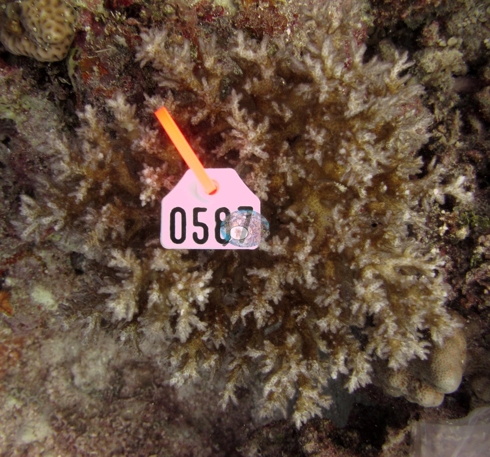
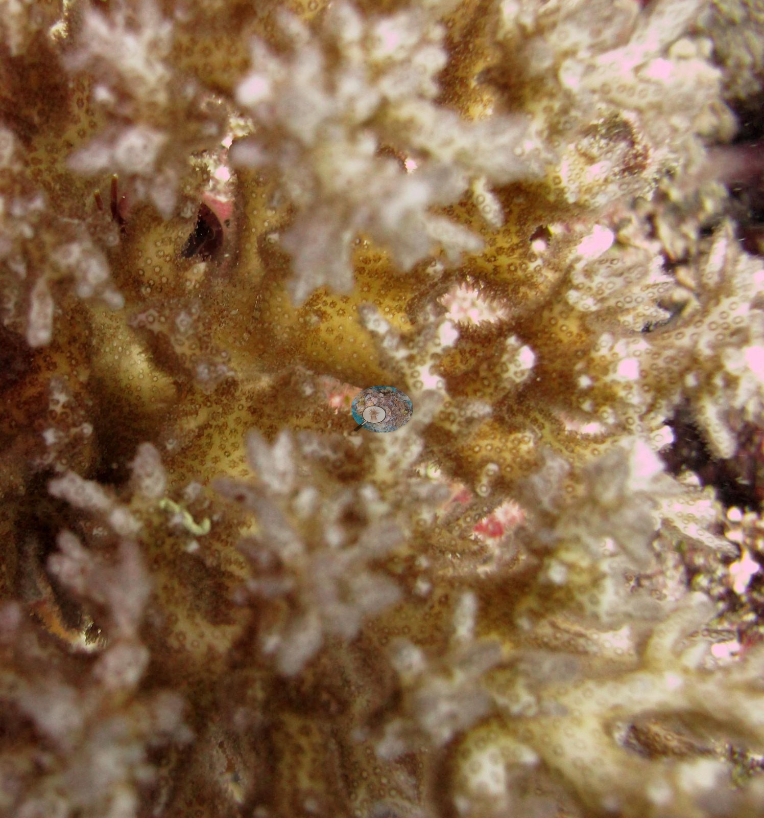
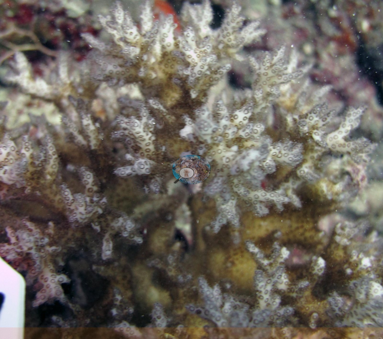
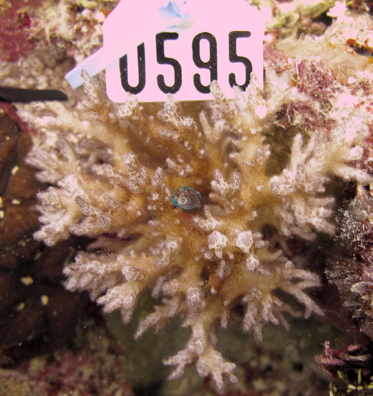
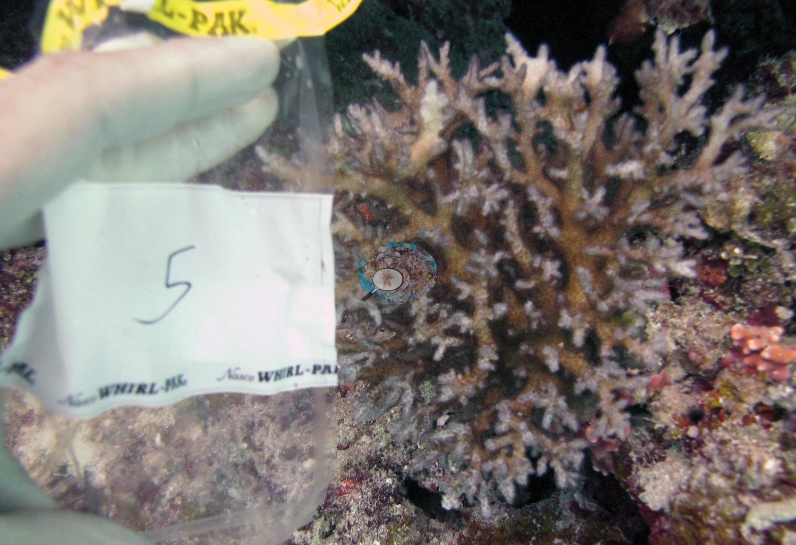
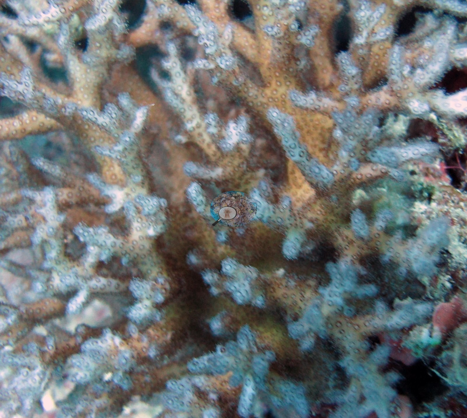
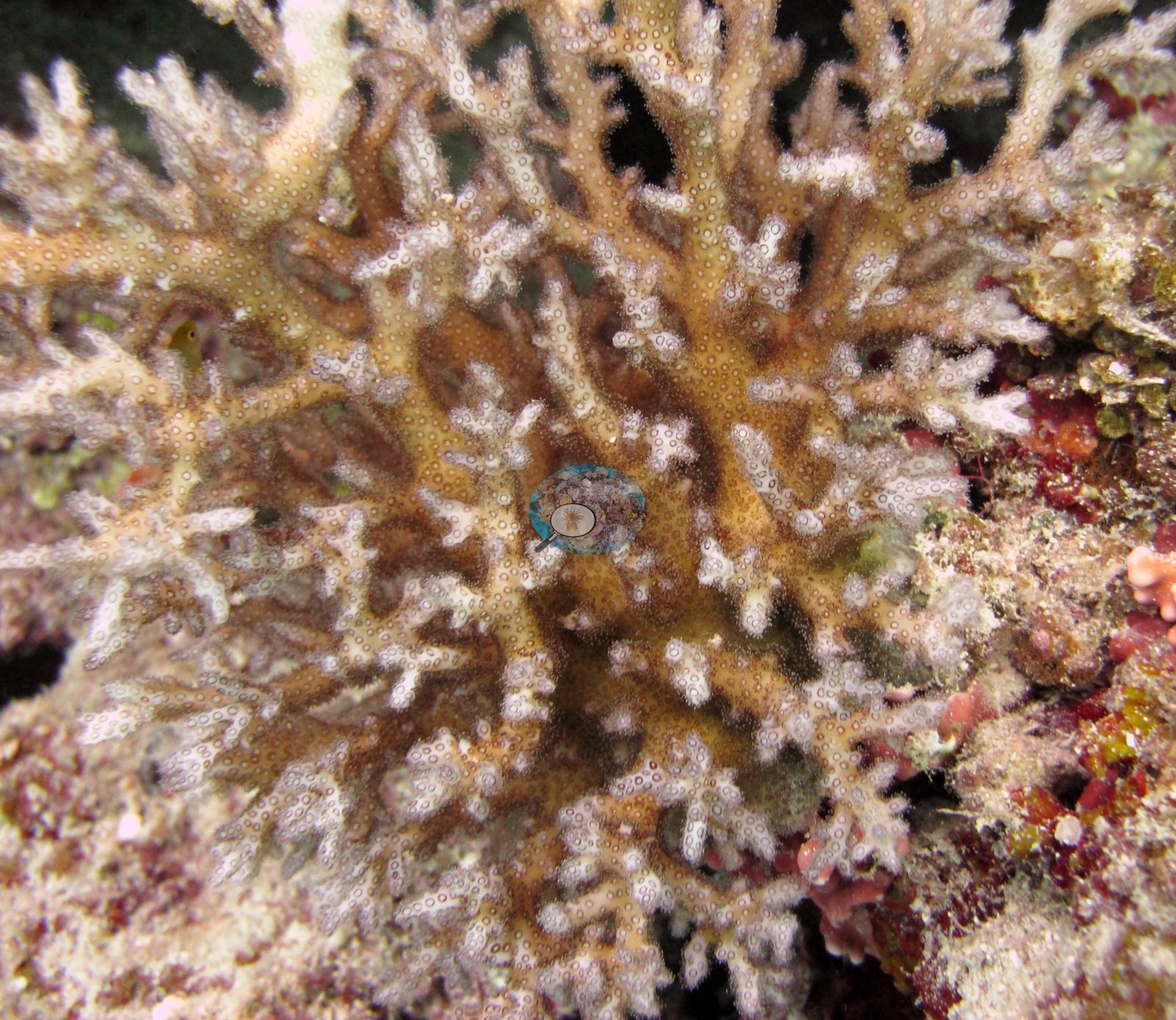
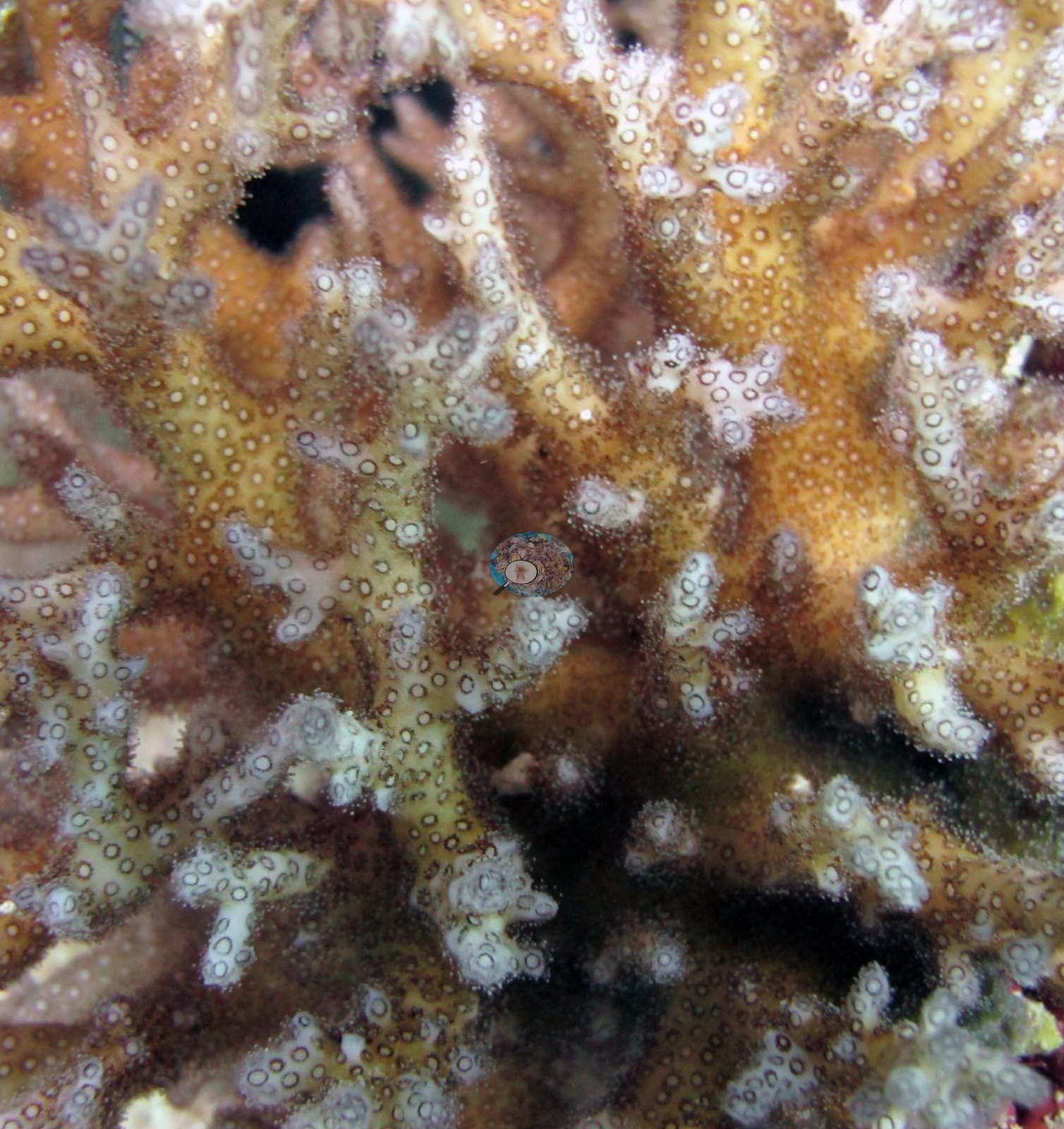
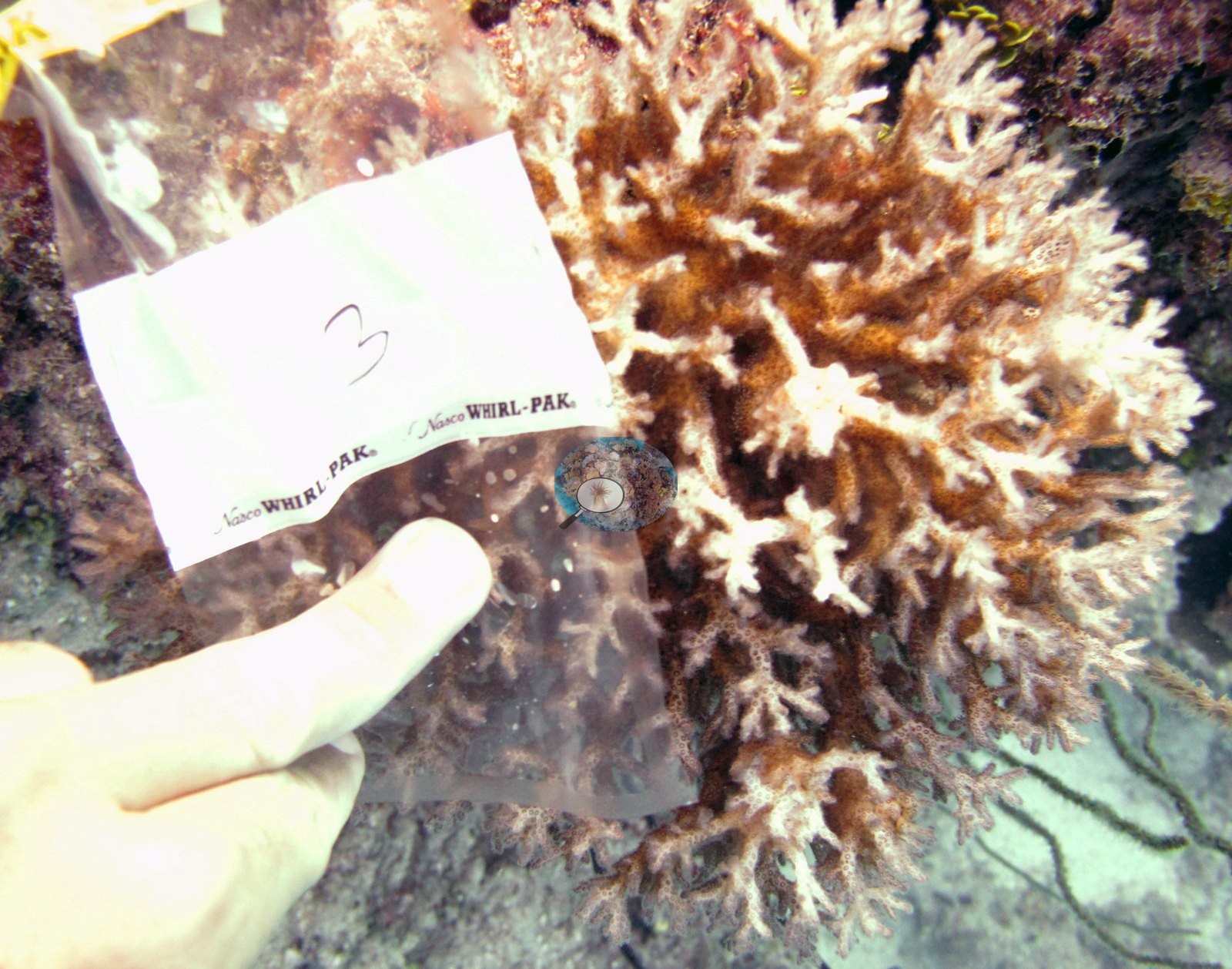
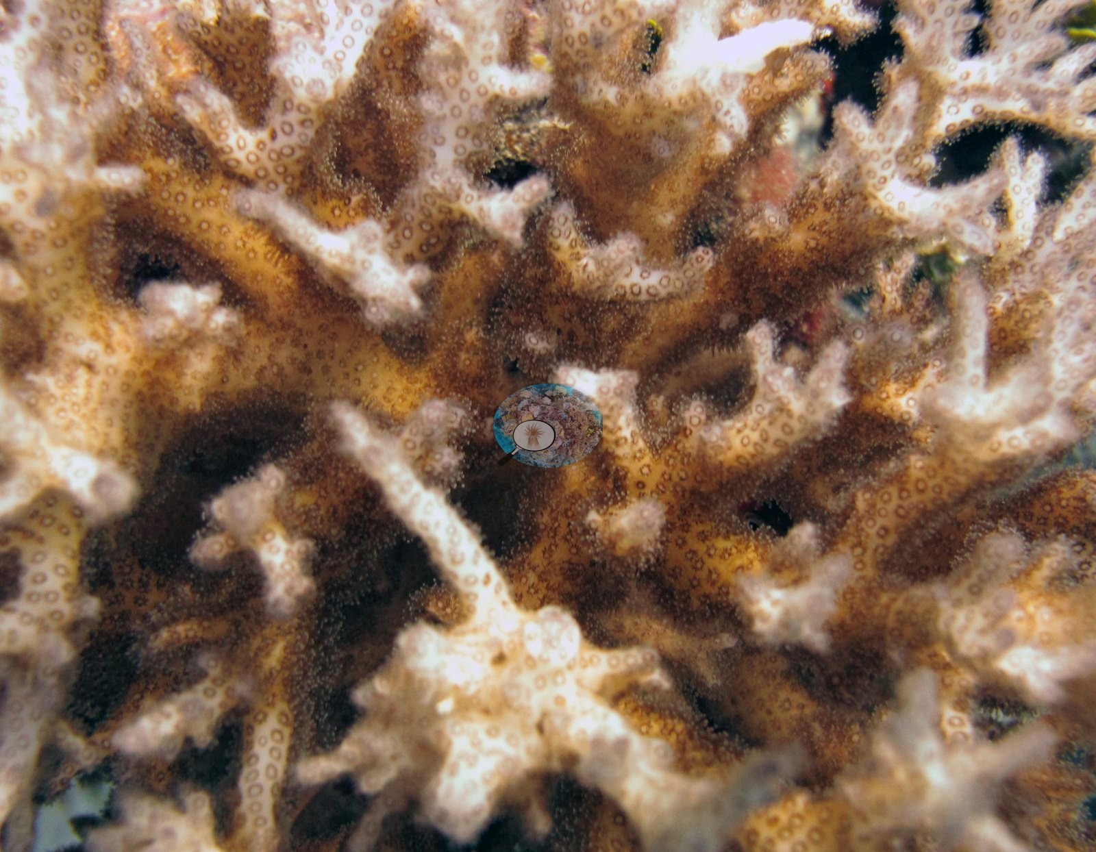
No lesions or crown-of-thorns damage appears on the corals in these frames, but a handful of subject portraits and two turbid wide shots cannot support a site-wide condition assessment. Anderson's sample sheet records two of the four colonies sampled here as partly pale (F136 '20% pale/bleached though likely healthy'; F137 '30% pale', its bleached part sampled), and the frames of F137 show that paling, with pale pink-white outer branches over brown interiors (e.g., Image 8, Image 9, Image 10).
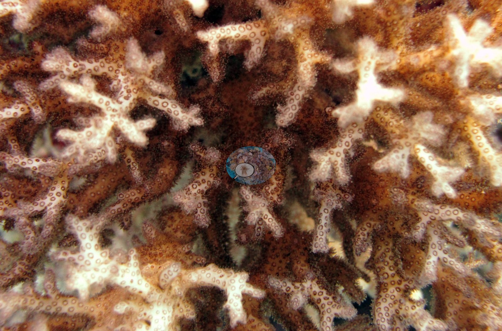
The one coral colony from this site with an outlier score was not flagged as a physiological outlier (Mayfield et al. 2017, PLoS ONE). The Living Oceans Foundation transects here recorded 8.0% cyanobacteria and no recently dead coral.
Fish and other fauna
Image 16, Image 19, Image 18, and Image 17 are four frames of one large fleshy anemone — Anderson's word for it — a smooth mauve-gray fluted disc with a raised central mouth ringed by blunt pale tentacles over a dark oral disc speckled with pale tubercles, worked from several angles and distances.
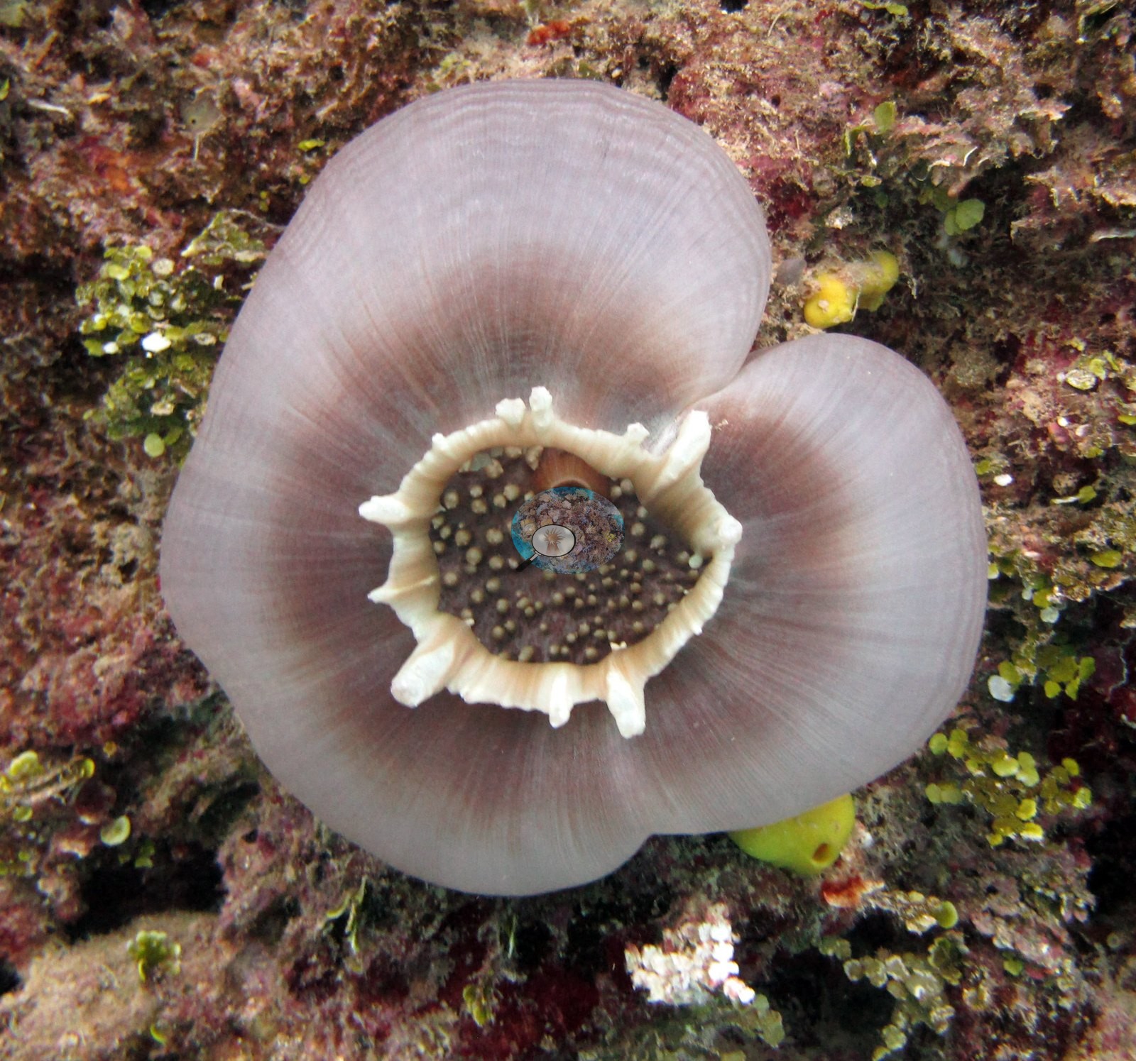
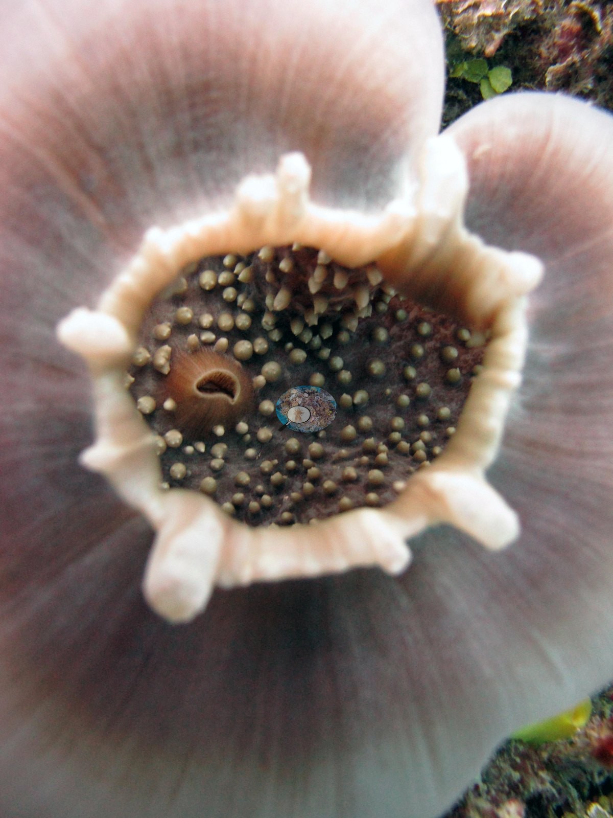
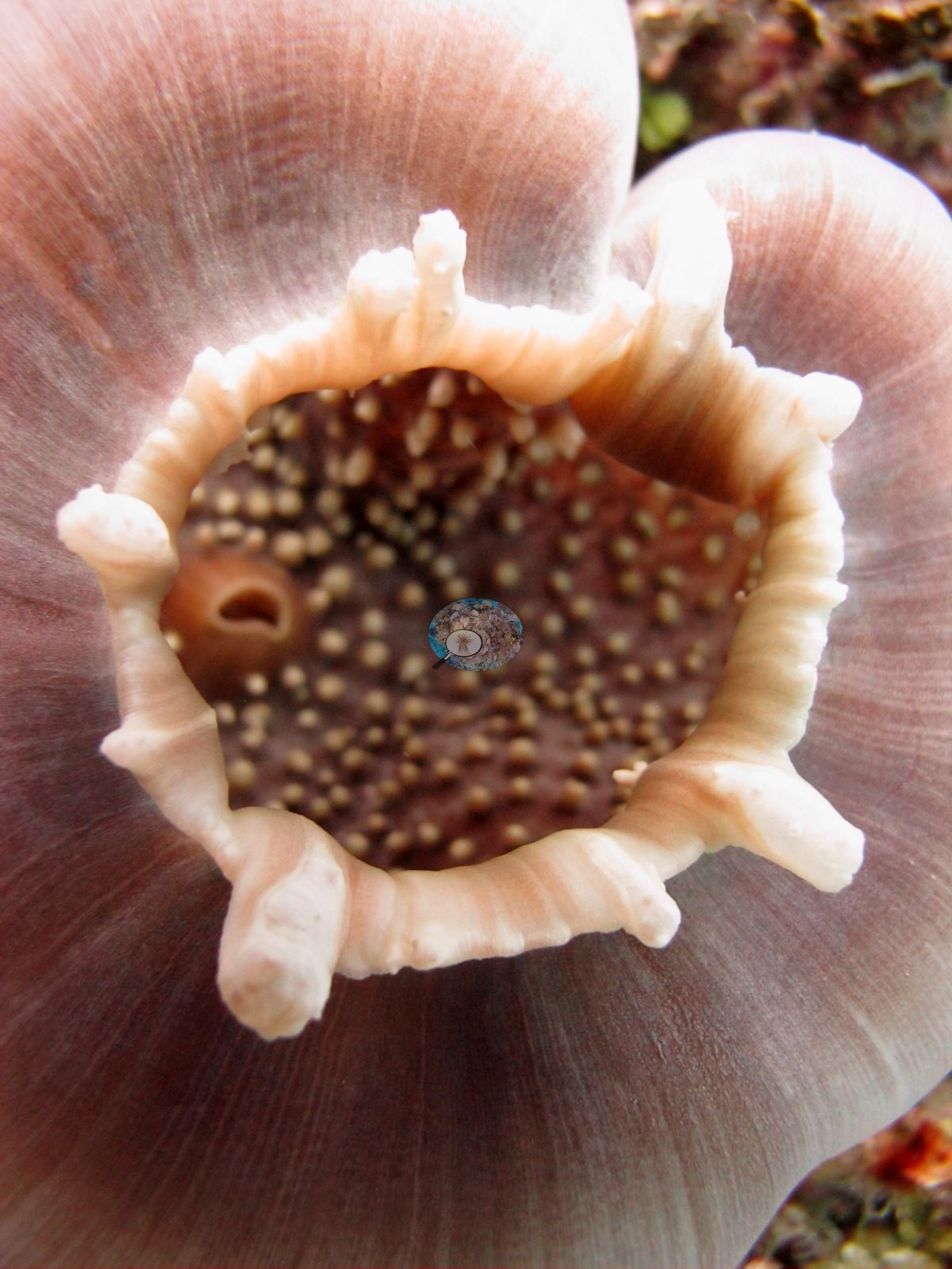
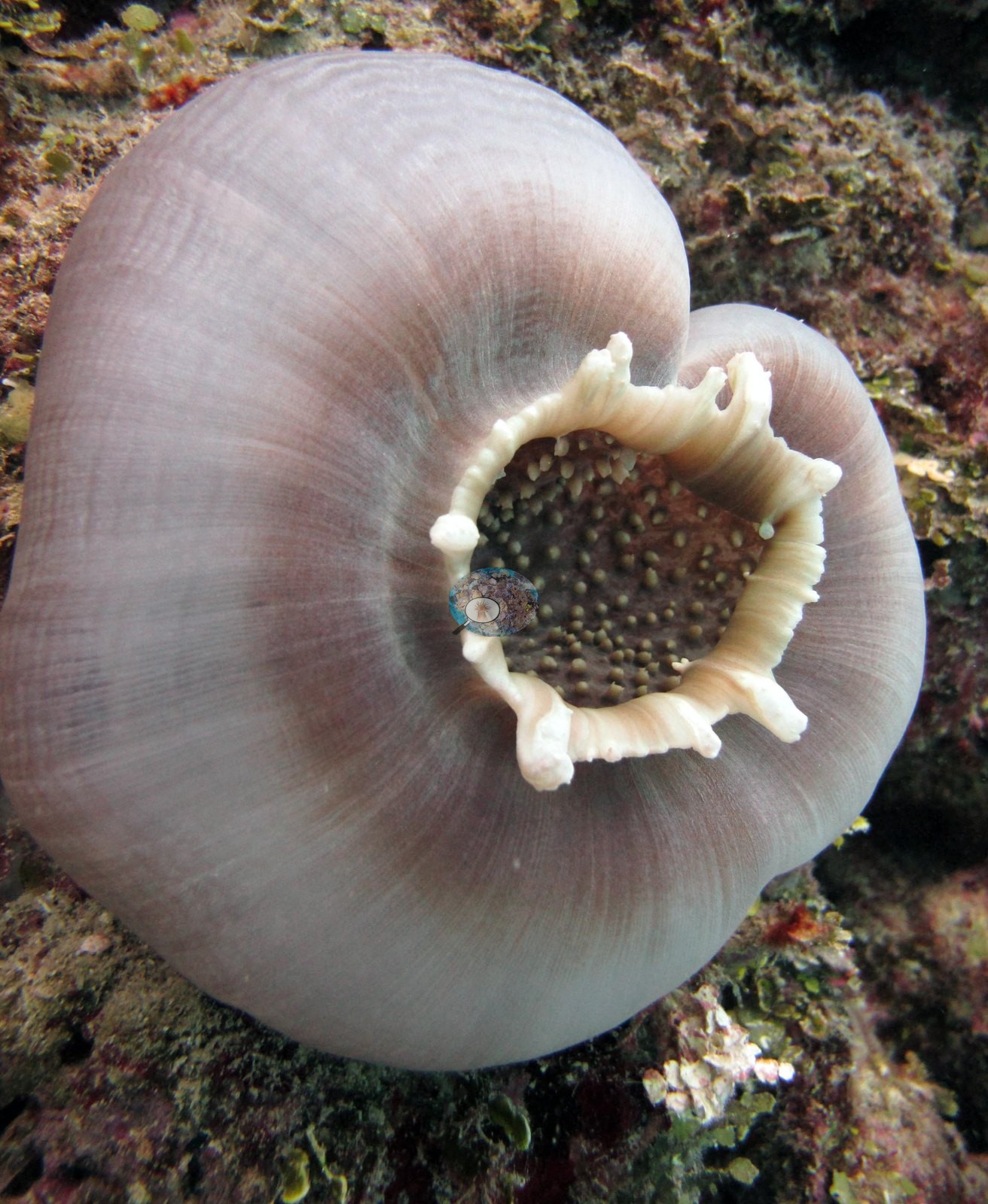
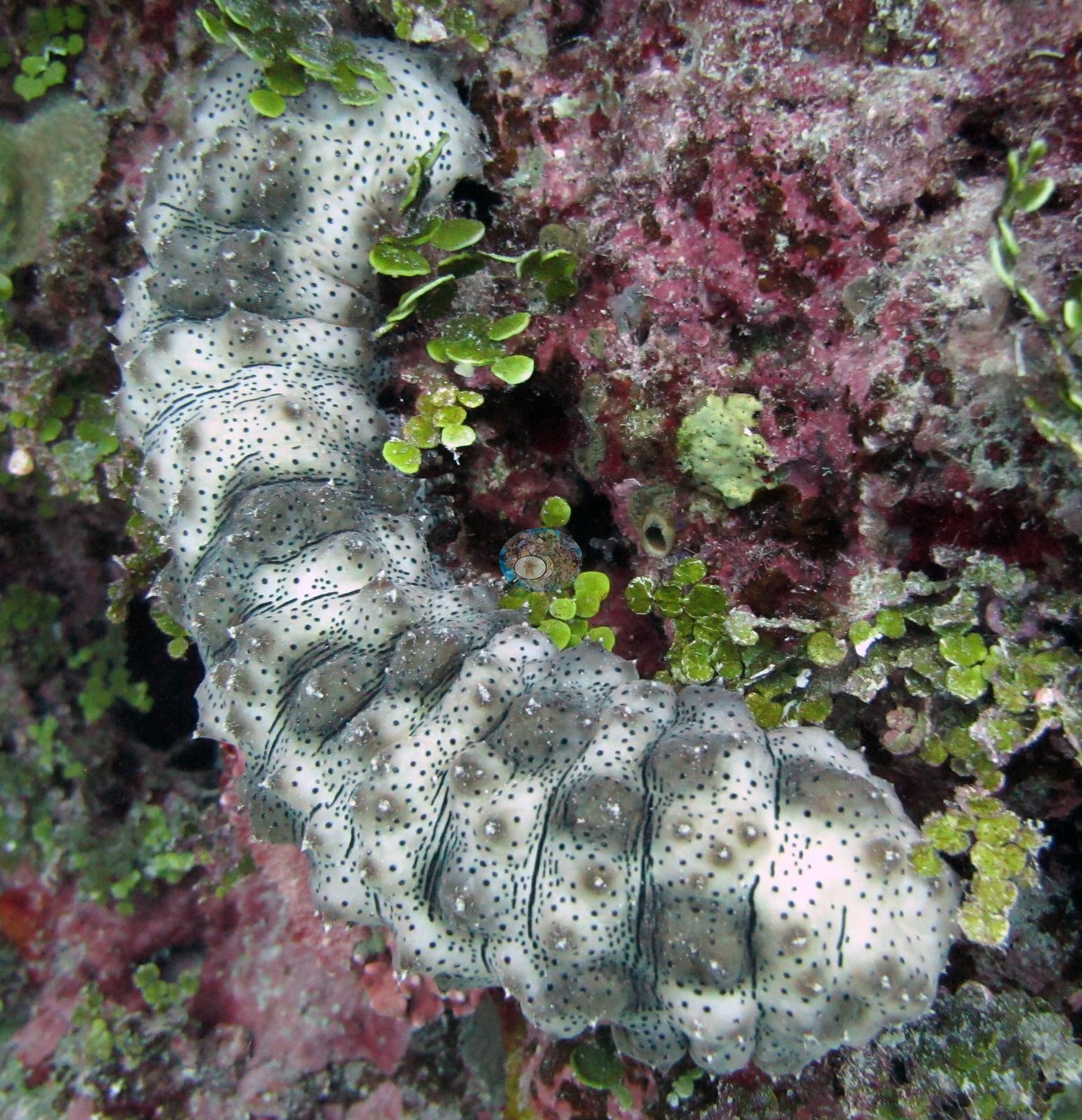
Image 24 is a spotted sea cucumber lying across red-crusted framework with green algal growth around it.
Comparisons with other data
Apart from the Living Oceans Foundation survey shown here, we found no other surveys at this site as of 2026. If you would like to share yours, please email us.
Dive record
FJVB62 was dived twice, and the photographs from it swing between very tight and very wide.
Other notes
The photographs are a macro set, not a survey set: near-identical anemone frames and a Diploastrea close-up would misrepresent the reef if read as a sample of it. Four Pocillopora colonies were sampled here, F135, F136, F137, and F138, the last labeled P. acuta in Anderson's own records; one of them, F136, was host to a small commensal crab photographed three times among its branches (e.g., Image 5, Image 6, Image 7).
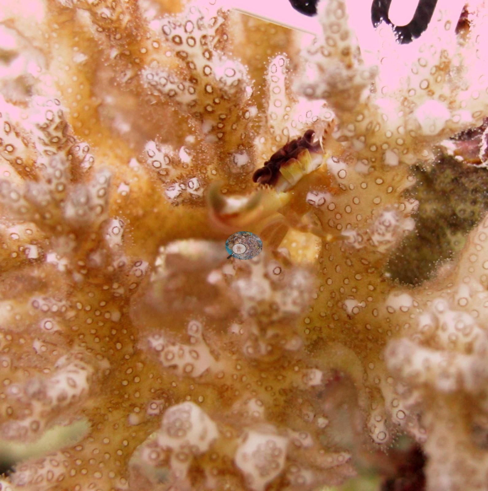
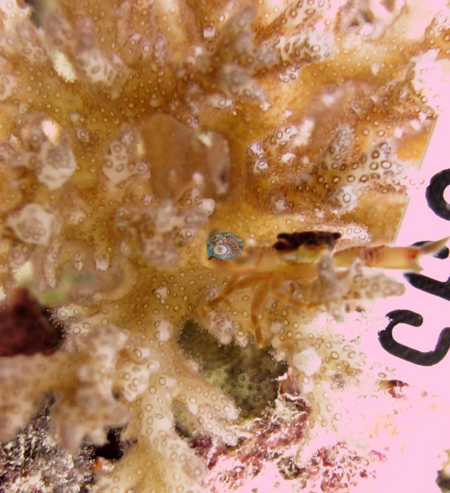
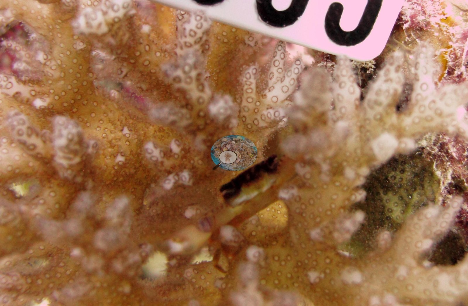
Those colony frames describe the colonies, not the reef.
Dive history
| Date | Time in | Max depth (m) | Bottom time (min) |
|---|---|---|---|
| 2013-06-23 | 15:40 | 18.3 | 57 |
| 2013-06-24 | 10:46 | — | — |
Data & links
- Download benthic survey data (CSV) ↗
- Download all LOF-GRE data (933 sites) ↗
- Photo portfolio ↗
- Coral Reef Diagnostics page for this area ↗
All images from Coral Reef Diagnostics. Data are from the Khaled bin Sultan Living Oceans Foundation Global Reef Expedition and Coral Reef Diagnostics.
 © Coral Reef Diagnostics.