
Part of the regional summaries: Fiji - Vanua Balavu, Fiji - Northern Lau (Vanua Balavu / Cicia / Nayau), and Fiji - Lau
More info
Fiji (NOAA CRW virtual station)A steep slope edge off Vanua Balavu whose shoulder is densely built with nearly every Acropora growth form, from pale tables jutting over the drop to staghorn stands and corymbose cushions, mixed with smooth tan massive boulders. Clouds of damselfish, chromis, and anthias hang off the face, and cover is 47%. Surveyed on the Living Oceans Foundation's Global Reef Expedition.
E dua na bati ni baba titobu mai Vanua Balavu, ka na kena tabana e buli vakaidina ena voleka ni veimataqali ivakarau ni tubu (growth forms) kece ni Acropora, mai na lase tepeli vulavula vakalailai e dremo tu ena dela ni titobu ki na staghorn kei na lase vakaveitabaki (corymbose) vakaiyavu, veiwaki kei na lase levu vakavatu (massive) savasava e roka (tan). Na ilawalawa levu ni ika damselfish, chromis, kei na anthias e tu ena bati ni cakau, ka 47% na ubi ni lase (coral cover).
AI translation of this summary; the English is authoritative. (low confidence)
CTD-cast midpoint; the cast read 27.2°C throughout
🗺 Detailed map of the Global Reef Expedition dive sites, Vanua Balavu, Fiji (Khaled bin Sultan Living Oceans Foundation; opens in a new window)
21 of the 25 photographs on this page are shown beside the text that discusses them under "Site description" below.
Site description
General benthic features
Nine frames look along and up a steep slope edge dropping into blue water (e.g., Image 21, Image 18, Image 19, Image 26, Image 25, Image 17, Image 23, Image 20, Image 22).
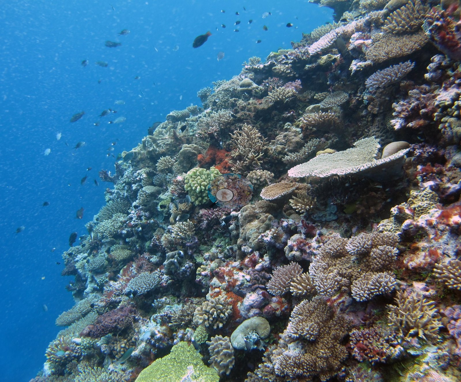
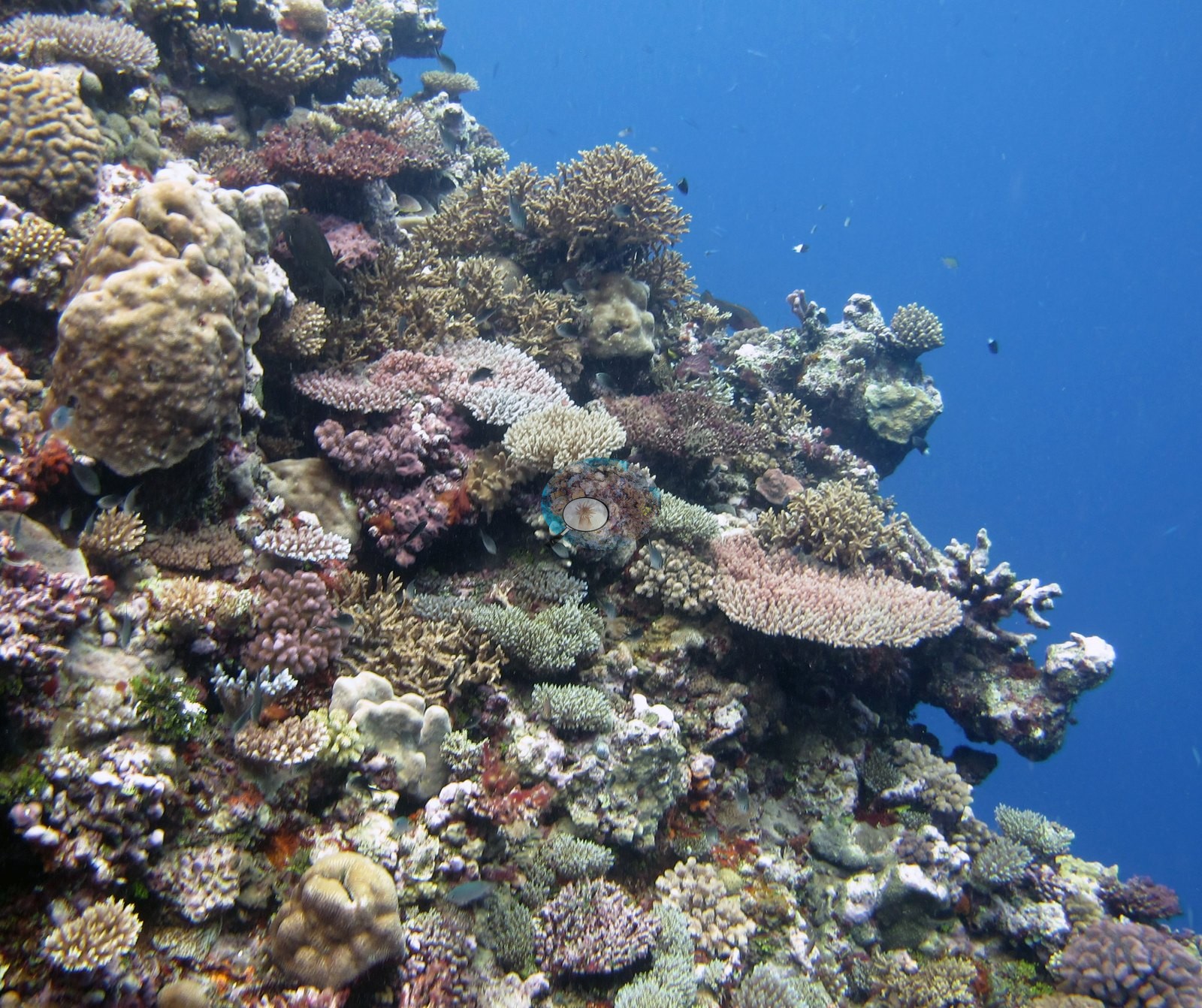
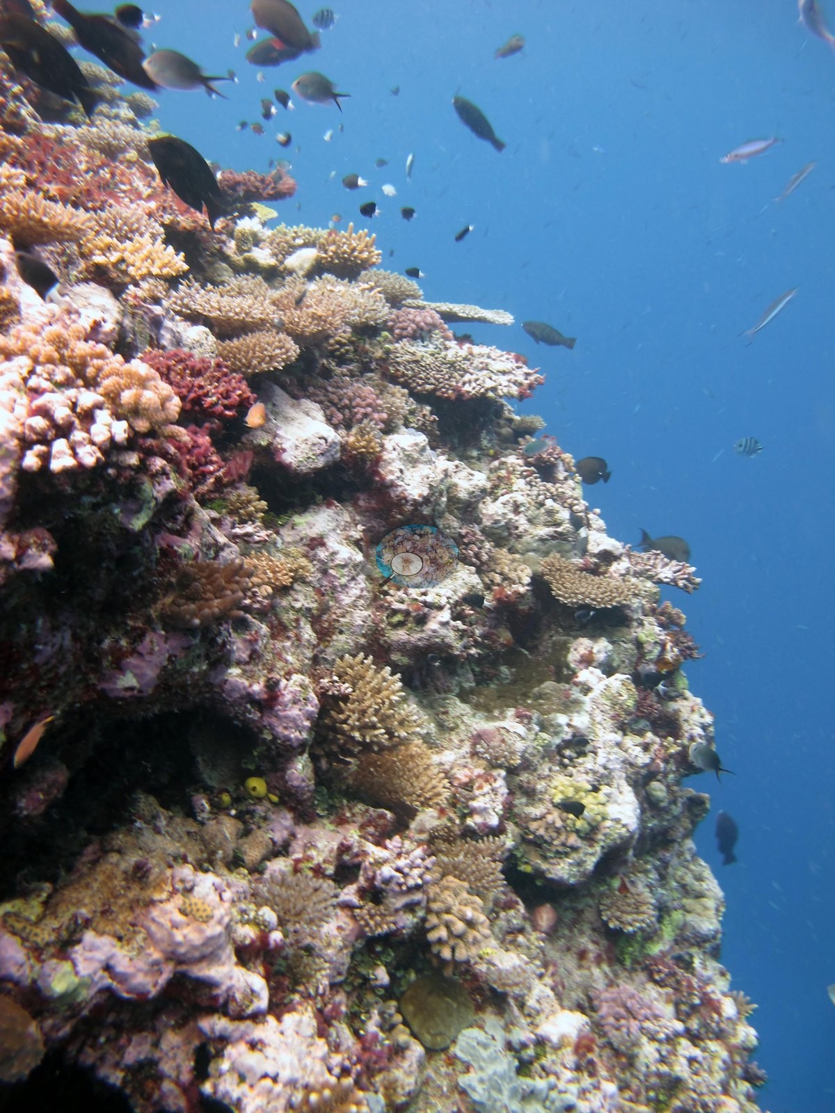
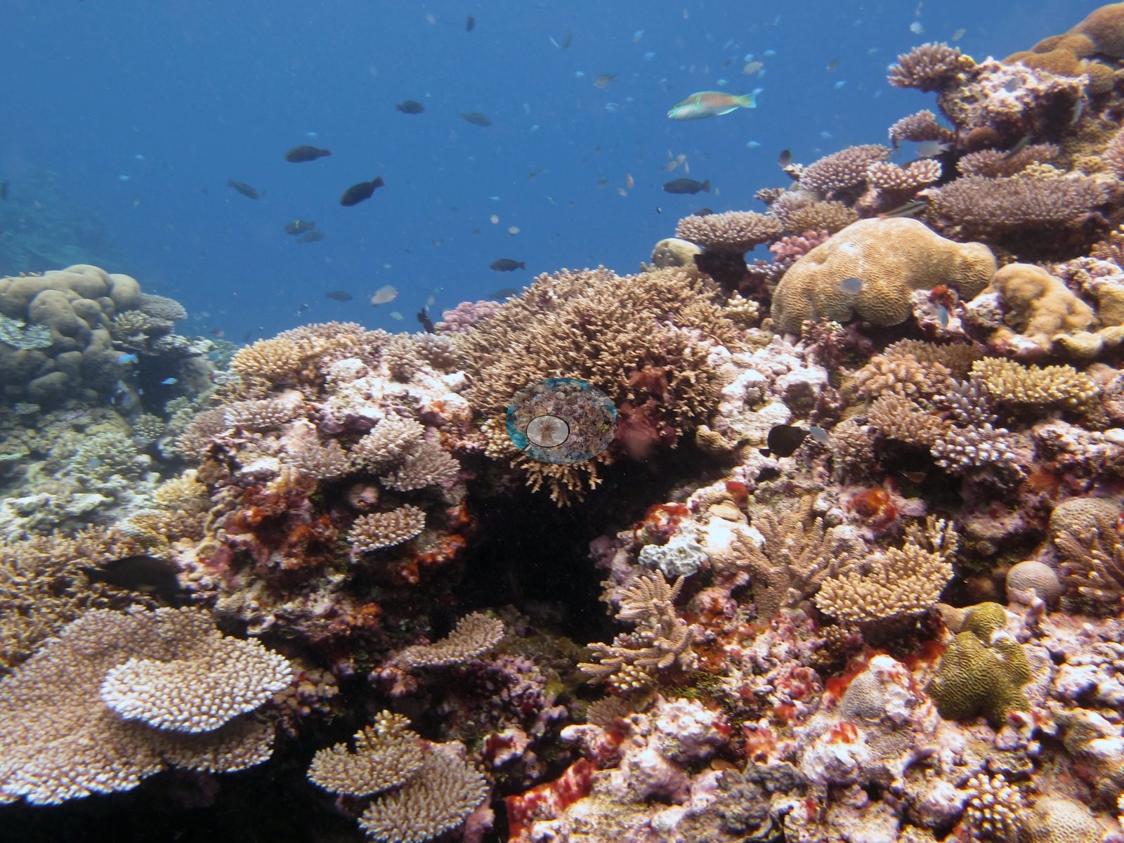
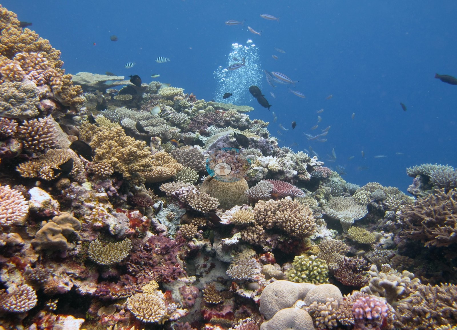
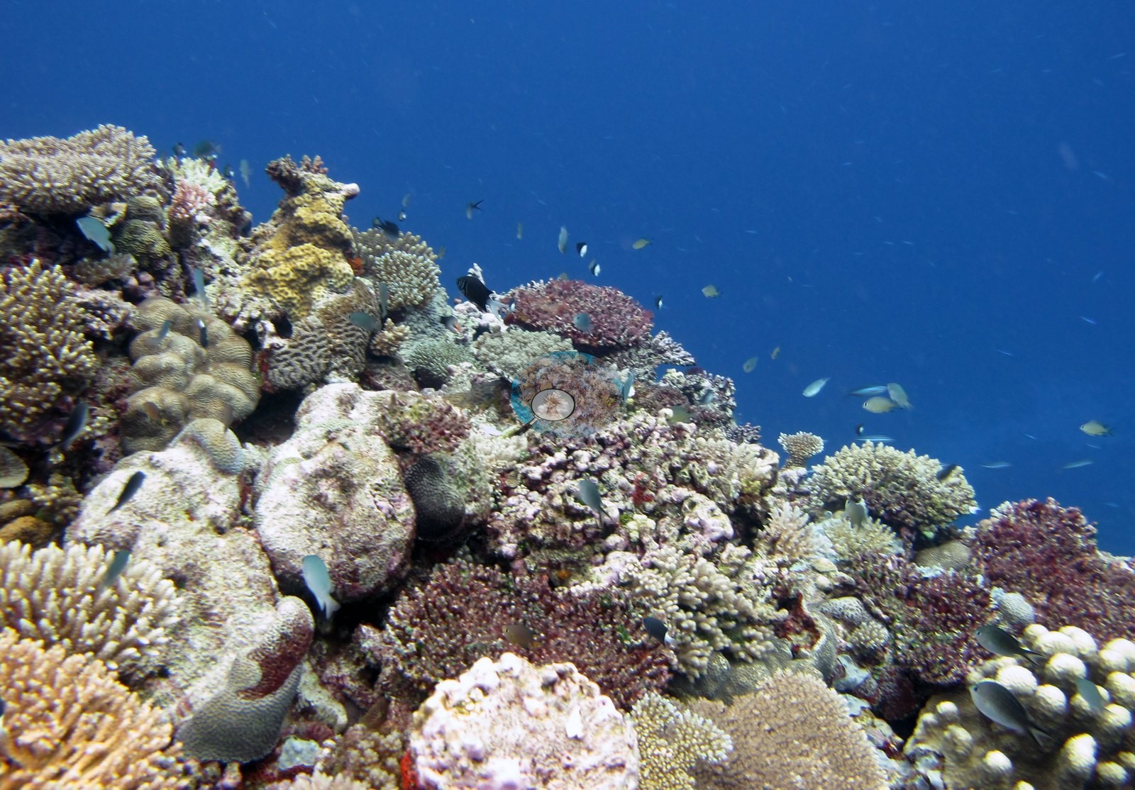
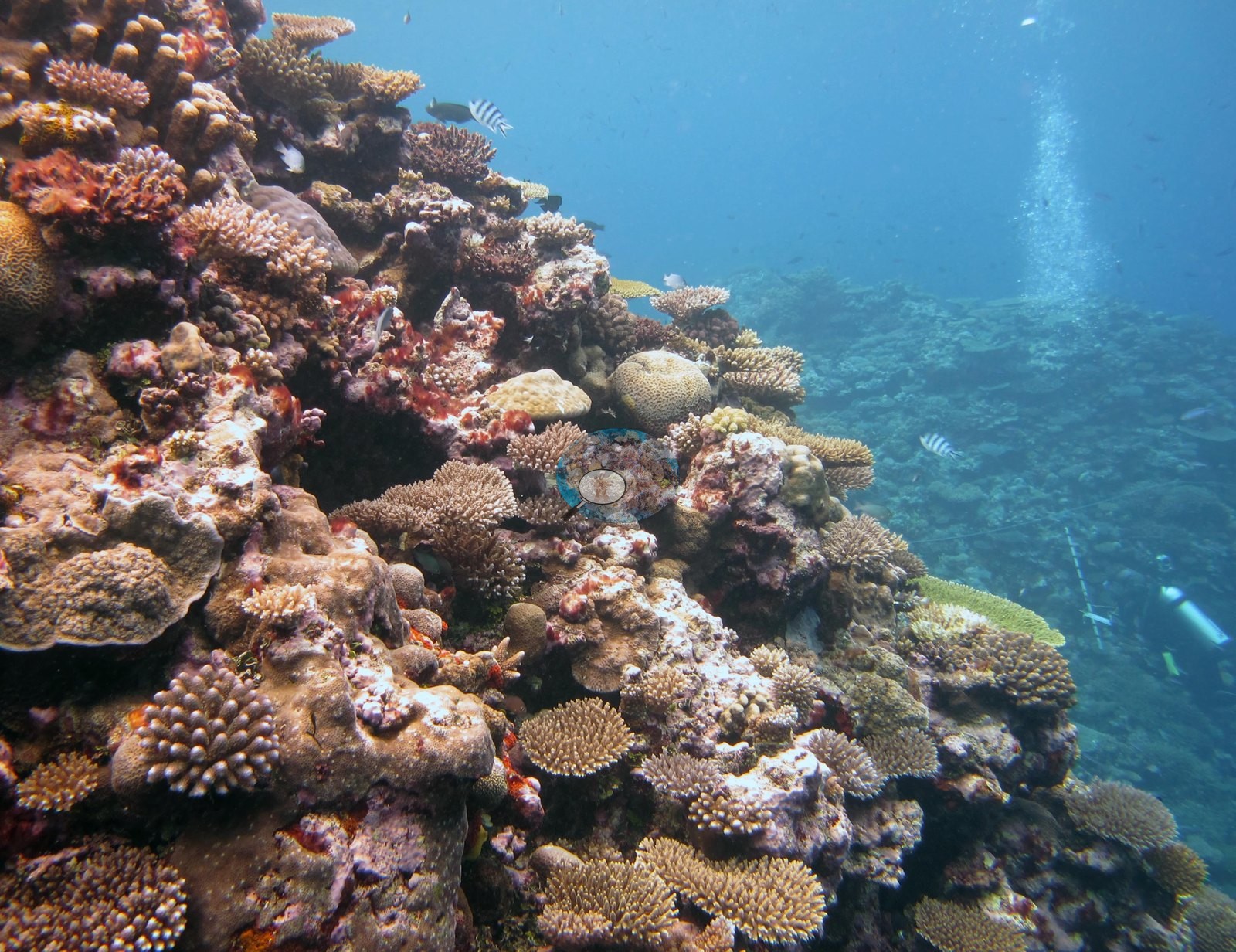
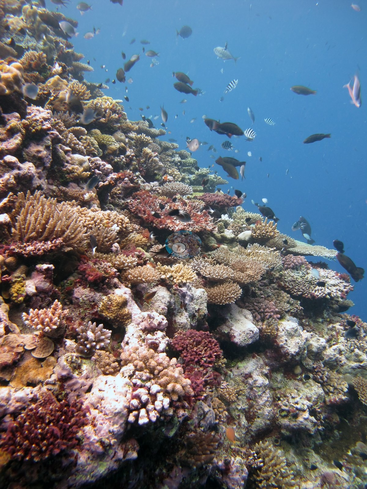
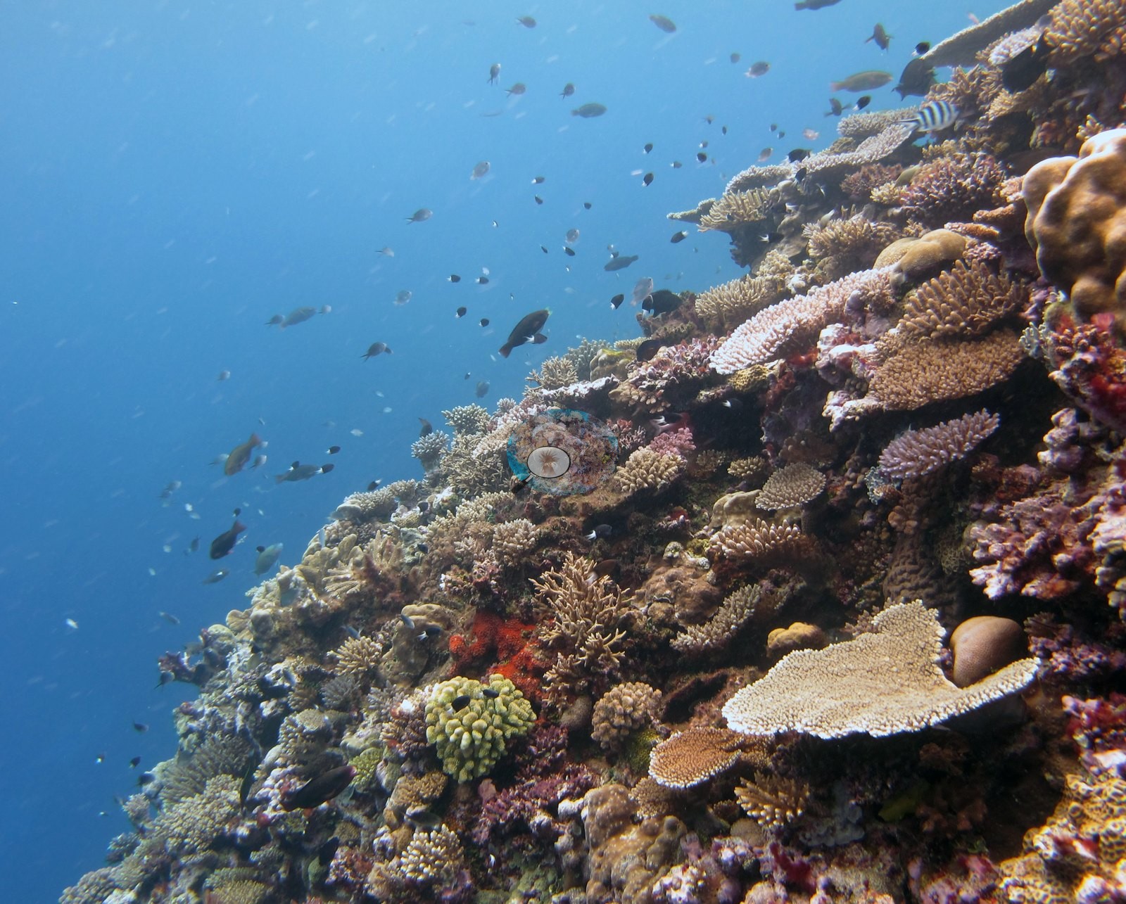
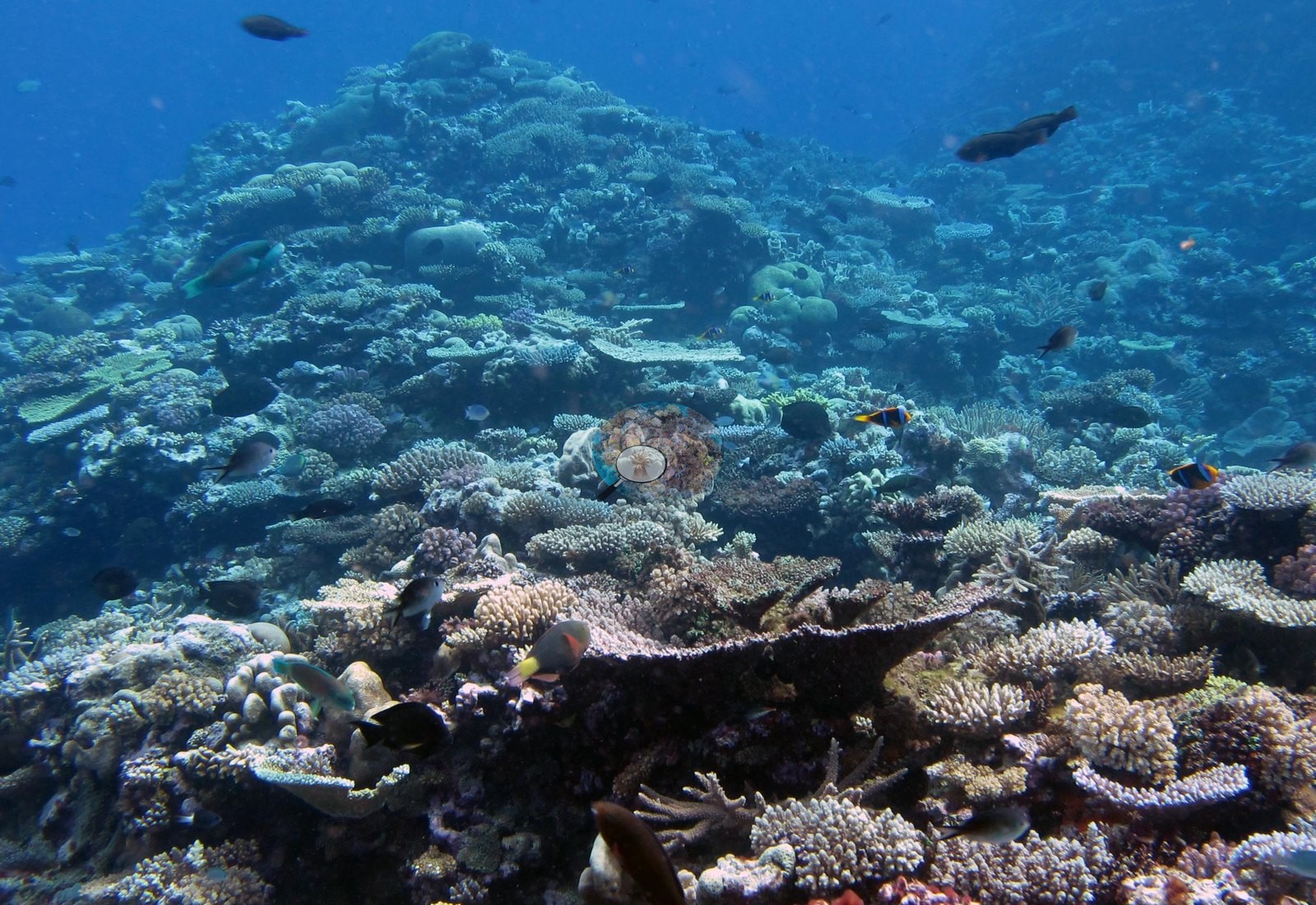
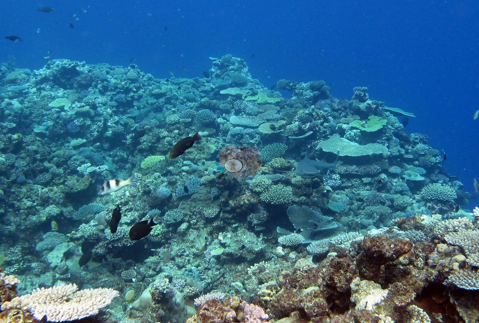
The slope shoulder is densely built: Acropora in most of the growth forms the genus manages, with pale tables cantilevered out over the drop (e.g., Image 26, Image 17, Image 22), corymbose cushions and staghorn stands packed against each other (e.g., Image 21, Image 18, Image 25, Image 23), and smooth tan massive boulders and knobbly mounding heads set among them (e.g., Image 18, Image 26, Image 25, Image 17). Color through this set runs pale lavender, cream, tan, and pink-brown, with green encrusting and plating growth, and maroon and red algal and coralline patches filling gaps (e.g., Image 21, Image 26, Image 25, Image 23). Bare rock is uncommon in the frames shot along the shoulder; the two frames looking across flatter ground (e.g., Image 12, Image 13) show a more open mosaic with rubble and pavement between colonies, so the reef is not uniform across the station.
Coral cover
- Reference: Khaled bin Sultan Living Oceans Foundation, Global Reef Expedition (in situ transect)
- Transect mean 47 ± 4.3% SE (n = 5 transects; Living Oceans Foundation per-transect benthic data); the headline above is this all-transect mean.
Stressors
No wide frame shows an obviously bleached or recently dead colony; a few dark, algae-covered dead branch sections are the only visible mortality, and no crown-of-thorns or disease lesion is identifiable; Anderson's sampling sheet records one of the four colonies sampled here (F112) as 5–10% bleached and the other three at 0%. Of the three coral colonies from this site with an outlier score, one (33%) was flagged as a physiological outlier (Mayfield et al. 2017, PLoS ONE). The Living Oceans Foundation transects here recorded 9.4% cyanobacteria and no recently dead coral.
Fish and other fauna
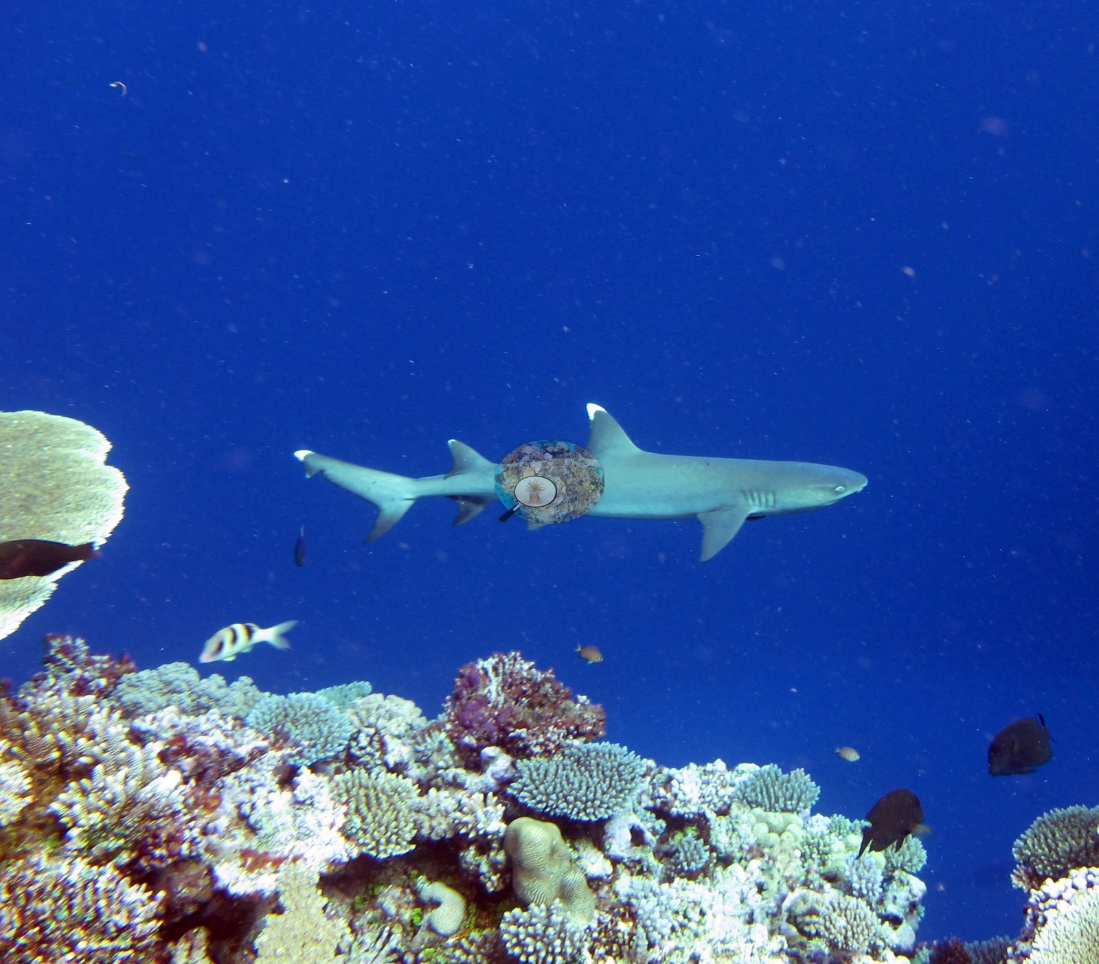
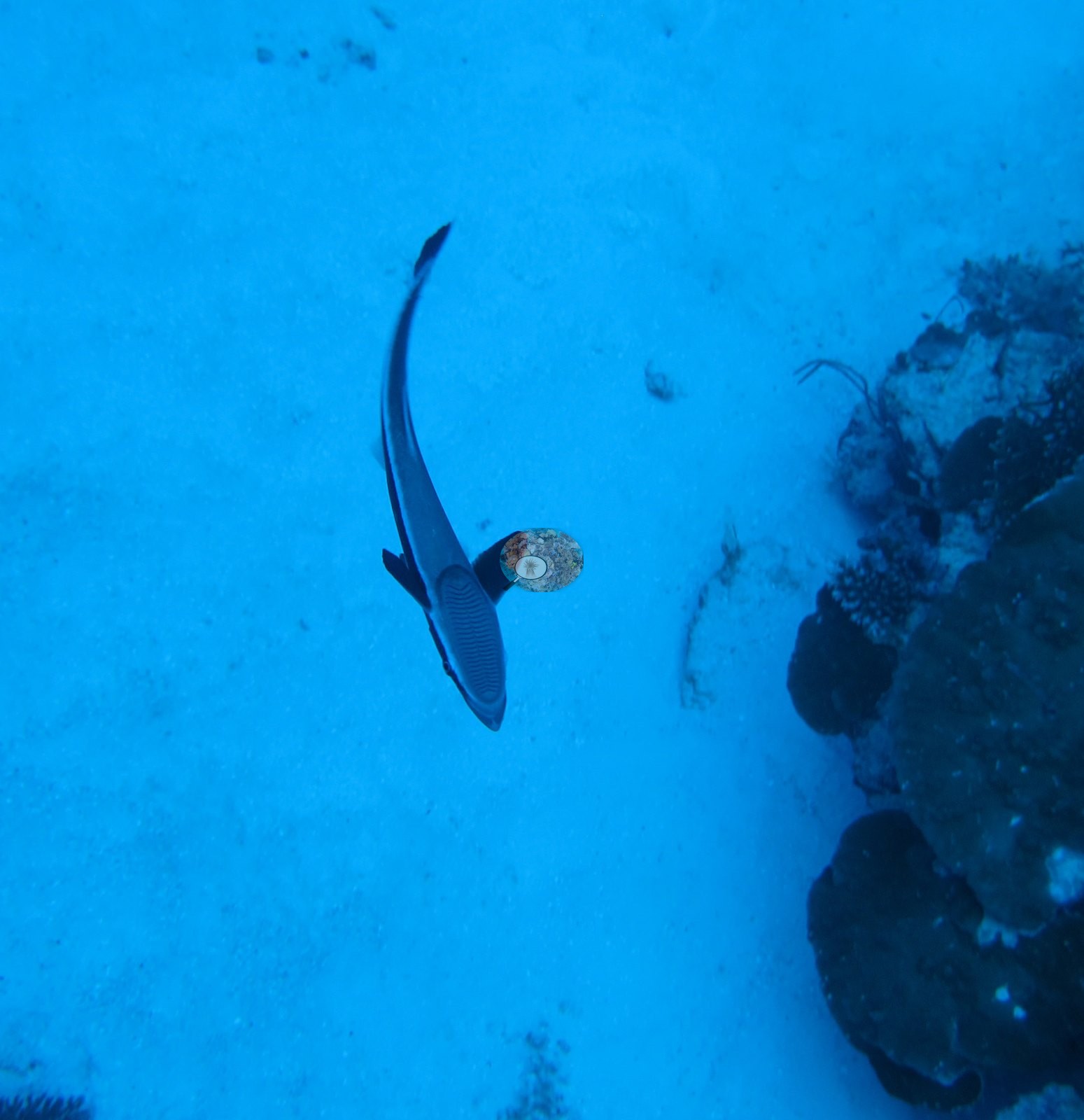
Planktivores — damselfishes, chromis, and anthias — hang off the slope face in almost every wide frame, densest in Image 20 and Image 25. A whitetip reef shark passes low over the reef top (e.g., Image 14) and a shark with a remora attached cruises over open sand in blue water (e.g., Image 16).
Comparisons with other data
Apart from the Living Oceans Foundation survey shown here, we found no other surveys at this site as of 2026. If you would like to share yours, please email us.
Other notes
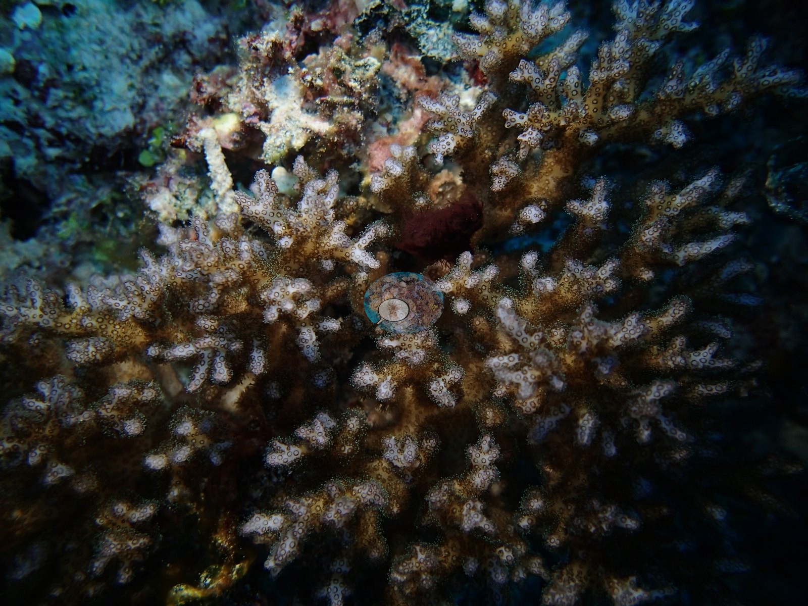
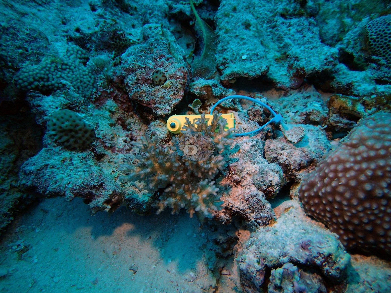
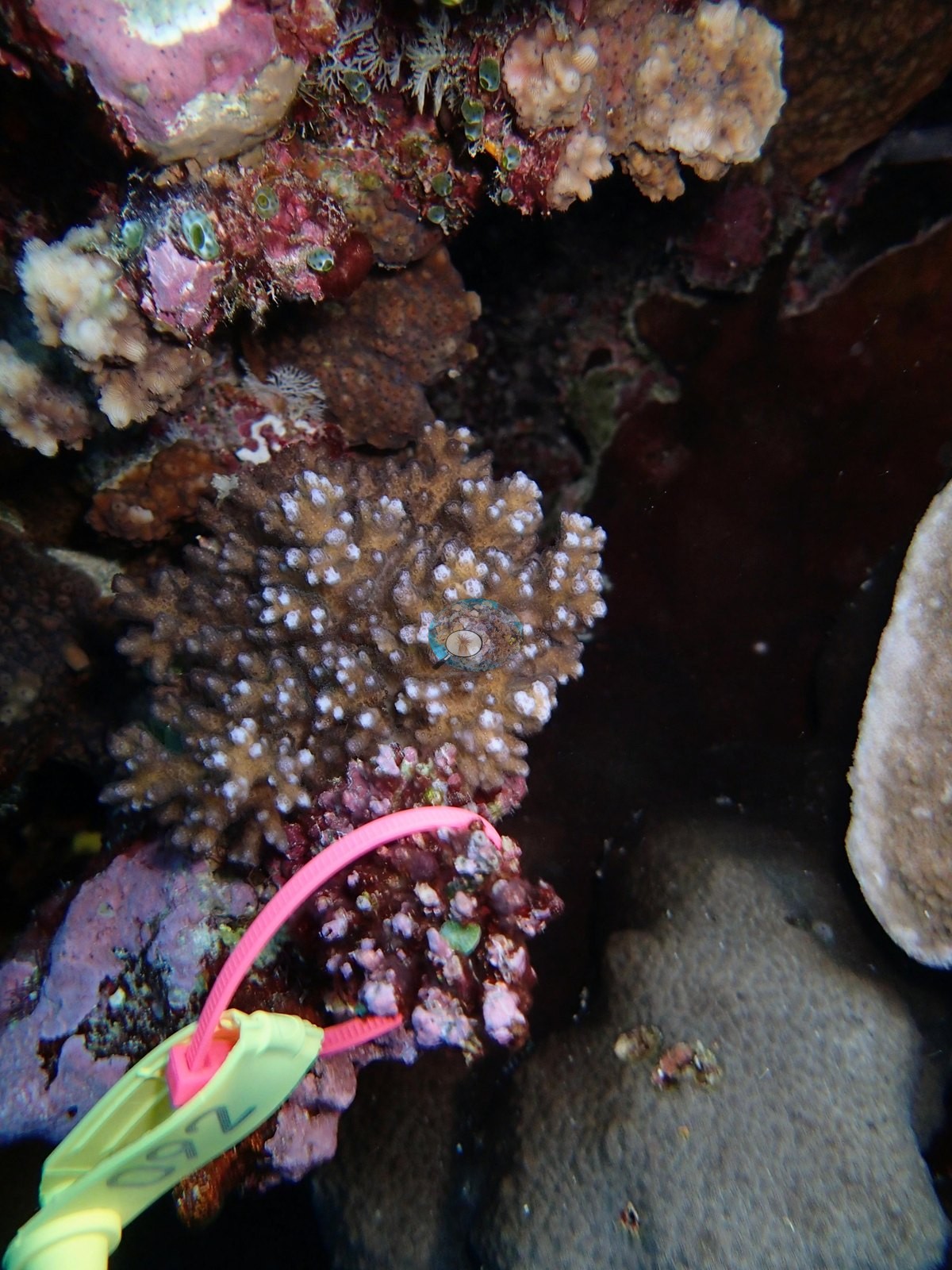
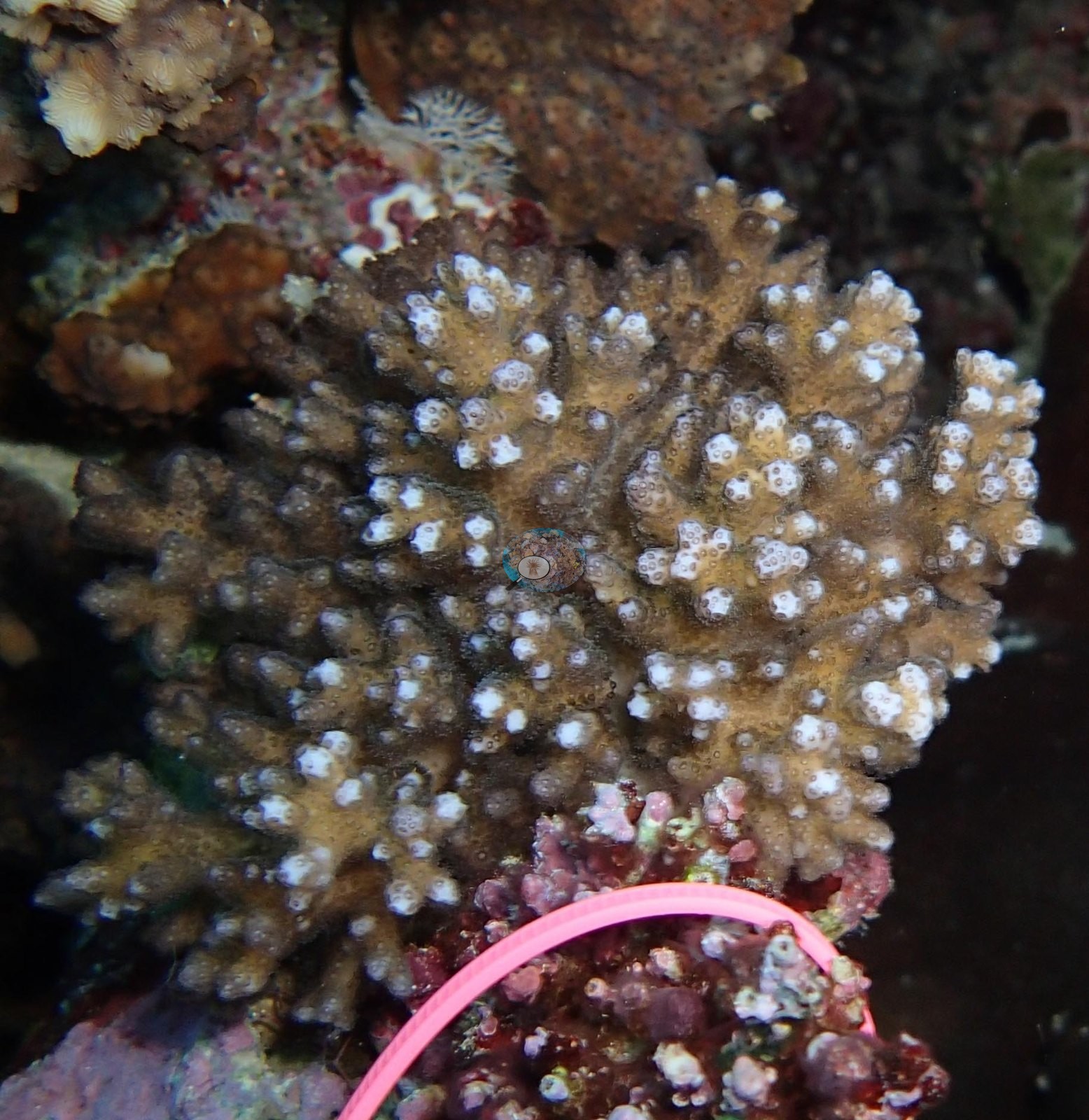
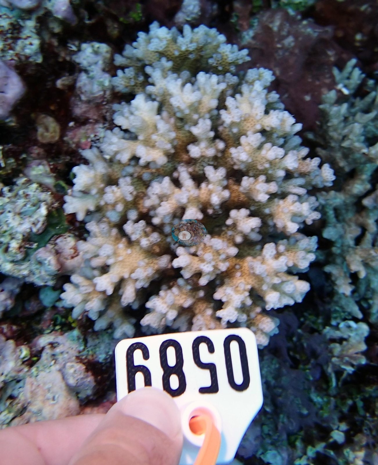
The complete set contains both the wide survey photography and the close-range colony photography from the physiological sampling on the same dive, and the two are easy to confuse. Six frames are shot at arm's length on individual branching colonies (e.g., Image 6, Image 4, Image 3, Image 9, Image 5, Image 11).
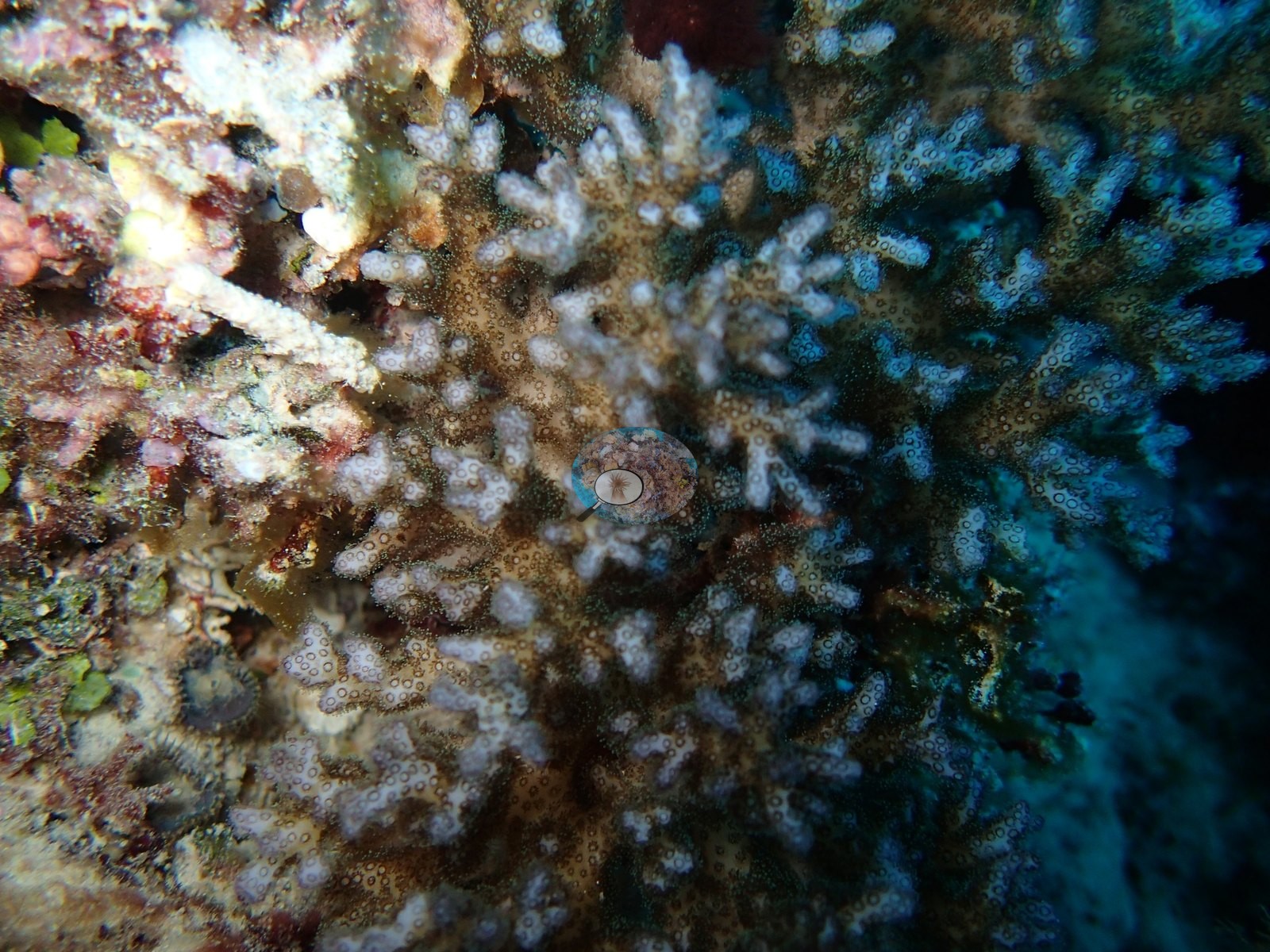
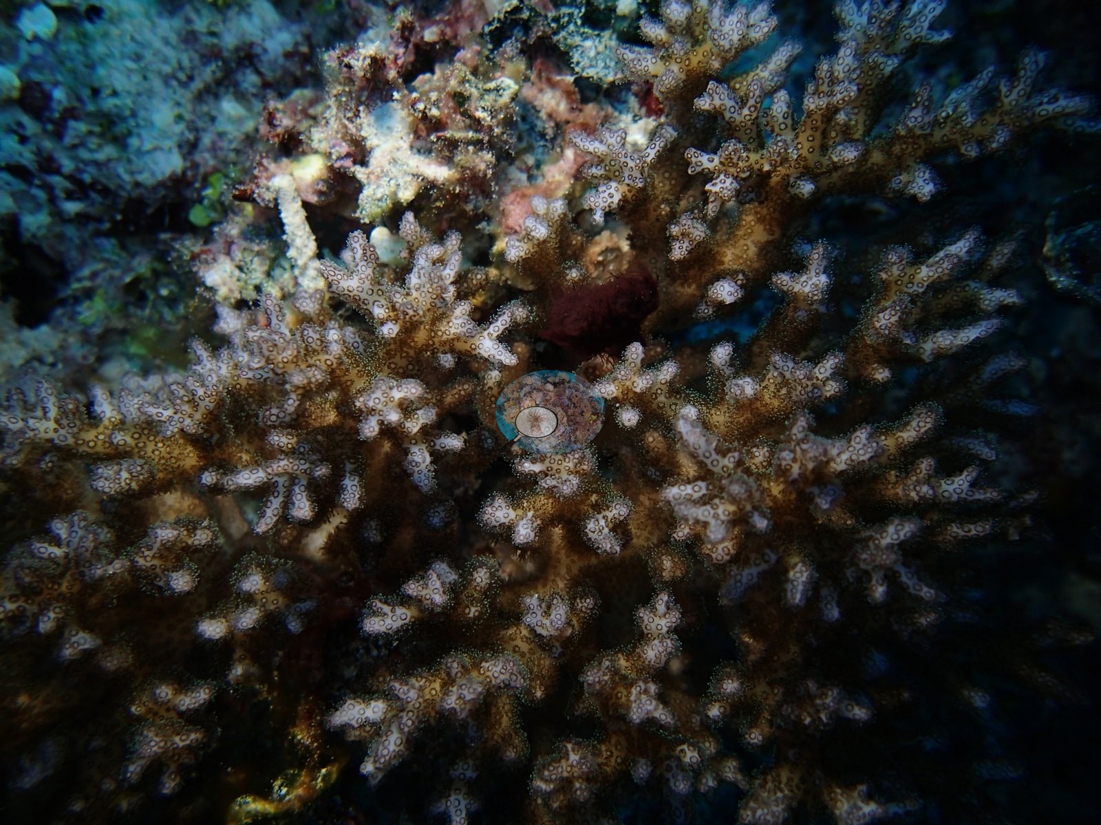
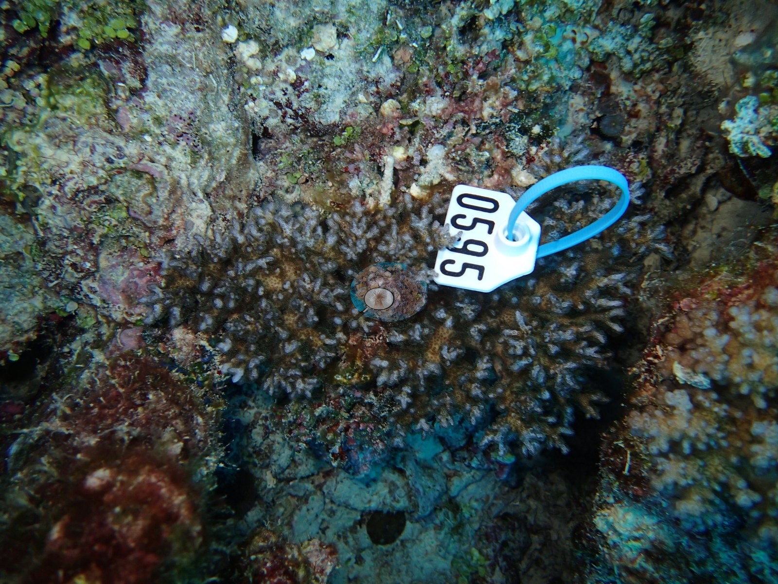
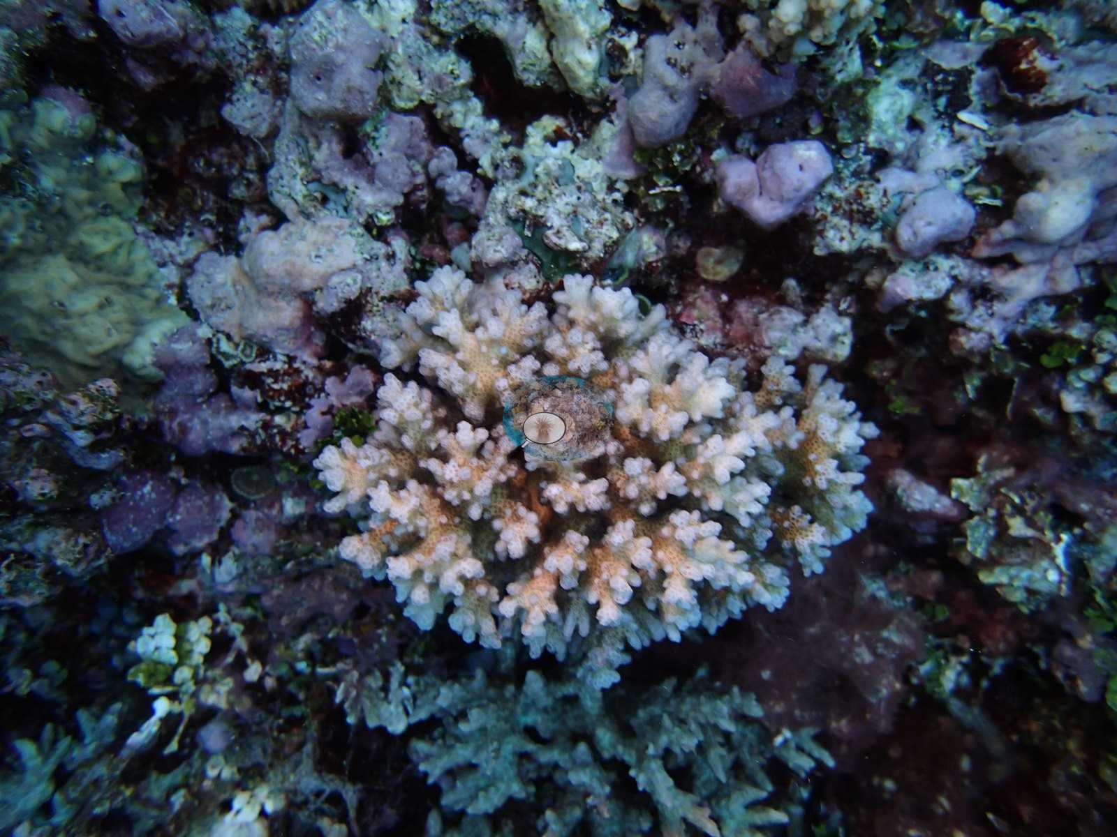
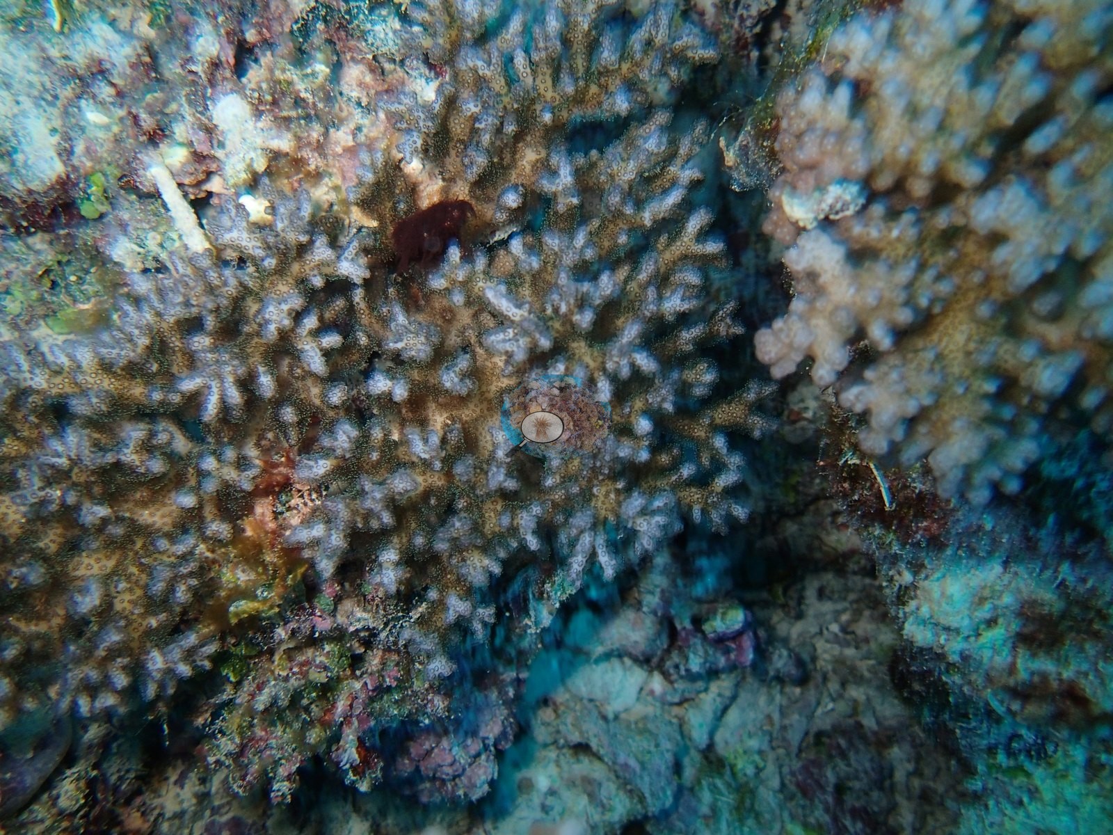
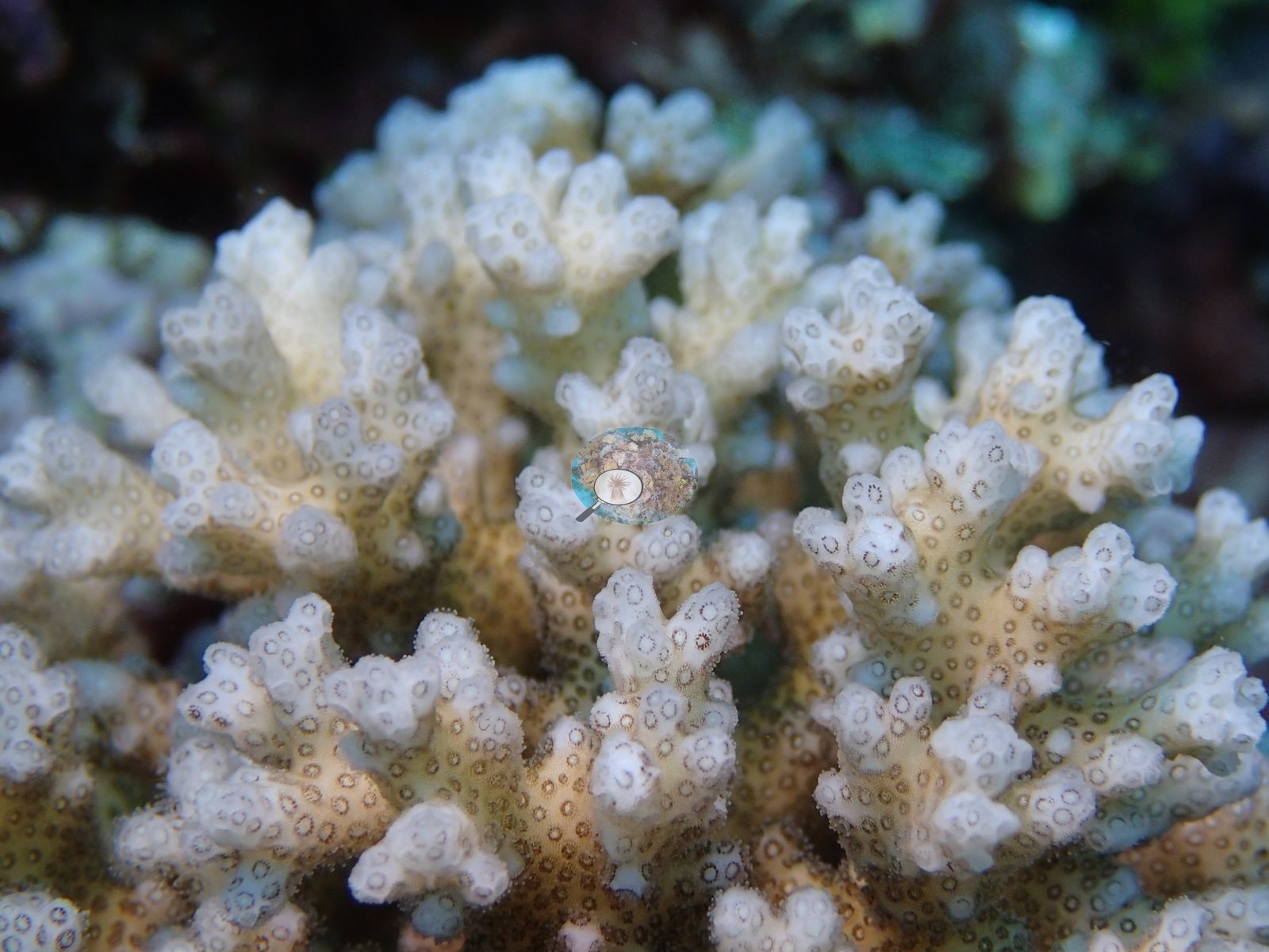
One of them (Image 3) shows a numbered plastic tag on a wire loop fixed into the framework beside a colony, which is the marker for a physiologically sampled colony; the number itself is not meaningful and is not recorded here. The tagged and photographed colonies are branching colonies in cream, pale tan, and gray-white (Anderson's sample sheet lists only Pocillopora here: P. acuta and P. damicornis), several with polyps fully extended (e.g., Image 9, Image 11). The colony in Image 11 is cream-white with tan bases and fills the frame. These are strobe-lit macro-range frames of individually selected colonies, and pale rendering at that distance cannot be separated from paling of the tissue; no wide frame in the set shows white colonies, and nothing here should be read as an extent of bleaching at this station. Their value is as a visual record tying this site to the physiological sampling: of the three colonies here with an outlier score, one was flagged as an outlier. Two frames carry a diver: a photographer on the slope face beside a coral Anderson identified as a mushroom coral (fungiid) (e.g., Image 24), and a diver working over a dark sand-and-rubble floor on a dive Anderson noted for its strong current (e.g., Image 15).
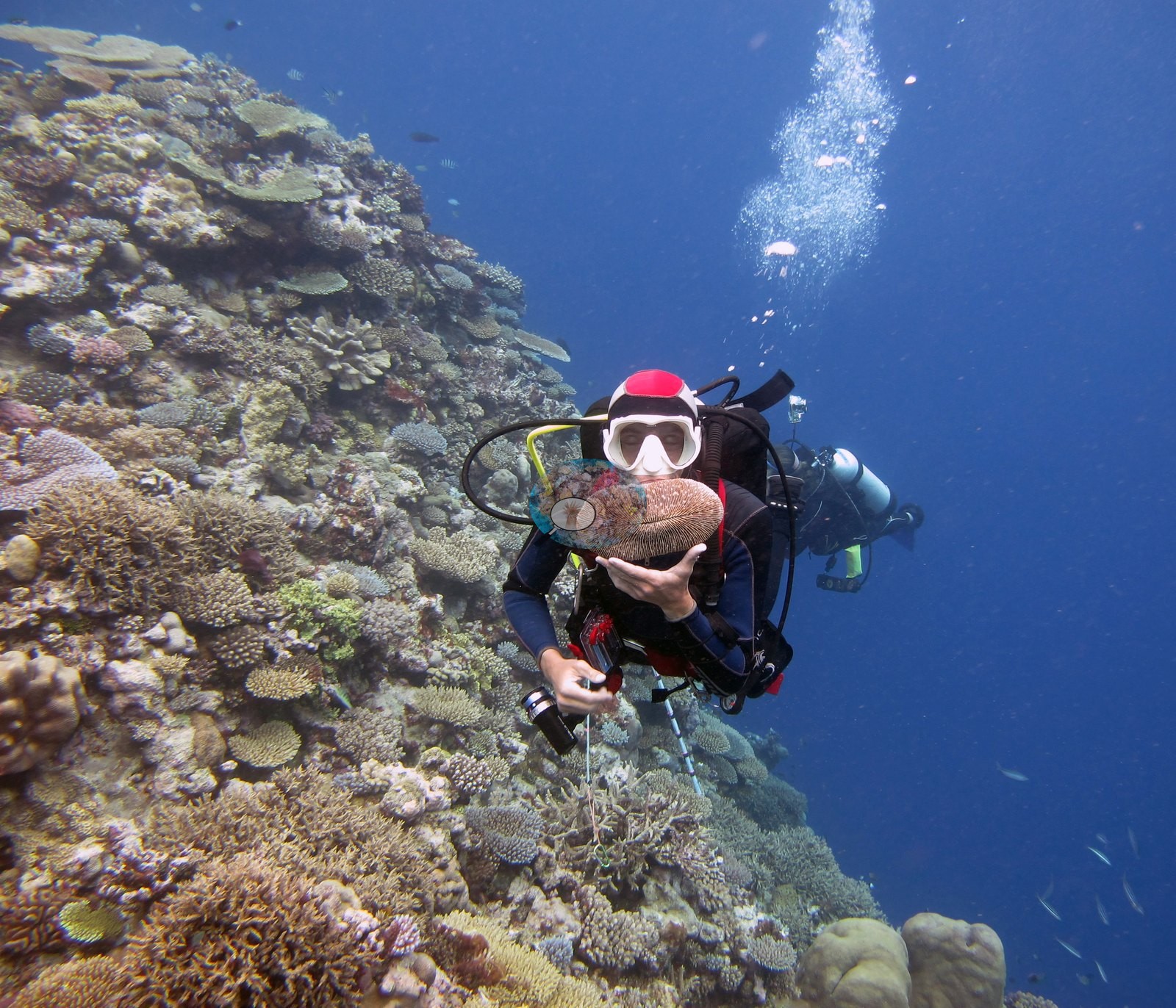
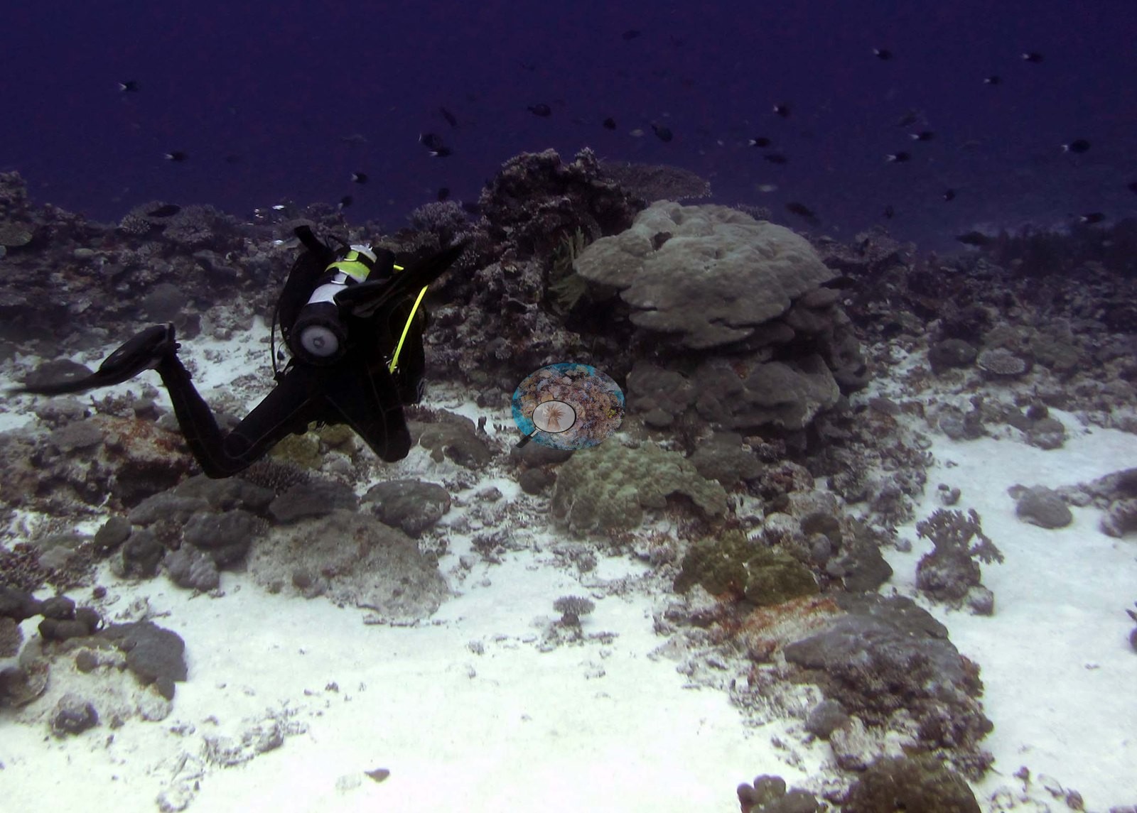
What the frames cannot settle: none of these is a planar downward view and the slope frames are strongly oblique, so nothing in the set supports a cover figure. The set is also weighted toward the best-built part of the slope — the two flatter-ground frames are a useful corrective. Depth is not resolvable from the imagery.
CV model comparison
More info
The photo row below is a methods comparison only: it is never averaged with the transect and never used as the headline. It compares site means only: the photos come from the same survey dive as the transect but not from the same points.| Method | Hard coral cover (%) | Photos (n) ± SE |
|---|---|---|
| Transect mean (all transects) | 47.4 | |
| CoralSCOP edited | 34.0* | n = 11 · SE 2.6 |
| Claude (edited photos) | 38.6* | n = 11 · SE 4.0 |
The two computer-vision methods agree within 4.6 pp (34.0–38.6%) and read 8.8–13.4 pp below the in-water transect (47.4%); the headline is the in-water transect because a diver transect is independent of the photographs.
*Based on only 11 reef-habitat photos; this estimate is less certain.
See a more detailed, stand-alone version of this table.
Data & links
- Download benthic survey data (CSV) ↗
- Download all LOF-GRE data (933 sites) ↗
- Photo portfolio ↗
- Coral Reef Diagnostics page for this area ↗
All images from Coral Reef Diagnostics. Data are from the Khaled bin Sultan Living Oceans Foundation Global Reef Expedition and Coral Reef Diagnostics.
 © Coral Reef Diagnostics.