
Part of the regional summaries: Fiji - Totoya, Fiji - Moala Group (Moala / Totoya / Matuku), and Fiji - Lau
More info
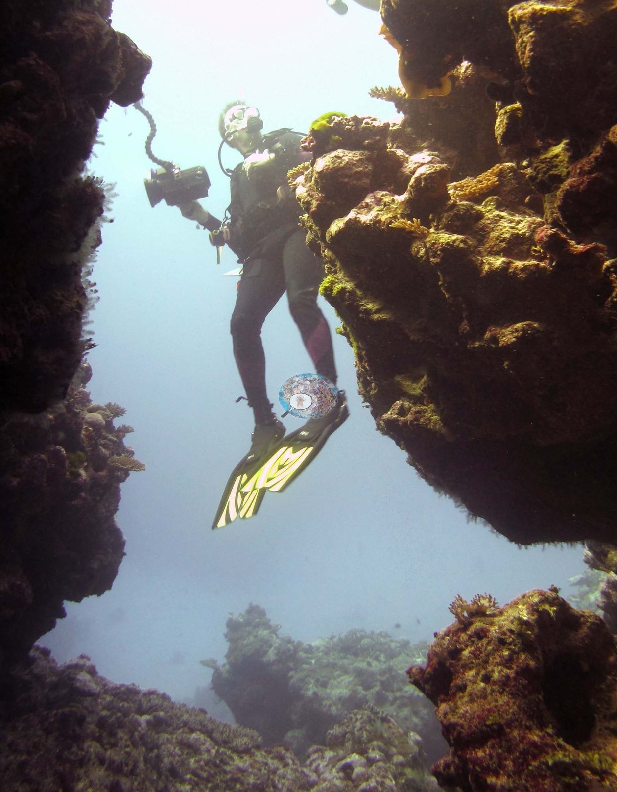
Fiji (NOAA CRW virtual station)A Totoya reef of steep spurs, walls, and a large overhung cave passage, its faces covered in a close mosaic of many small branching, plating, and massive colonies over pink and lilac coralline crust. Several of the photographs record the survey team at work, including a diver framed in a coral archway. Surveyed on the Living Oceans Foundation's Global Reef Expedition.
E dua na cakau e Totoya ni spur titobu, na bati, kei na dua na qara levu e dremo e rawa ni curu kina (cave passage), ka na kena bati e ubi ena dua na veiwaki voleka ni lase lalai vakatabataba, vakaiteba (plating), kei na lase levu (massive) e levu sara ena dela ni lumi vakalase (coralline crust) e roka (pink, lilac). E vica na itaba e vakaraitaka na ilawalawa ni vakadidike ena nodra cakacaka, okati kina e dua na dau nunu e tu ena loma ni dua na matamata ni lase (coral archway).
AI translation of this summary; the English is authoritative. (low confidence)
CTD-cast midpoint of 27.1–27.7°C
🗺 Detailed map of the Global Reef Expedition dive sites, Totoya, Fiji (Khaled bin Sultan Living Oceans Foundation; opens in a new window)
18 of the 19 photographs on this page are shown beside the text that discusses them under "Site description" below.
Site description
General benthic features
The reef frames confirm and extend the spur-and-wall picture. Image 7, Image 8, Image 11, and Image 14 look along steep faces and promontories that drop into blue: their surfaces are a close mosaic of small corymbose and digitate Acropora, yellow-brown and olive plating and foliose colonies, small massives, and a great deal of pink, lilac, and maroon coralline-crusted framework between them — real cover made of many small colonies rather than a few large ones.
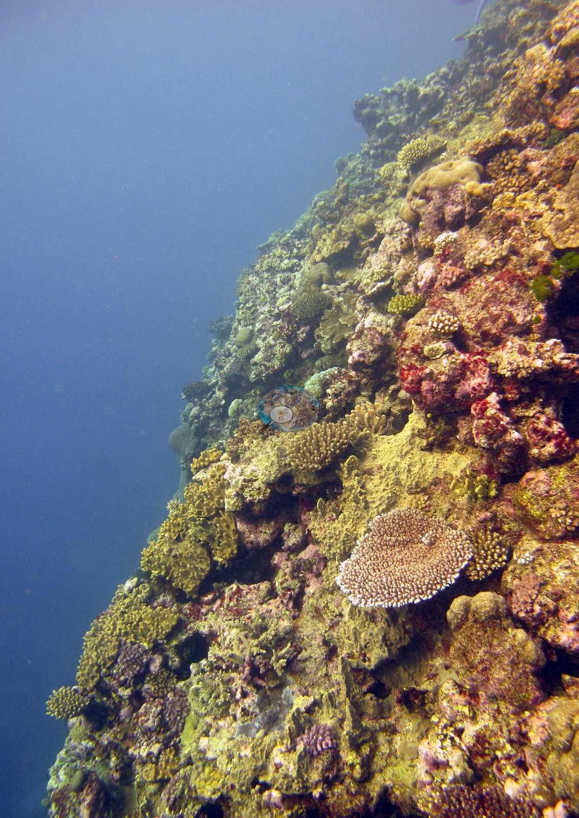
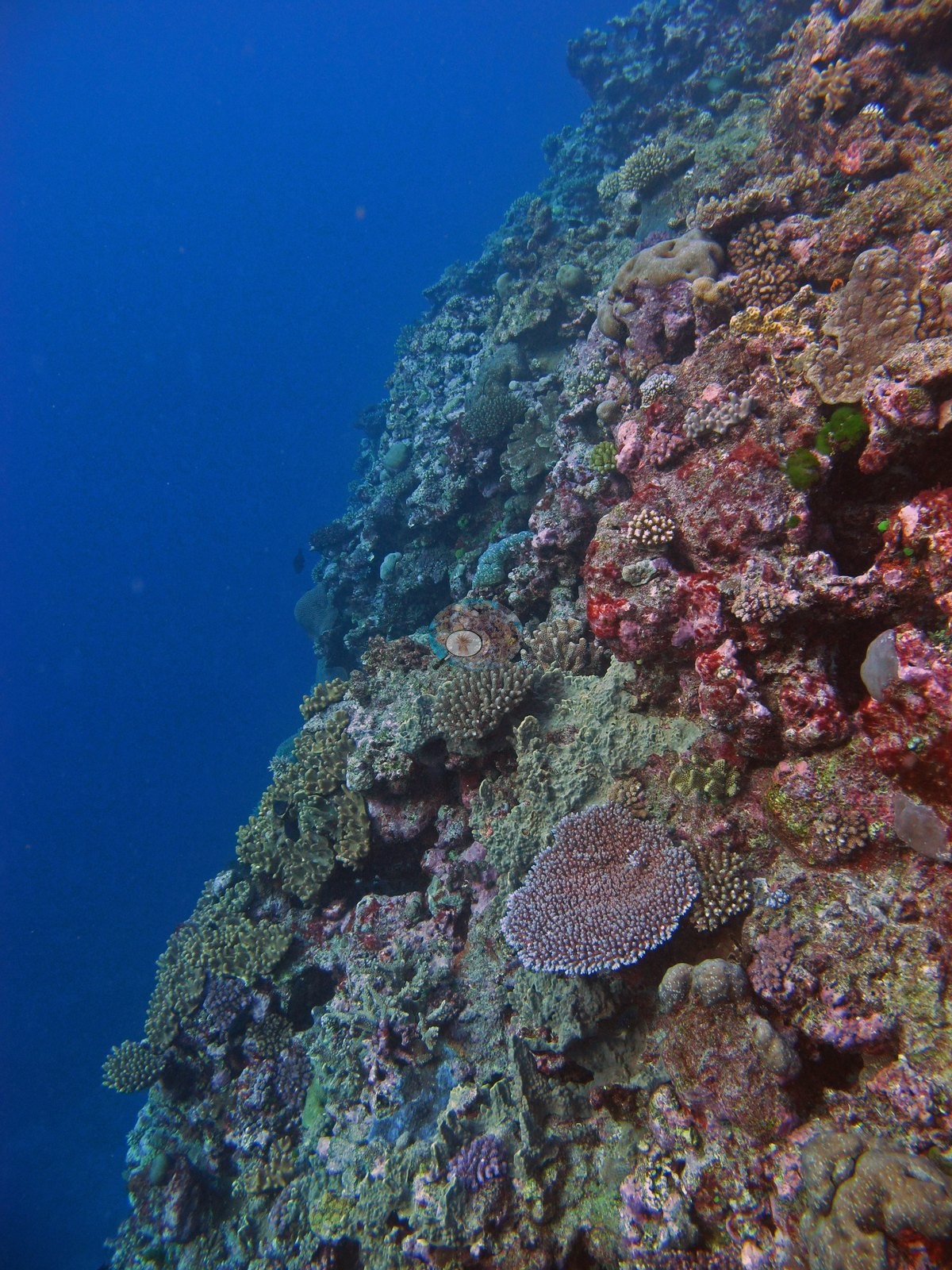
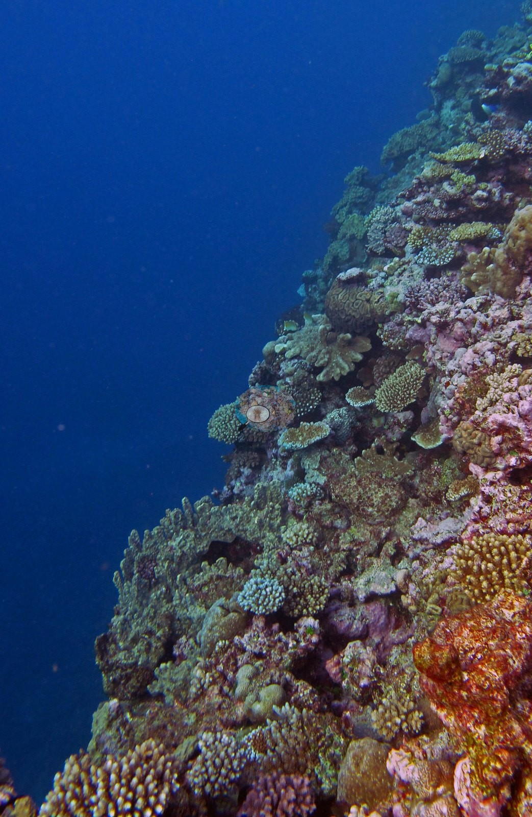
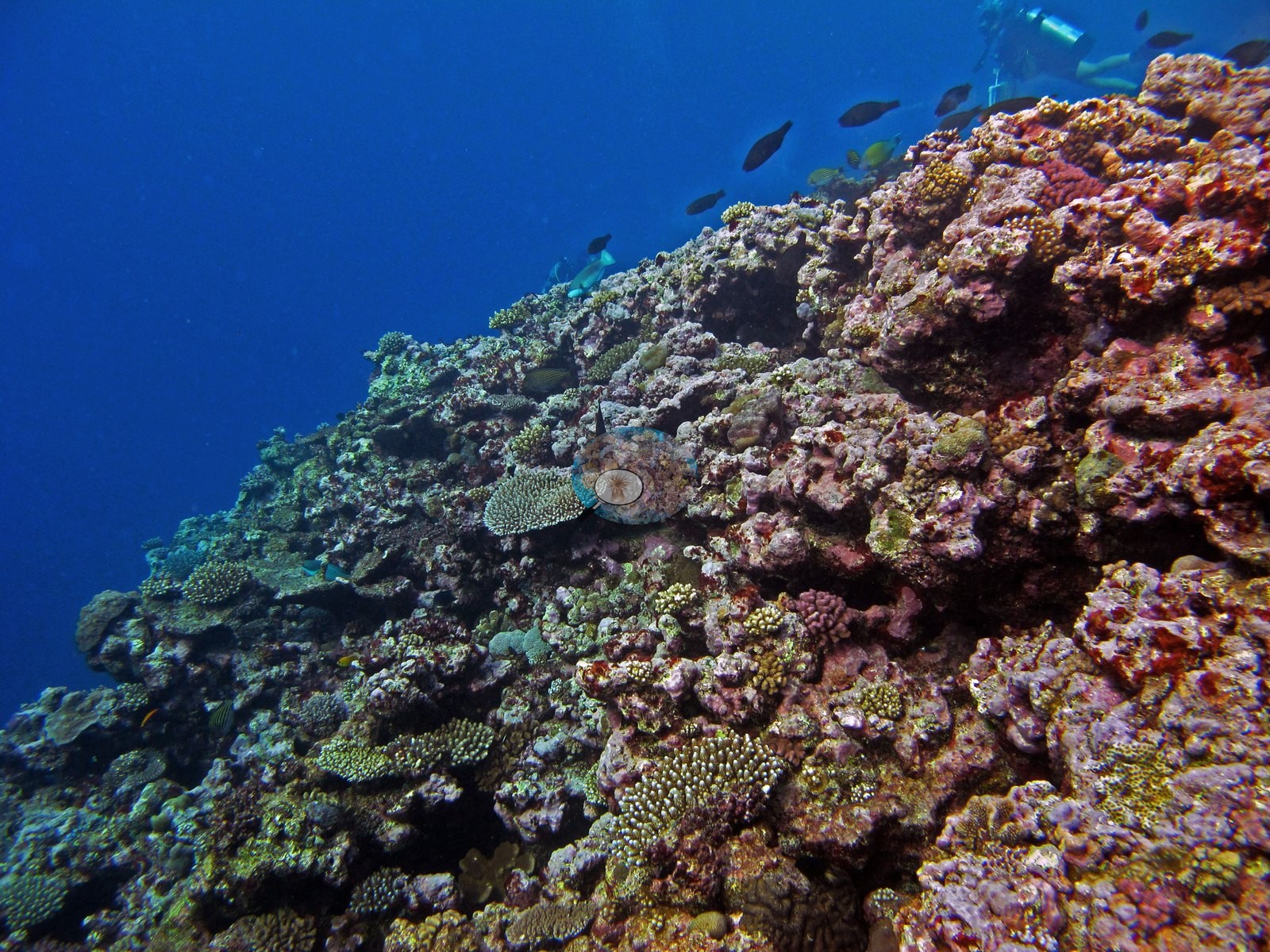
Image 5 and Image 16 are the cave frames, looking out from and into a large overhung passage with blue water beyond and the passage walls carrying plating and knobby colonies, and they establish that this site has genuine cavernous relief, not just grooves.
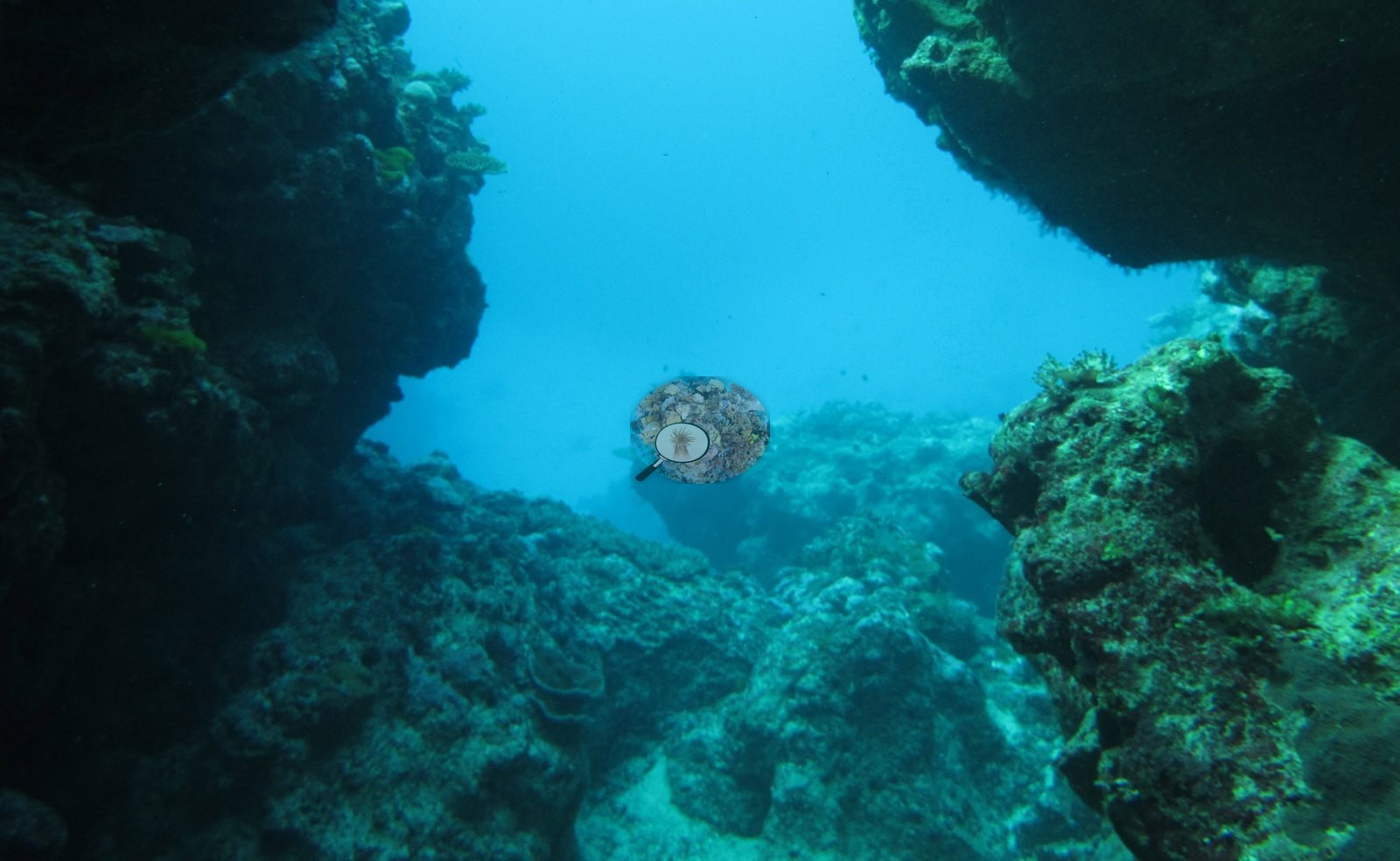
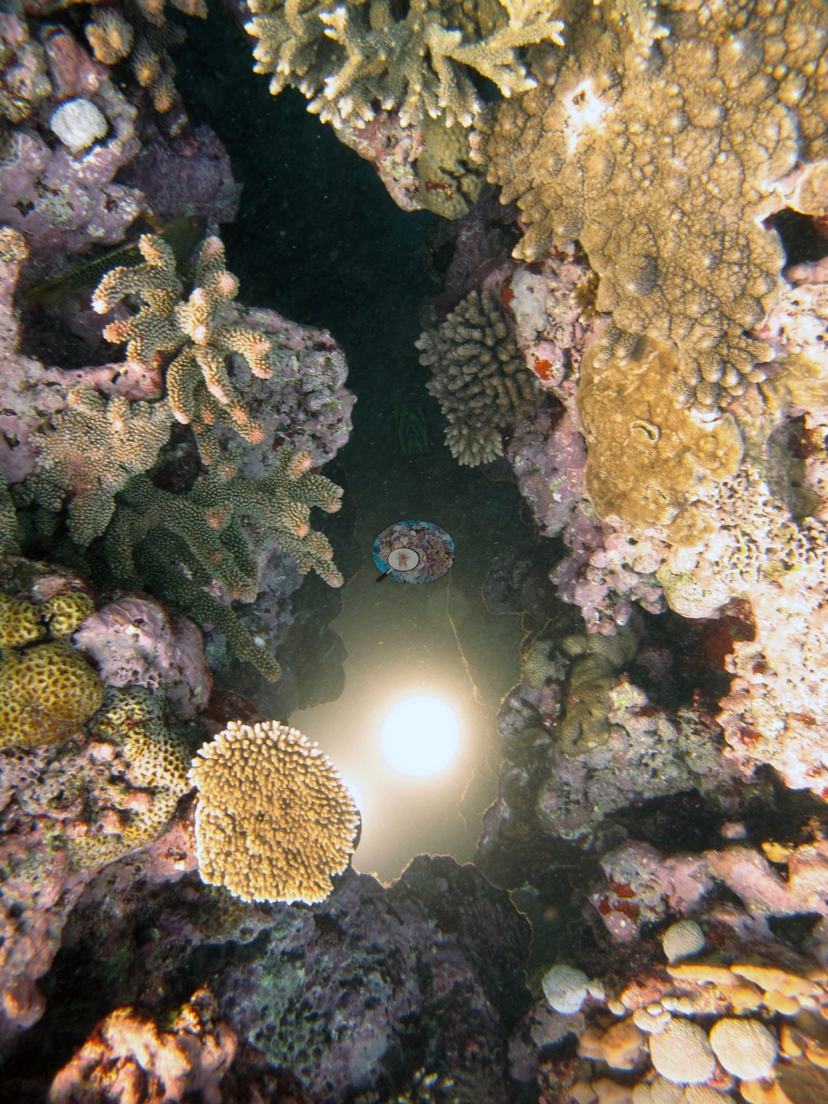
Image 12, which Anderson calls the Acropora garden, and Image 13 and Image 15 are the closest thing in the set to a reef-flat view: densely packed tabulate, corymbose, digitate, and bottlebrush acroporids with tan massive Porites and small faviids wedged among them.
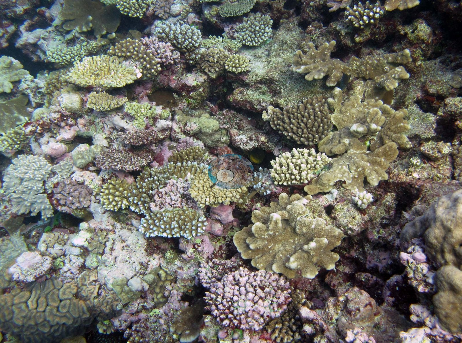
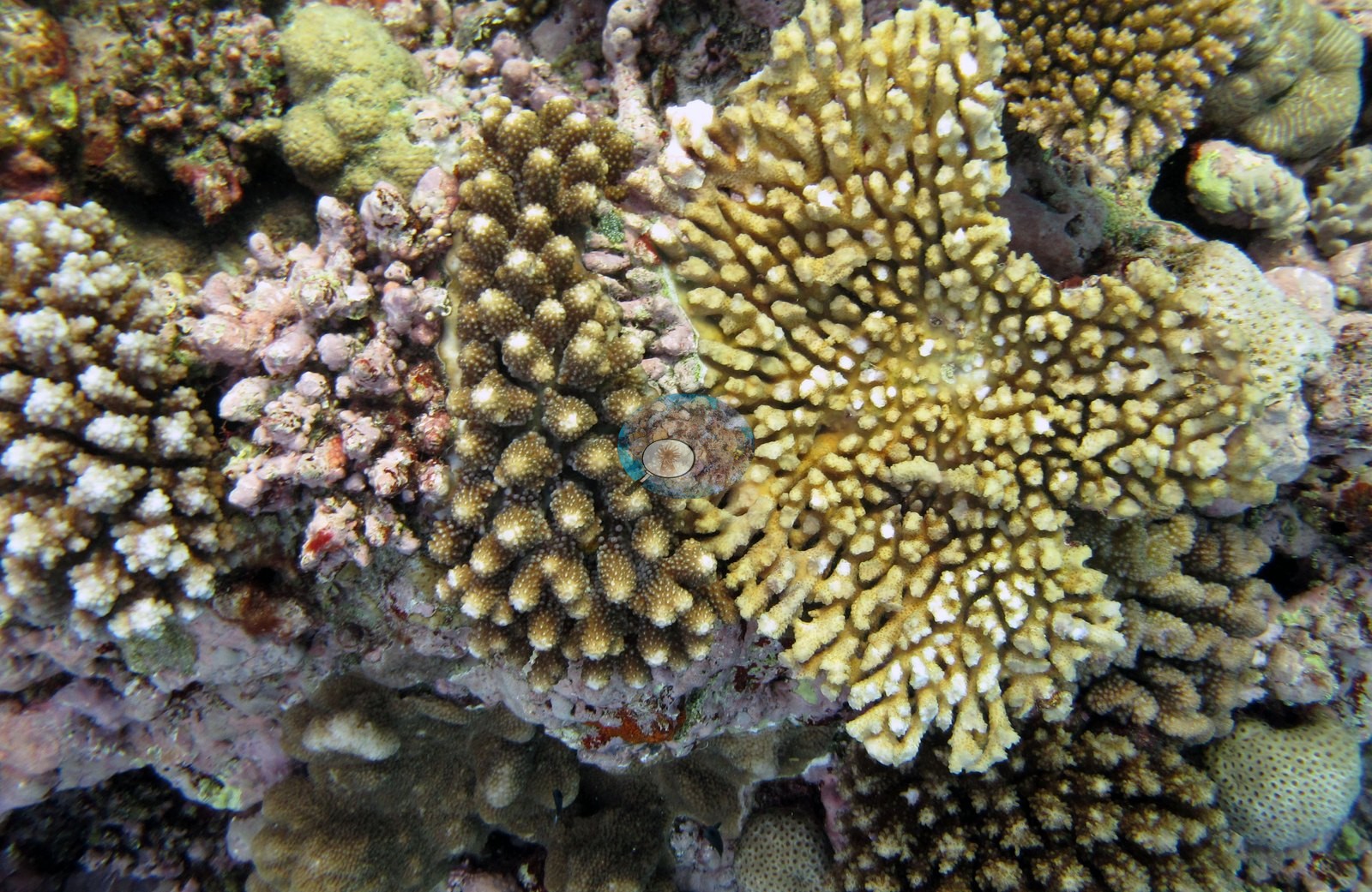
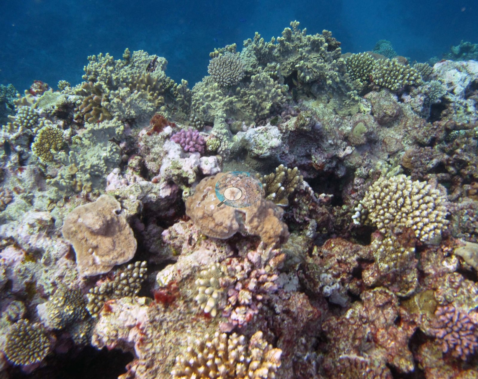
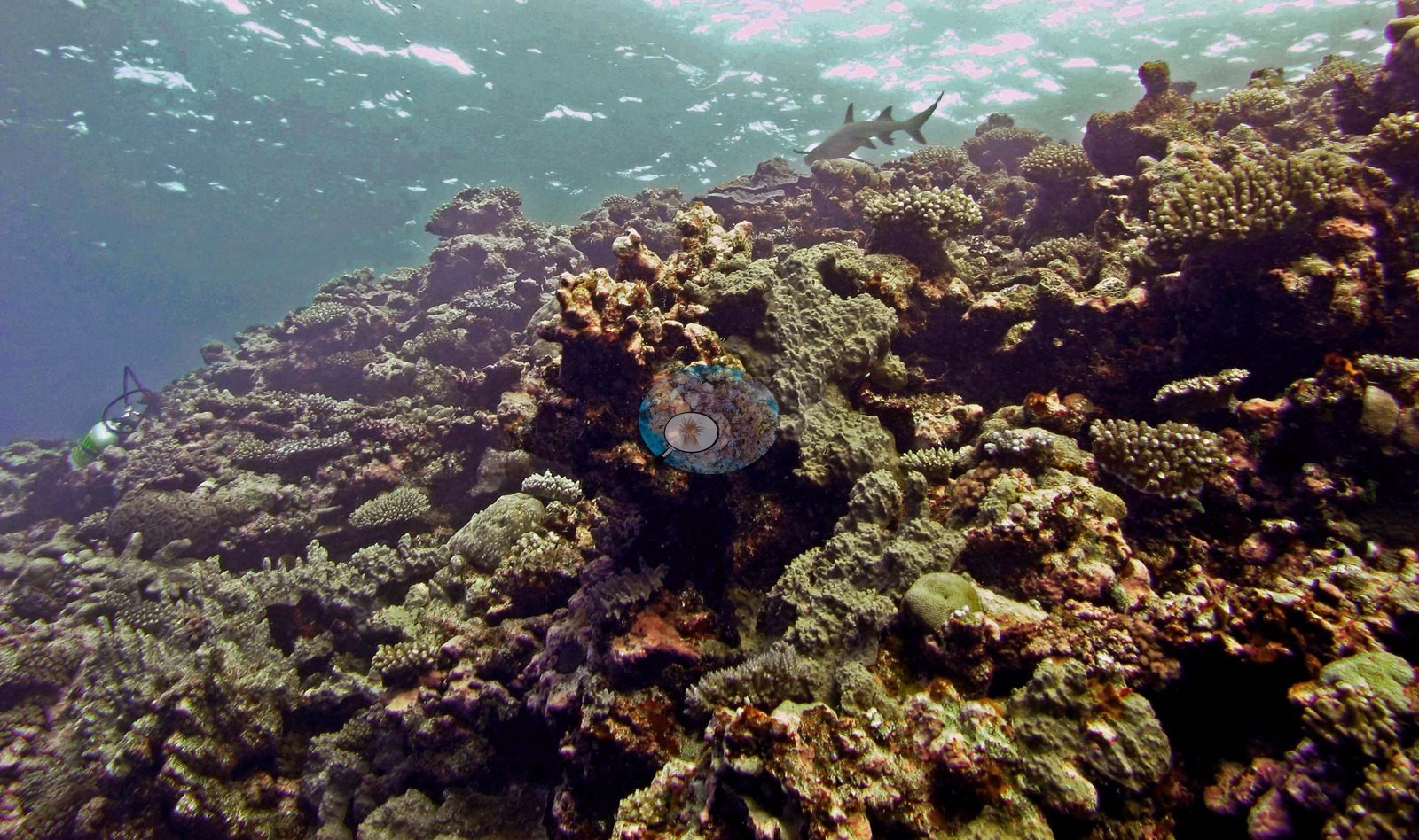
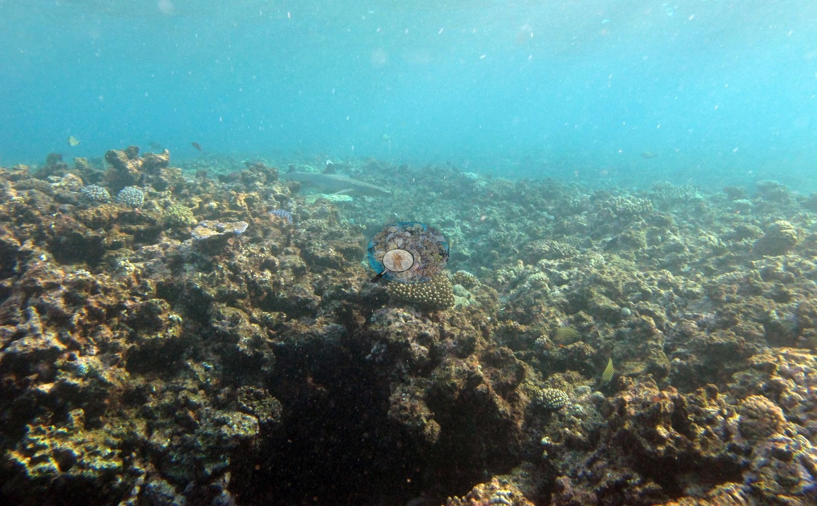
Image 18, Image 3, and Image 14 are the shallow end, labeled at 2–3 m, where the framework is lower, more coralline-crusted, and more algal, with a reef shark passing through two of them (e.g., Image 3, Image 18).
Coral cover
- Reference: Khaled bin Sultan Living Oceans Foundation, Global Reef Expedition (in situ transect)
- Coral cover 29%, from the LOF-GRE in situ benthic transect (the expedition's Fiji coral data workbook, cover sheet) — a diver transect, and the authoritative figure for this site; it is not derived from these photographs. Transect mean 29 ± 6.4% SE (n = 4 transects; Living Oceans Foundation per-transect benthic data, which lists these four transects, labeled FJTO09, under FJTO08's station code).
Stressors
Nothing in these frames shows unambiguous bleaching, disease lesions, or crown-of-thorns feeding scars, and no bleaching extent is estimated. Color rendering varies enough between the flash frames and the ambient-light frames that color-based condition calls would not be safe here in any case. The Living Oceans Foundation transects here recorded 7.3% cyanobacteria and no recently dead coral.
Fish and other fauna
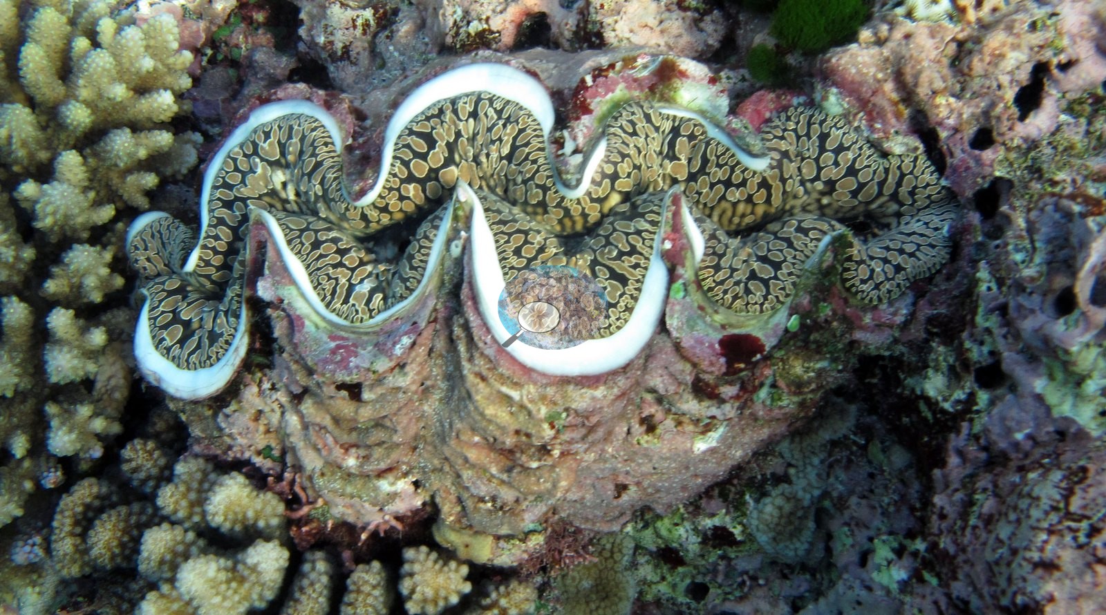
Image 17 is a giant clam set into branching Acropora, its mantle patterned olive and cream.
Comparisons with other data
Apart from the Living Oceans Foundation survey shown here, we found no other surveys at this site as of 2026. If you would like to share yours, please email us.
Other notes
The full FJTO09 photo set is as much a record of the survey team at work as of the reef, and both halves are worth having. Several frames are people and method: a diver with a slate working over the reef top (e.g., Image 10), two divers on the wall in poor visibility (e.g., Image 2), and a silhouetted diver with bubble column (e.g., Image 21), plus a bubble curtain rising up the reef face (e.g., Image 19).
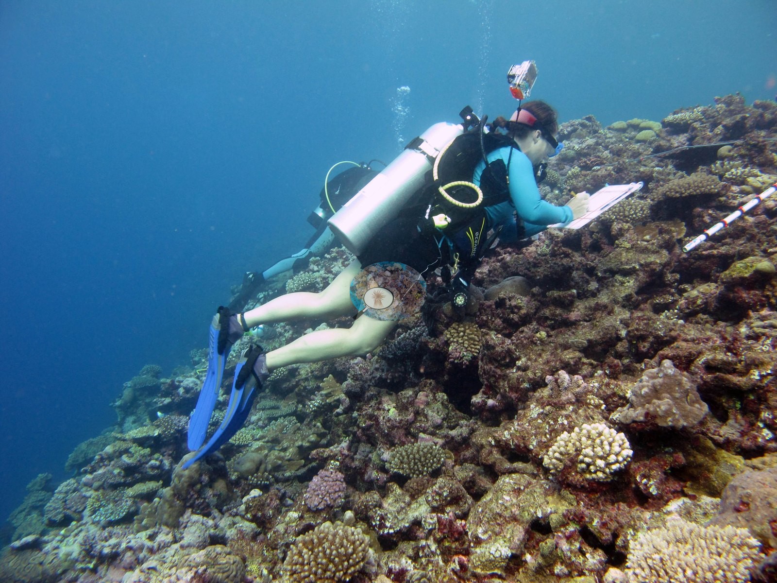
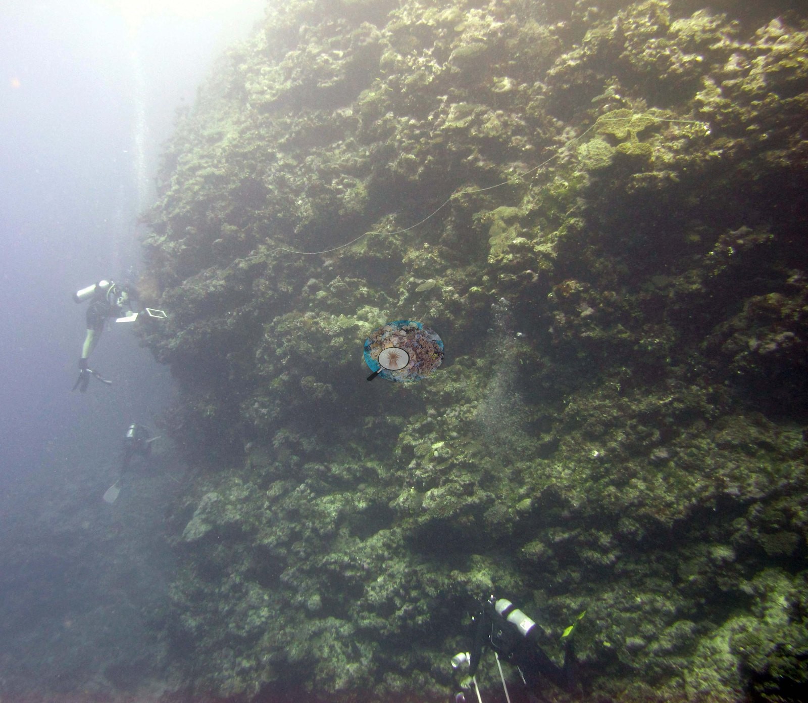
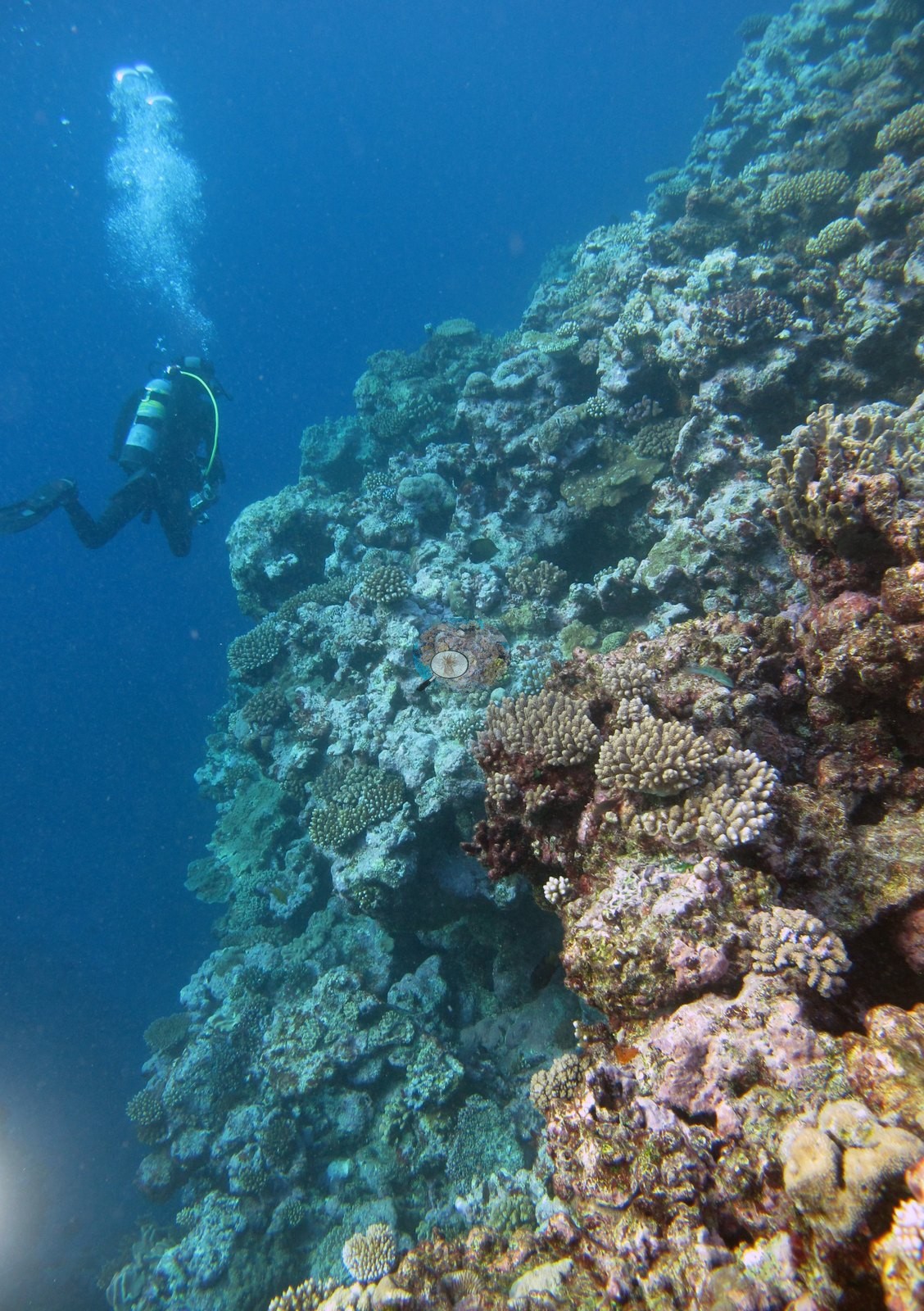
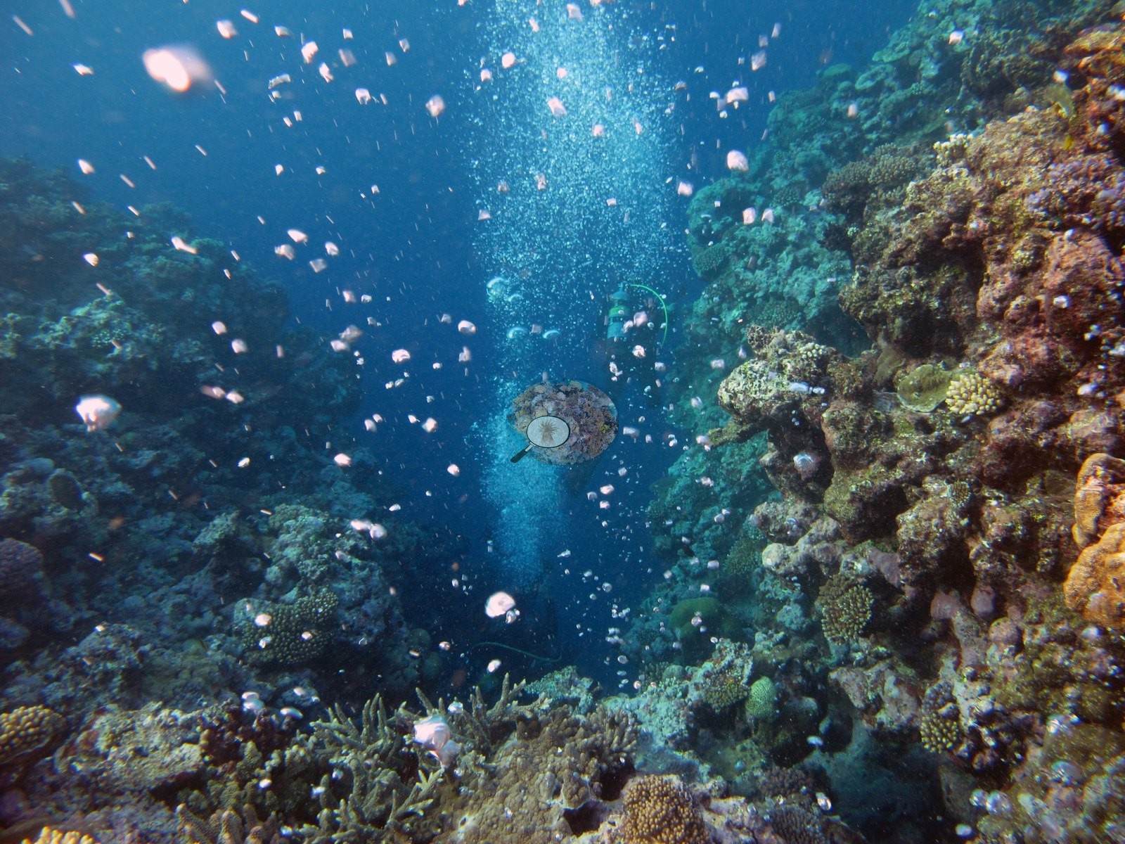
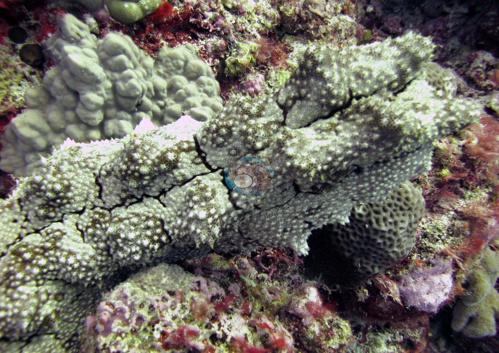
These carry the site's working context and must not be read as benthic cover. Two close frames carry Anderson's own field notes: Image 9 is a pale plating/tabular acroporid left unidentified, and Image 1 a large warty sea cucumber sprawled across the reef surface.
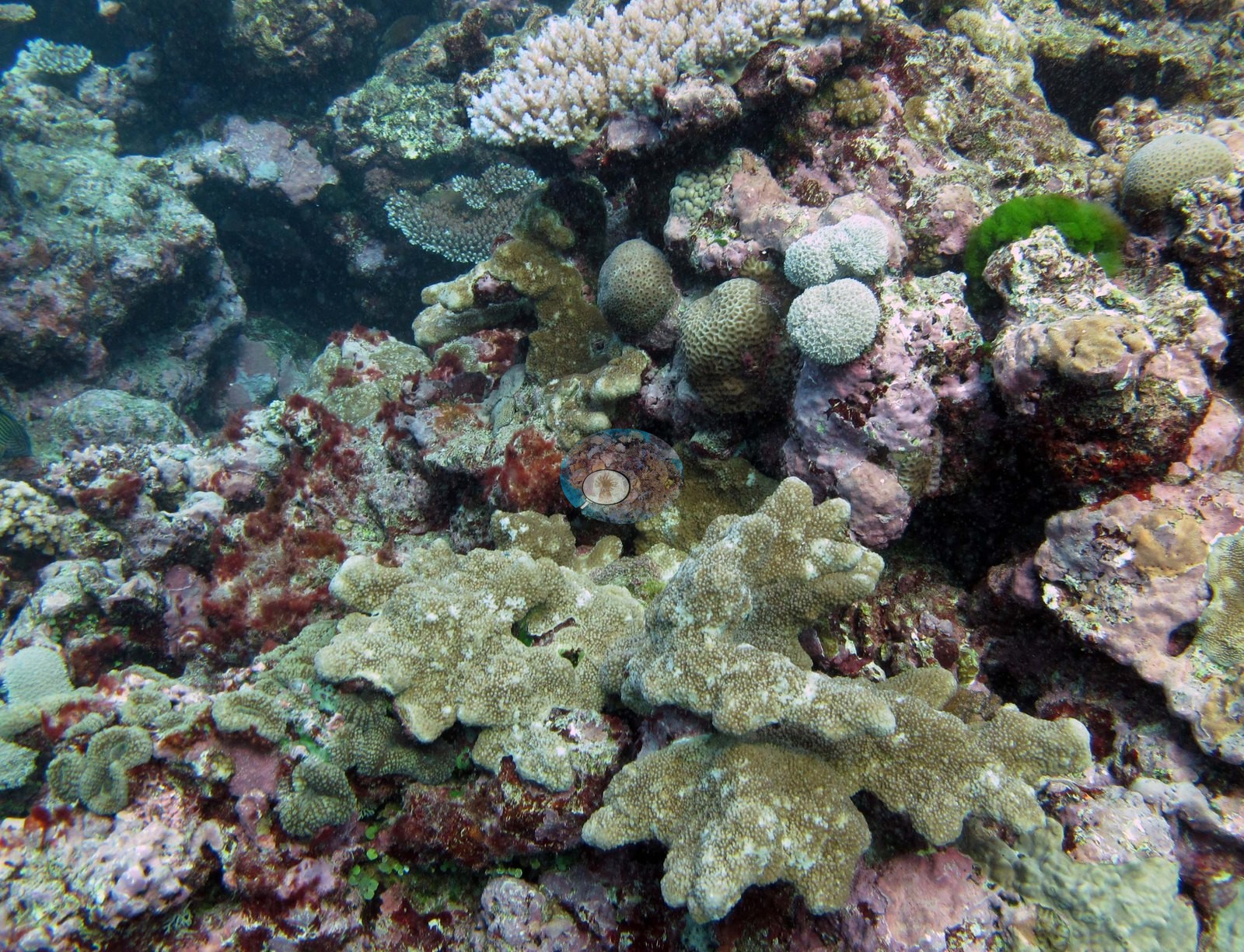
Neither is a habitat frame. What the frames cannot settle: depth. Only Image 14 carries a depth in its label, and that is Anderson's annotation rather than a gauge in shot — so the wall, cave, and garden frames cannot be placed on the depth profile in the survey narrative below, and no identifiable frame shows the deepest part of the dive (29.6 m; the 33.6 m figure is the station CTD cast).
Survey team's site notes: A diverse benthic community with the dominant scleractinian community differing by location. Shallower areas may be dominated by branching acroporids and (sub)massive poritids with encrusting and plating corals also present. Small mounds of hosting diverse mixed coral assemblages may be present. At greater depths, colonies take on plating morphologies and form a series of shingle-like structures. Prominent macroalgae include Halimeda, Caulerpa, and Dictyota. Sediment filled grooves, with large pieces of rubble. Shallower depths are dominated with small Acropora and Pocillopora colonies. Fore reef sand flats benthic cover is usually very low because the substrate is mobile and largely unconsolidated thus inhibiting colonization by benthic organisms. In some cases, large Porites lobata colonies and tabular acroporids occur within the sand flats, especially in deeper areas (30-50 m). Macroalgae become more common with depth due to the abatement of wave energy with increasing depth. The deeper fore reef habitats at the base of the reef were generally low relief sand flats, mostly uncolonized. Often there were sparse assemblages of cyanobacteria and small patches of macroalgae, along with isolated larger corals.
CV model comparison
More info
The photo row below is a methods comparison only: it is never averaged with the transect and never used as the headline. It compares site means only: the photos come from the same survey dive as the transect but not from the same points.| Method | Hard coral cover (%) | Photos (n) ± SE |
|---|---|---|
| Transect mean (all transects) | 28.8 | |
| CoralSCOP edited | 24.7* | n = 13 · SE 3.8 |
| Claude (edited photos) | 18.4* | n = 13 · SE 4.1 |
The two computer-vision methods agree within 6.3 pp (18.4–24.7%) and read 4.1–10.4 pp below the in-water transect (28.8%); the headline is the in-water transect because a diver transect is independent of the photographs.
*Based on only 13 reef-habitat photos; this estimate is less certain.
See a more detailed, stand-alone version of this table.
Data & links
All images from Coral Reef Diagnostics. Data are from the Khaled bin Sultan Living Oceans Foundation Global Reef Expedition and Coral Reef Diagnostics.
 © Coral Reef Diagnostics.