
Part of the regional summaries: Fiji - Totoya, Fiji - Moala Group (Moala / Totoya / Matuku), and Fiji - Lau
More info
Fiji (NOAA CRW virtual station)A well-built fore-reef crest off Totoya with big table Acropora over a meter across and continuous staghorn thickets full of small planktivores, crossed by a school of yellow-striped snapper. Chunky golden Pocillopora eydouxi and a purple pocilloporid are among the colonies Anderson photographed and labeled, at 46% cover. Surveyed on the Living Oceans Foundation's Global Reef Expedition.
E dua na dakuna ni yasana i tuba ni cakau (fore-reef crest) e buli vinaka mai Totoya, e tu kina na lase tepeli levu ni Acropora e sivia na dua na mita na kena raba kei na veikau ni staghorn e tomani tiko ka vonu ena ika lalai e kania na plankton (planktivores), ka qalo sivita e dua na ilawalawa ni ika (yellow-striped snapper). Na Pocillopora eydouxi levu ka roka koula kei na dua na pocilloporid e roka (purple) e okati ena lase a taba ka vakaiyacataka ko Anderson, ka 46% na ubi ni lase (coral cover).
AI translation of this summary; the English is authoritative. (low confidence)
CTD-cast midpoint; the cast read 27.1°C throughout
Two other CRD dive sites with their own pages within 5 km shown in orange — click a marker to open its page.
🗺 Detailed map of the Global Reef Expedition dive sites, Totoya, Fiji (Khaled bin Sultan Living Oceans Foundation; opens in a new window)
18 of the 20 photographs on this page are shown beside the text that discusses them under "Site description" below.
Site description
General benthic features
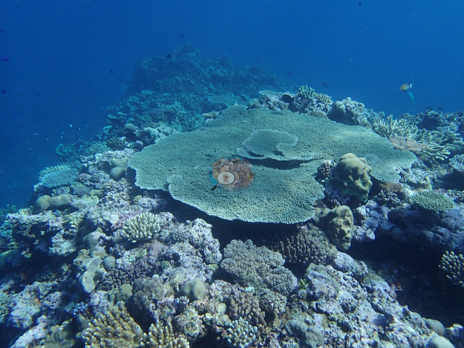
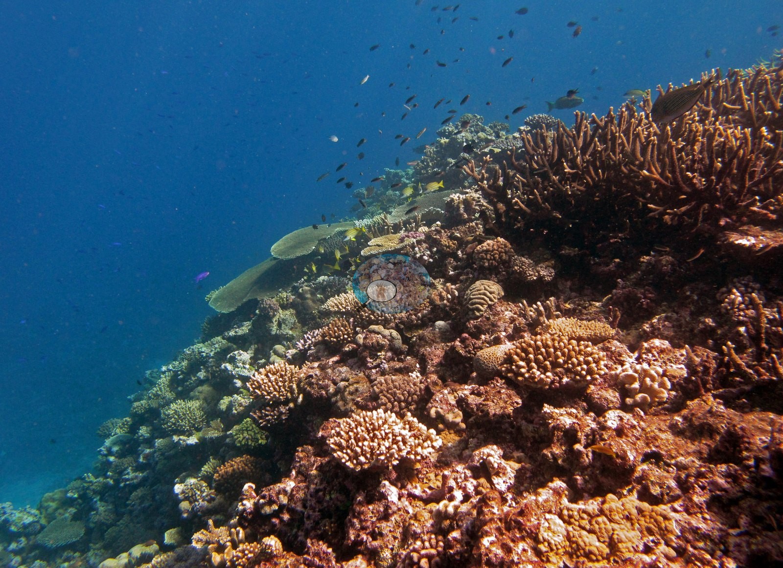
This is a well-built fore-reef crest and the wider set makes its two signature elements plain. The first is tabulate Acropora: at least three frames are built around large tables, one broad gray-green table over a meter across standing clear on its stalk above a mixed crest (e.g., Image 18), the same or a sister table photographed from a lower angle with corymbose and branching colonies stacked behind it (e.g., Image 25), and a further tan table stepping along the crest into blue water (e.g., Image 26).
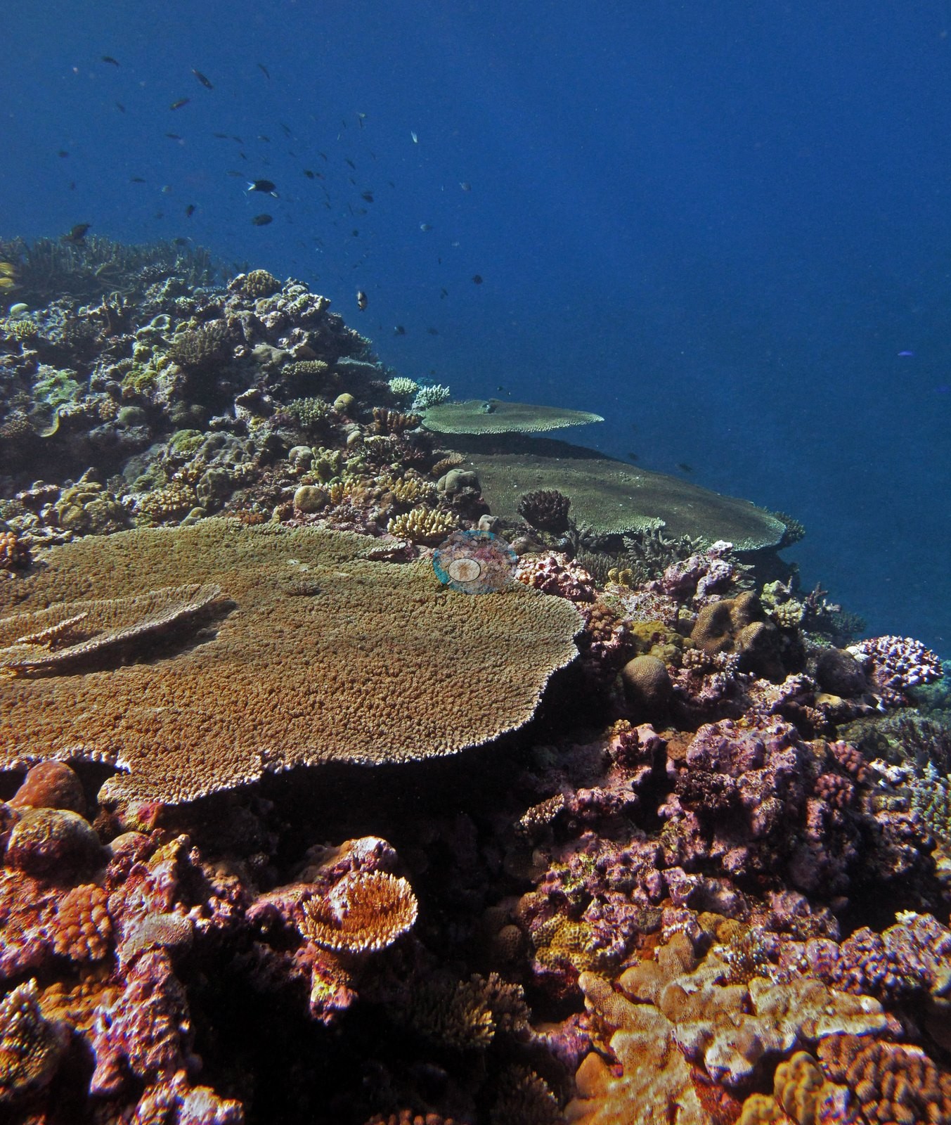
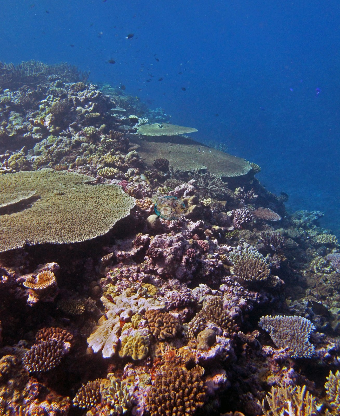
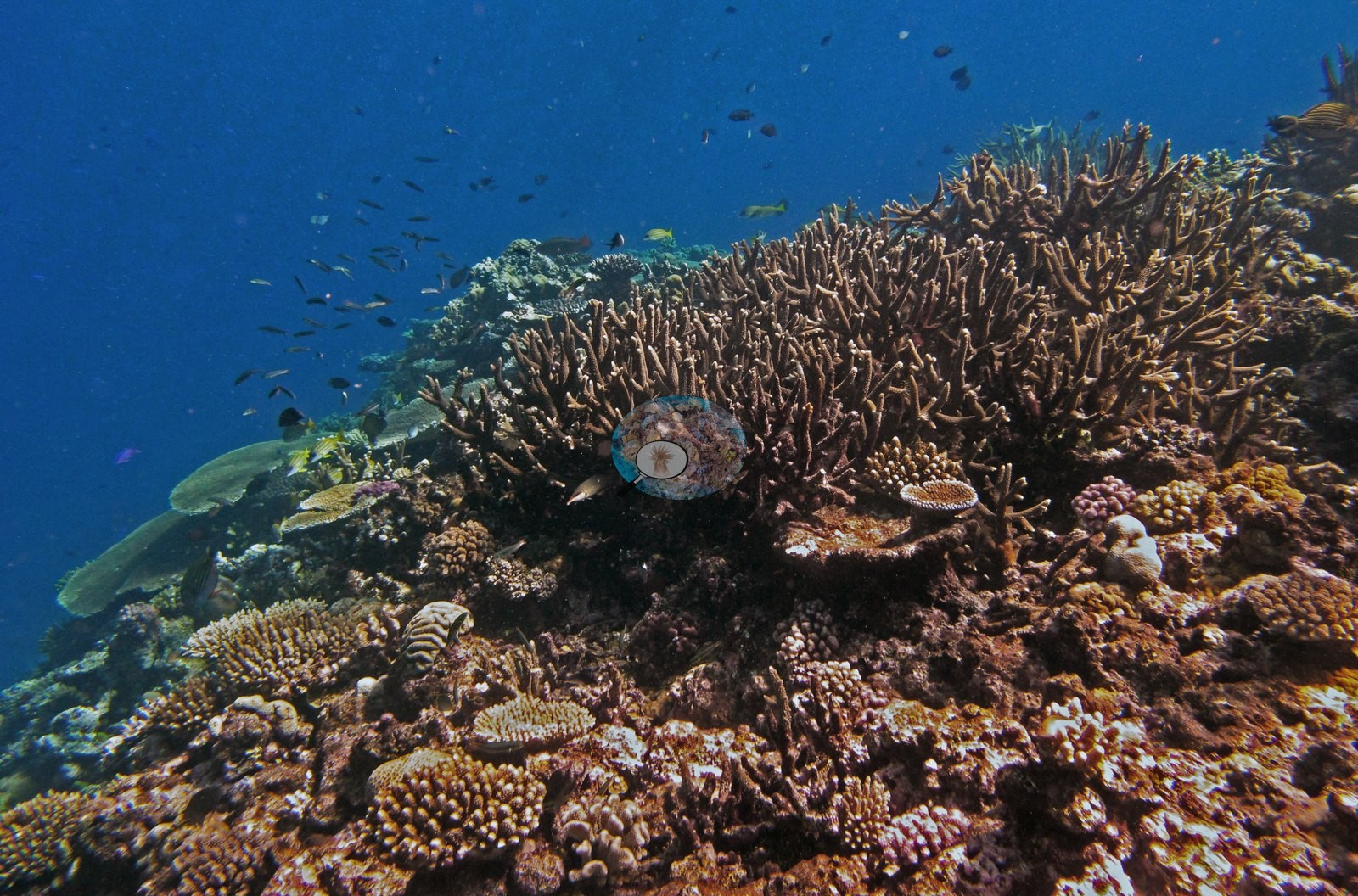
The second is branching and staghorn Acropora, which forms continuous stands rather than isolated colonies — open staghorn thickets of long, fine, upward-angled branches carrying clouds of small dark planktivores (e.g., Image 16, Image 17), and a dense brown-and-tan crest of shorter arborescent and corymbose colonies packed edge to edge (e.g., Image 23, Image 24).
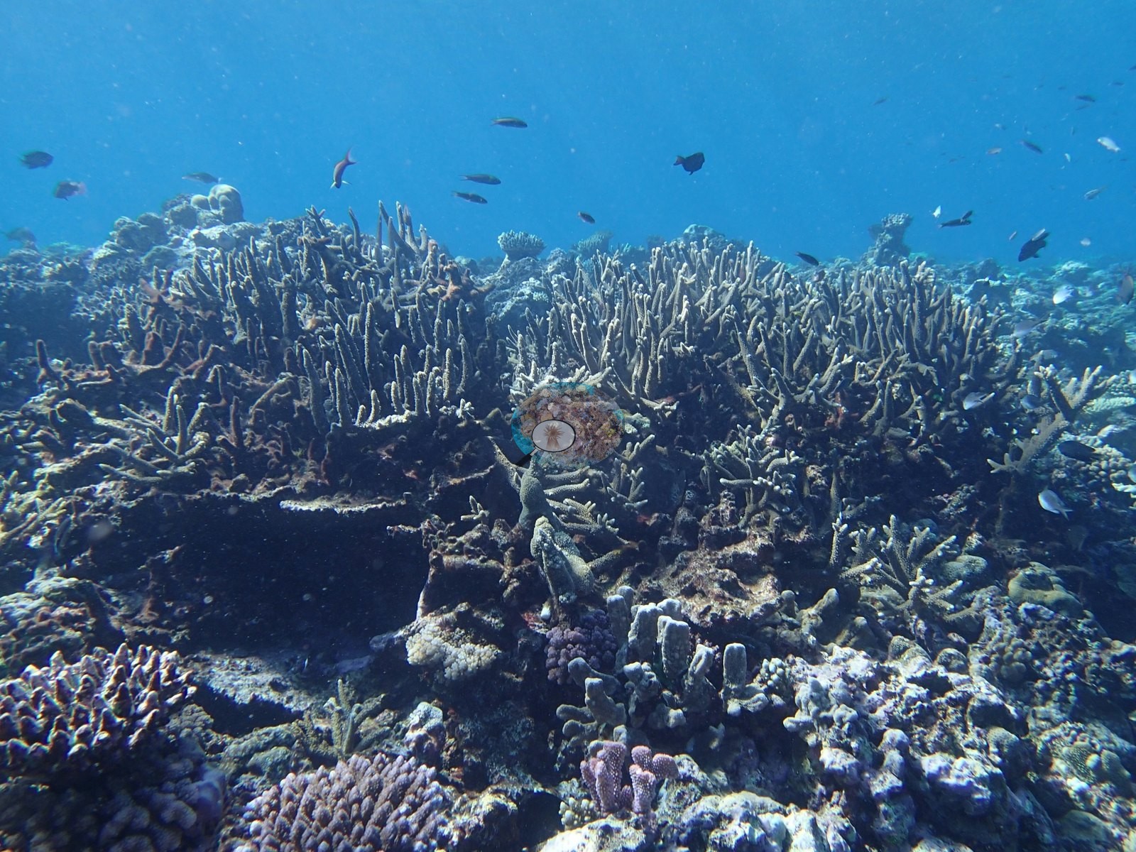
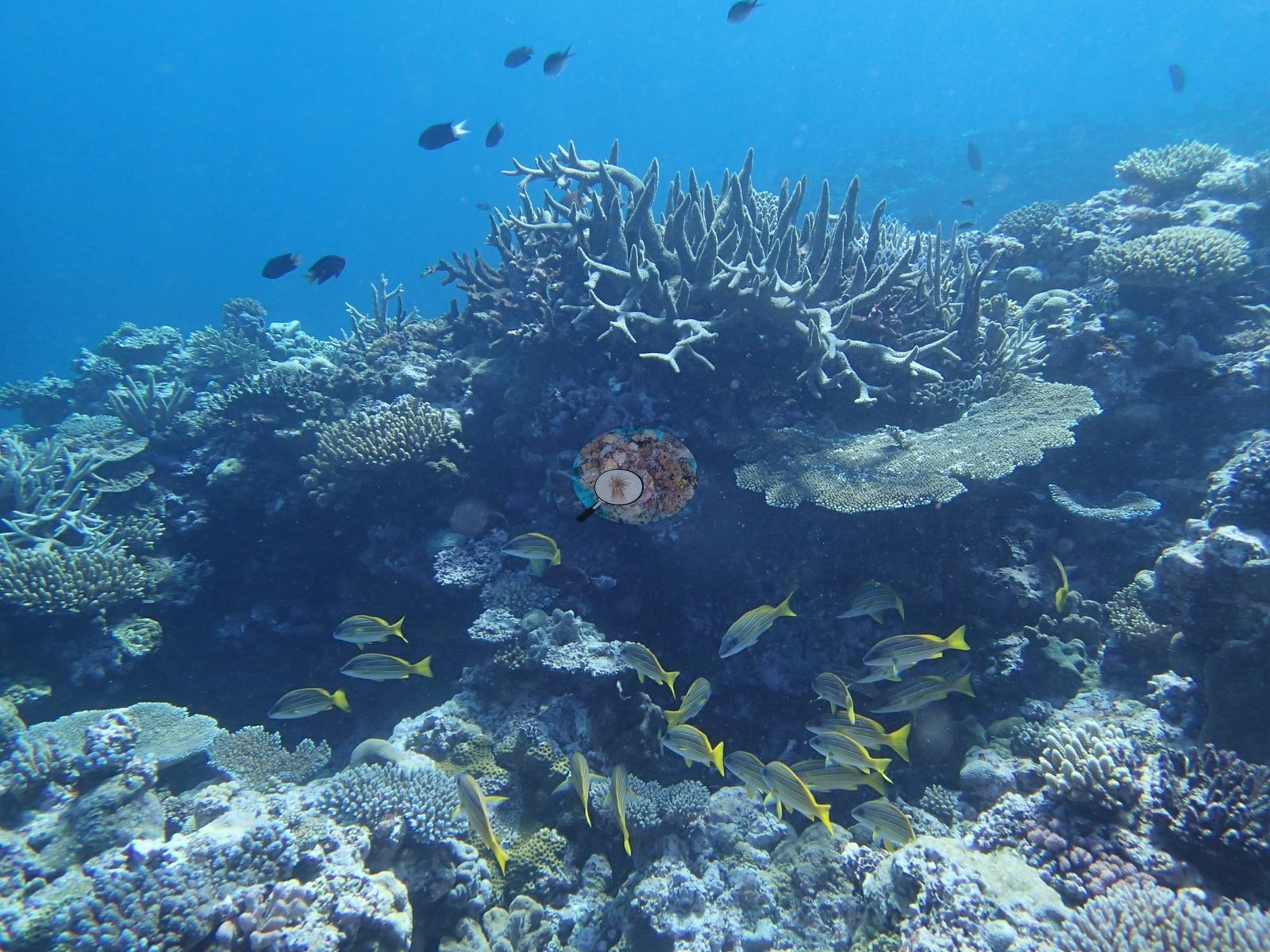
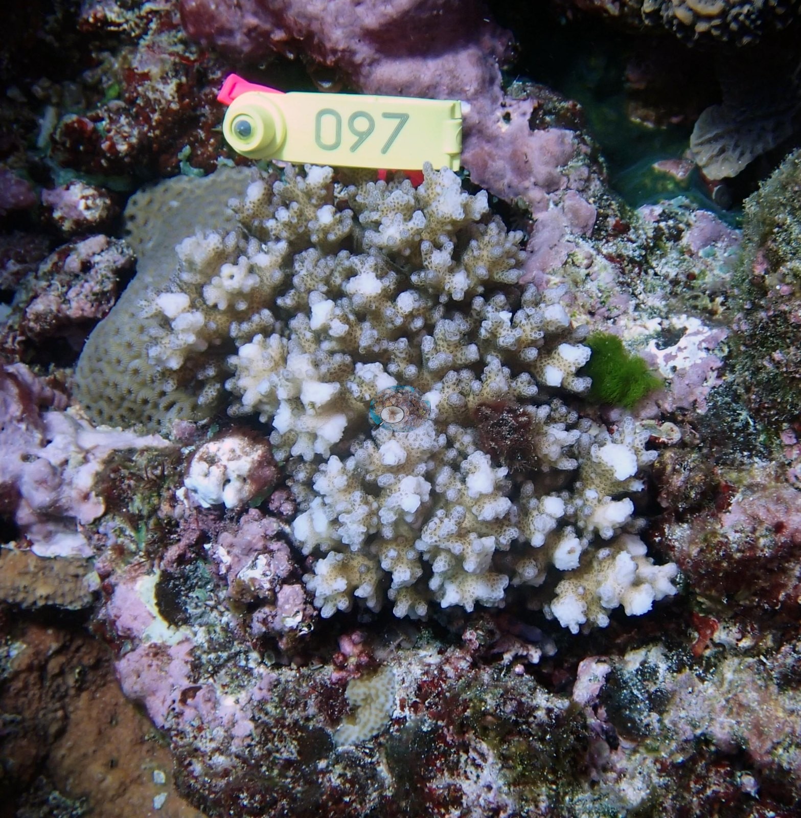
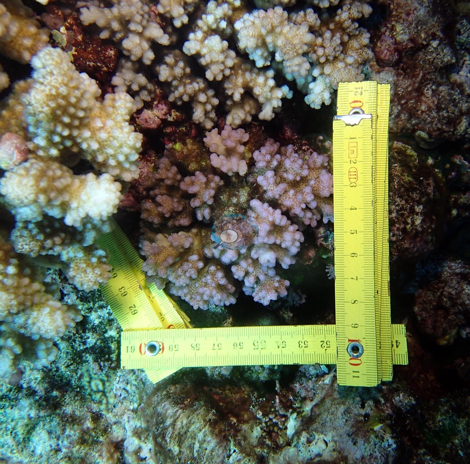
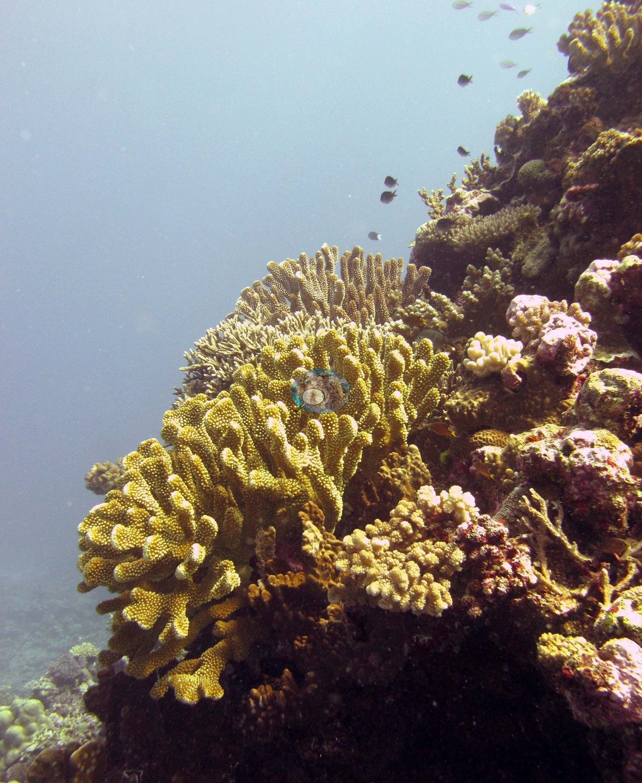
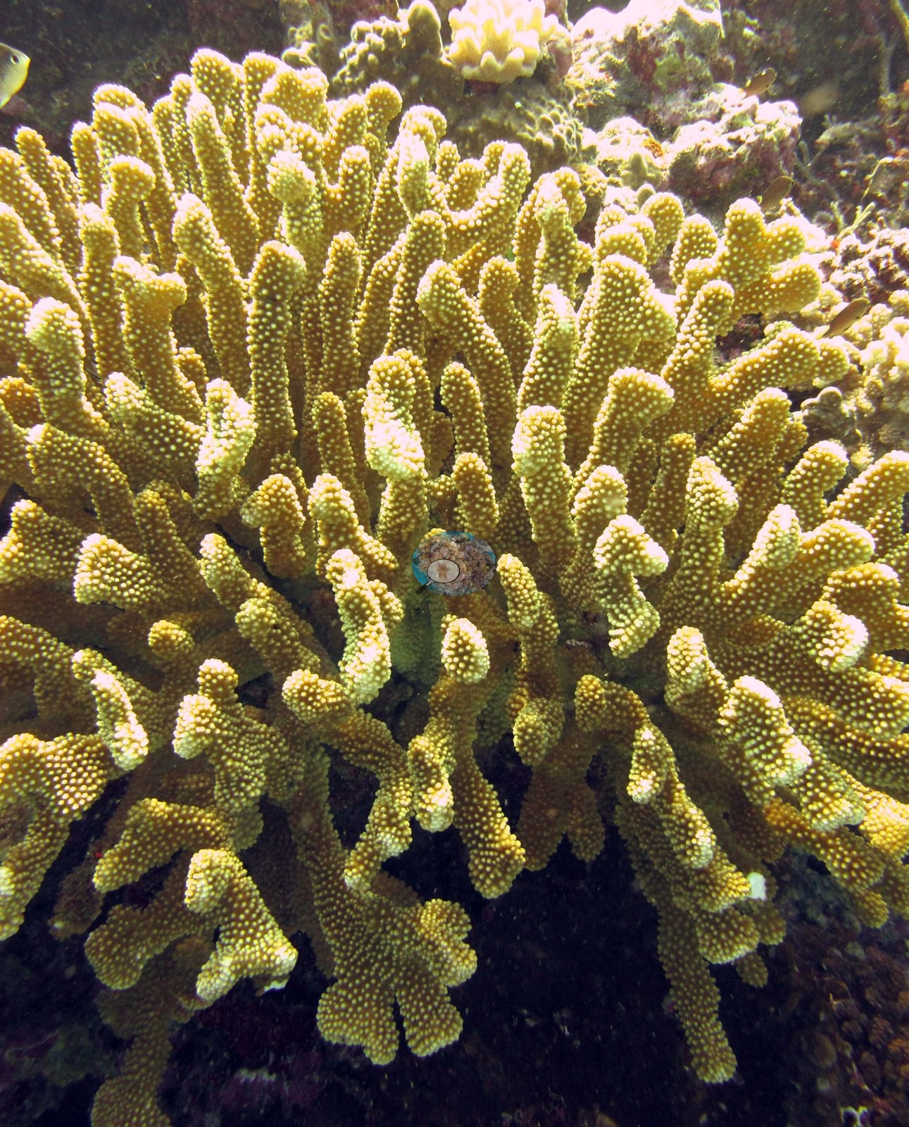
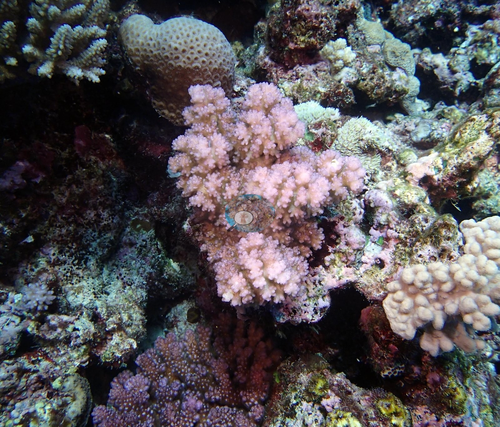
Pocilloporids are the recurring close subject, with Anderson's own identifications: a mustard-yellow colony with thick, blunt, densely corallite-studded branches identified as P. eydouxi, shot both in habitat (e.g., Image 21) and close (e.g., Image 20), and a pale pink-mauve colony with fine rounded branchlets recorded as a purple pocilloporid, sitting among cream corymbose Acropora and pink coralline crust (e.g., Image 12).
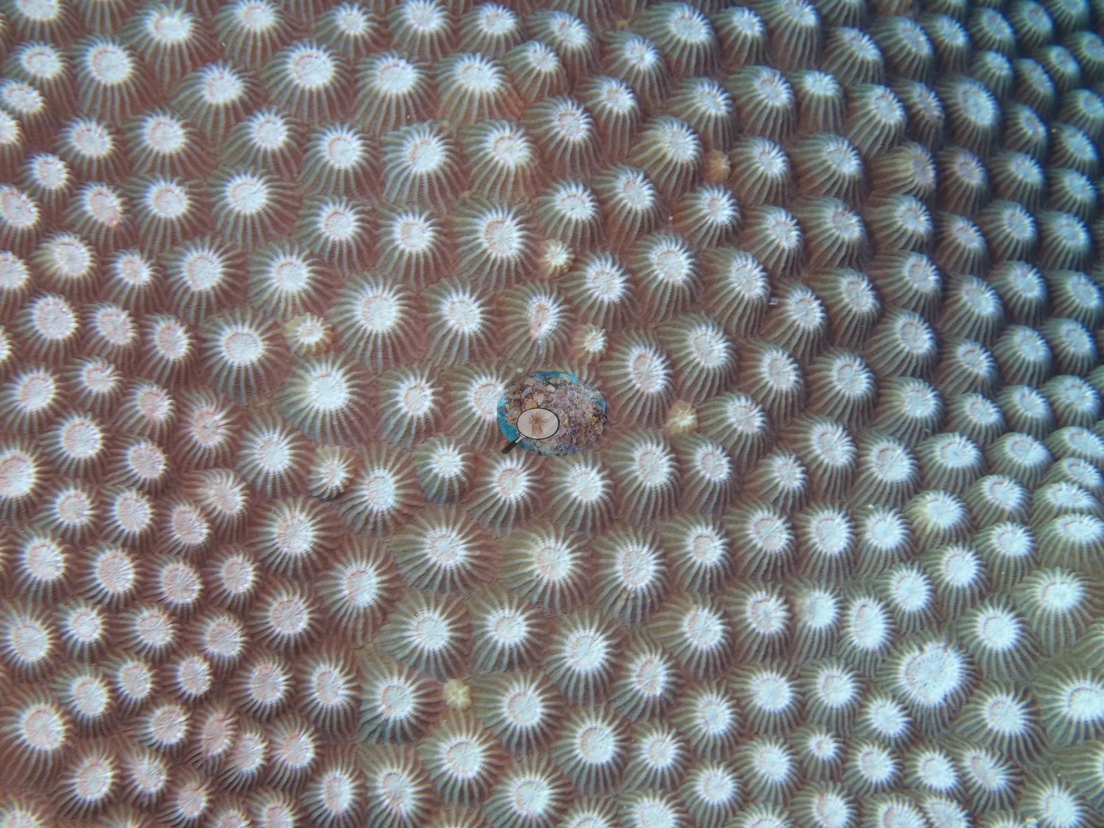
Massive corals appear as both habitat and macro: one frame fills the field with a single massive's surface, showing widely and evenly spaced cream corallites raised from a tan coenosteum (e.g., Image 3) — a distinctive, regularly spaced massive that the photographs describe well but that is not named here — and a large pale blue-gray massive with a fish tucked against its base occupies Image 1.
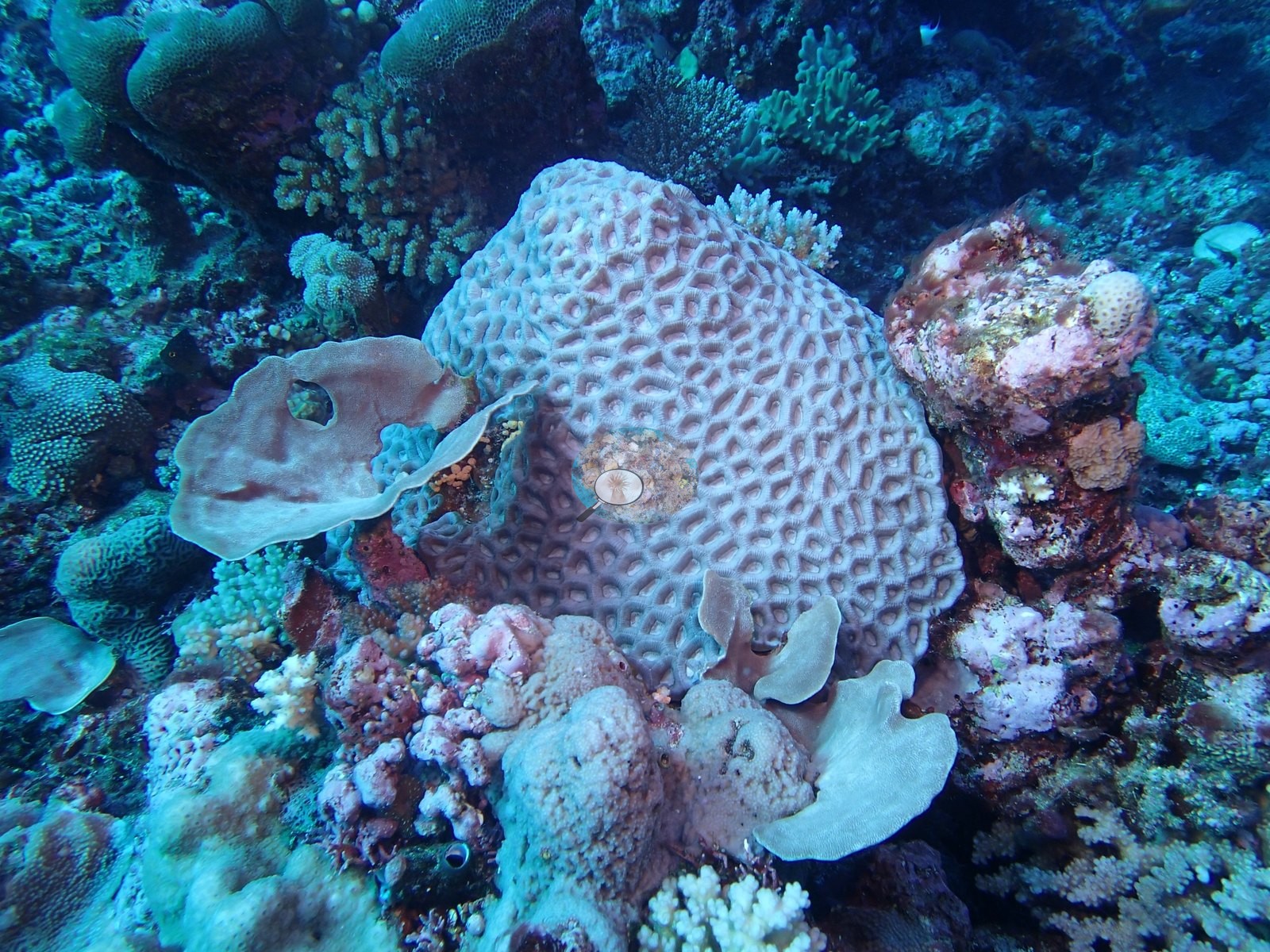
One colony in Image 13, Image 14, and Image 15 has thick, contorted, knobby lobes with fine pale polyps over the whole surface; the photographs do not settle whether this is a heavily thickened stony colony or a leather soft coral.
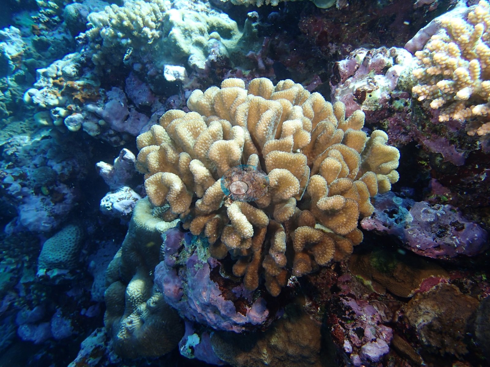
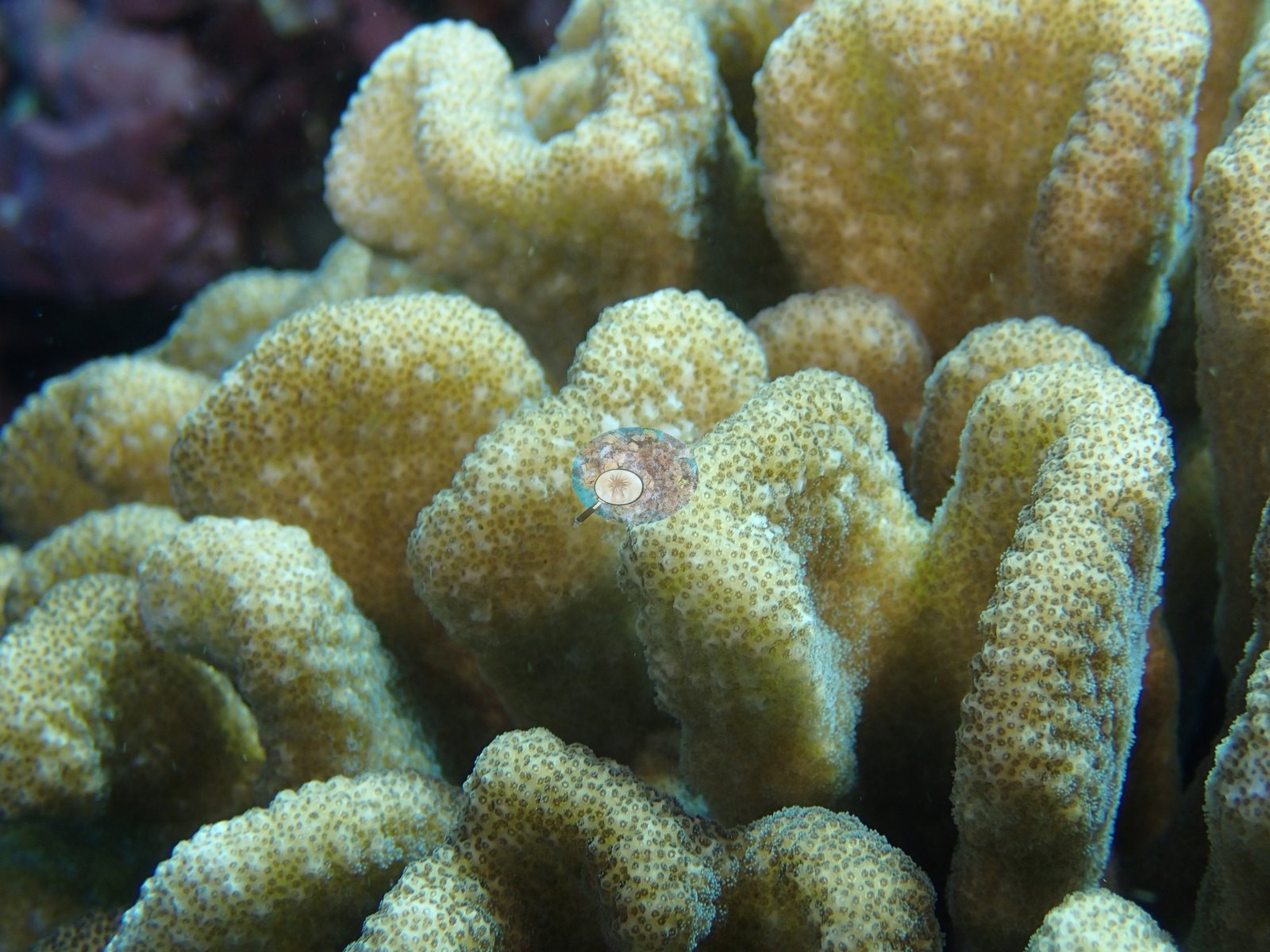
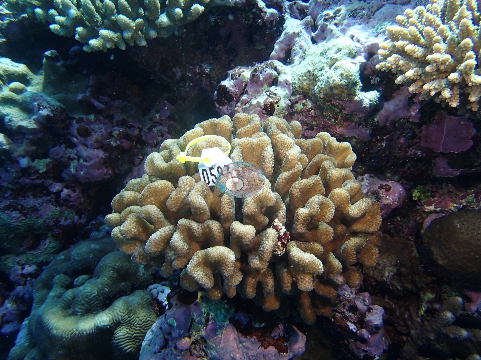
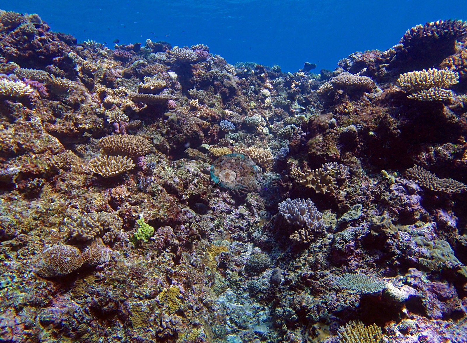
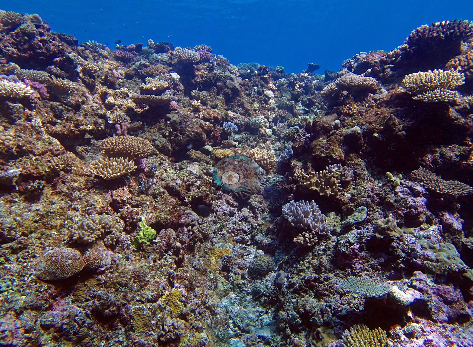
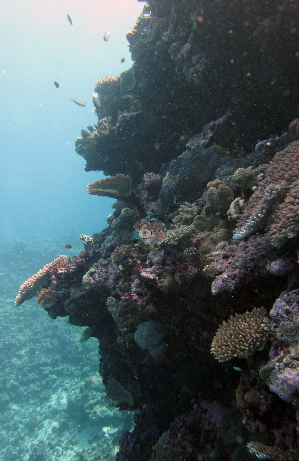
Image 8 looks across a reef top heavily crusted in mauve and pink coralline algae with small yellow-green colonies set into it, and Image 22, which Anderson noted as the wall, looks up a steep, shadowed reef face against open blue.
Coral cover
- Reference: Khaled bin Sultan Living Oceans Foundation, Global Reef Expedition (in situ transect)
- Coral cover 46%, from the LOF-GRE in situ benthic transect (the mean of all of this station's own transects in the expedition's per-transect benthic data) — a diver transect, and the authoritative figure for this site.
- Transect mean 46 ± 2.5% SE (n = 5 transects; Living Oceans Foundation per-transect benthic data); the headline above is this all-transect mean.
Stressors
Across the frames there is no visible bleaching, no disease lesion, and no crown-of-thorns feeding scar; the white margins on the tables and branching colonies are growing edges. The one sign of physical disturbance in the set is a detached, tipped plate fragment lying beside an intact table, which is localized rather than general. Anderson's sampling sheet records numerous fish bites on one of the Pocillopora damicornis colonies sampled here, which was otherwise darkly pigmented. The Living Oceans Foundation transects here recorded 6.6% cyanobacteria and no recently dead coral; they carry no bleaching, disease, or crown-of-thorns field.
Fish and other fauna
A school of yellow-striped snapper (bluestripe-snapper-type) drifts across the staghorn ground in Image 17.
Comparisons with other data
Apart from the Living Oceans Foundation survey shown here, we found no other surveys at this site as of 2026. If you would like to share yours, please email us.
Other notes
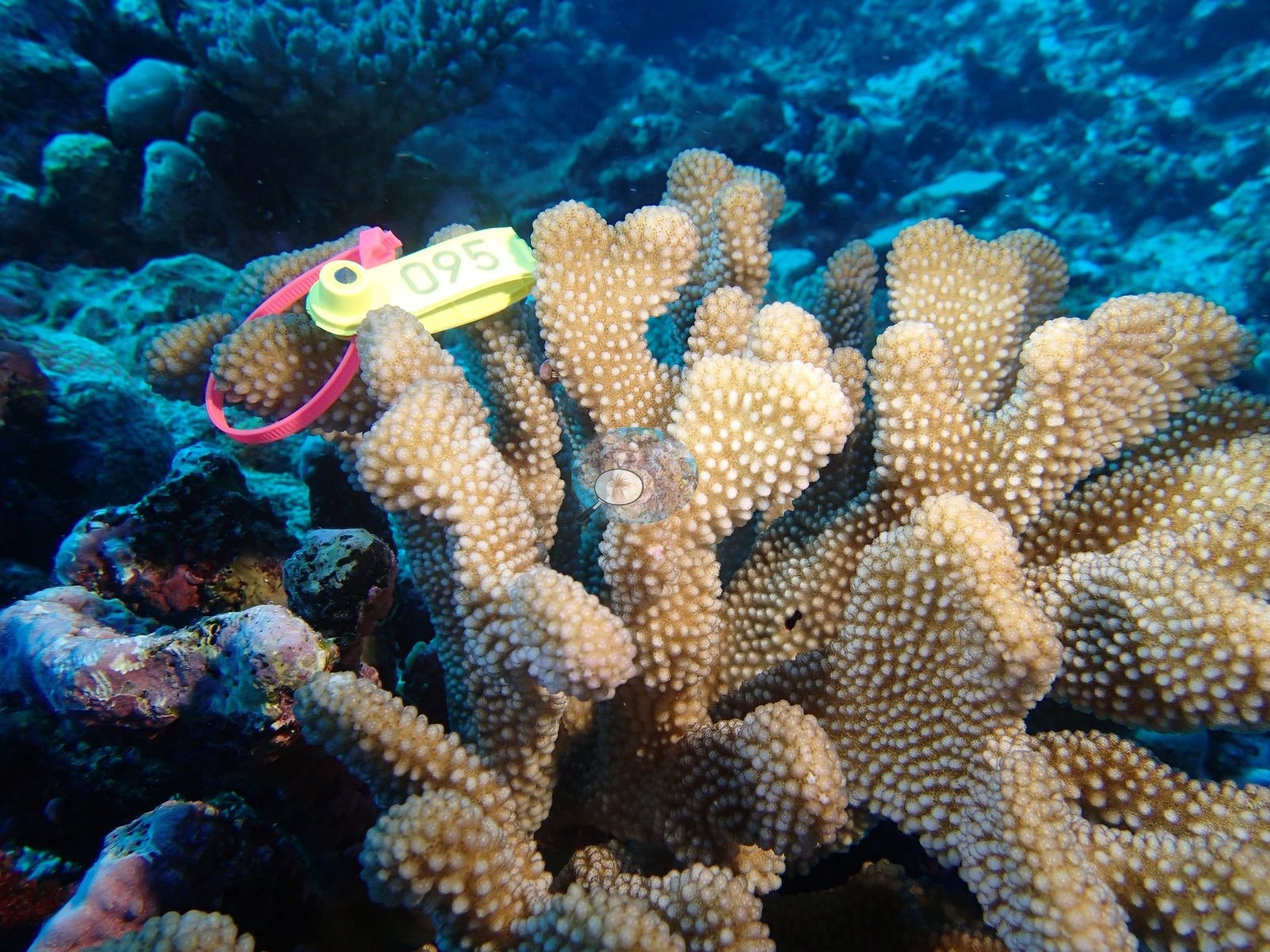
Image 2 shows a golden-tan branching colony with thick, blunt, wart-covered branches.
Worth noting separately: the one coral colony from this site with an outlier score (of three sampled) was flagged as a physiological outlier (Mayfield et al. 2017), which is a tissue-level result and is not something these photographs show or contradict.
Survey team's site notes: A diverse benthic community with the dominant scleractinian community differing by location. Shallower areas may be dominated by branching acroporids and (sub)massive poritids with encrusting and plating corals also present. Small mounds of hosting diverse mixed coral assemblages may be present. At greater depths, colonies take on plating morphologies and form a series of shingle-like structures. Prominent macroalgae include Halimeda, Caulerpa, and Dictyota. High coral cover, with large spur and groove ridges. Sediment filled grooves, with large pieces of rubble. Fore reef sand flats benthic cover is usually very low because the substrate is mobile and largely unconsolidated thus inhibiting colonization by benthic organisms. In some cases, large Porites lobata colonies and tabular acroporids occur within the sand flats, especially in deeper areas (30-50 m). Macroalgae become more common with depth due to the abatement of wave energy with increasing depth. The deeper fore reef habitats at the base of the reef were generally low relief sand flats, mostly uncolonized. Often there were sparse assemblages of cyanobacteria and small patches of macroalgae, along with isolated larger corals.
Data & links
- Download benthic survey data (CSV) ↗
- Download all LOF-GRE data (933 sites) ↗
- Photo portfolio ↗
- Coral Reef Diagnostics region page ↗
All images from Coral Reef Diagnostics. Data are from the Khaled bin Sultan Living Oceans Foundation Global Reef Expedition and Coral Reef Diagnostics.
 © Coral Reef Diagnostics.