
Part of the regional summaries: Fiji - Totoya, Fiji - Moala Group (Moala / Totoya / Matuku), and Fiji - Lau
More info
Fiji (NOAA CRW virtual station)A Totoya station whose photos mostly document the physiology sampling work, with tagged colonies (Anderson's sampling sheet records one sampled colony here) and divers at work. The reef itself ranges from a densely occupied coral wall of small colonies over pink coralline framework to a flatter, rubbly reef top with scattered corals, at 25% cover. Surveyed on the Living Oceans Foundation's Global Reef Expedition.
E dua na vanua ni vakadidike e Totoya ka levu na kena itaba e vakaraitaka na cakacaka ni taura ivakaraitaki vakayago (physiology sampling), kei na lase e vakatakilakilataki (tagged) (na ivola ni ivakaraitaki nei Anderson e vola e dua na lase a taura kina na ivakaraitaki eke) kei na dau nunu ena nodra cakacaka. Na cakau e veitalia mai na dua na bati ni lase (coral wall) e vonu ena lase lalai ena dela ni ivakatudei ni lumi vakalase (coralline) e roka (pink) ki na dakuna ni cakau e savasava cake ka levu na tikitiki ni lase mate (rubble) kei na vica na lase veitawasei, ka 25% na ubi ni lase (coral cover).
AI translation of this summary; the English is authoritative. (low confidence)
CTD-cast midpoint; the cast read 27.0°C throughout
Two other CRD dive sites with their own pages within 5 km shown in orange — click a marker to open its page.
🗺 Detailed map of the Global Reef Expedition dive sites, Totoya, Fiji (Khaled bin Sultan Living Oceans Foundation; opens in a new window)
22 of the 24 photographs on this page are shown beside the text that discusses them under "Site description" below.
Site description
Setting
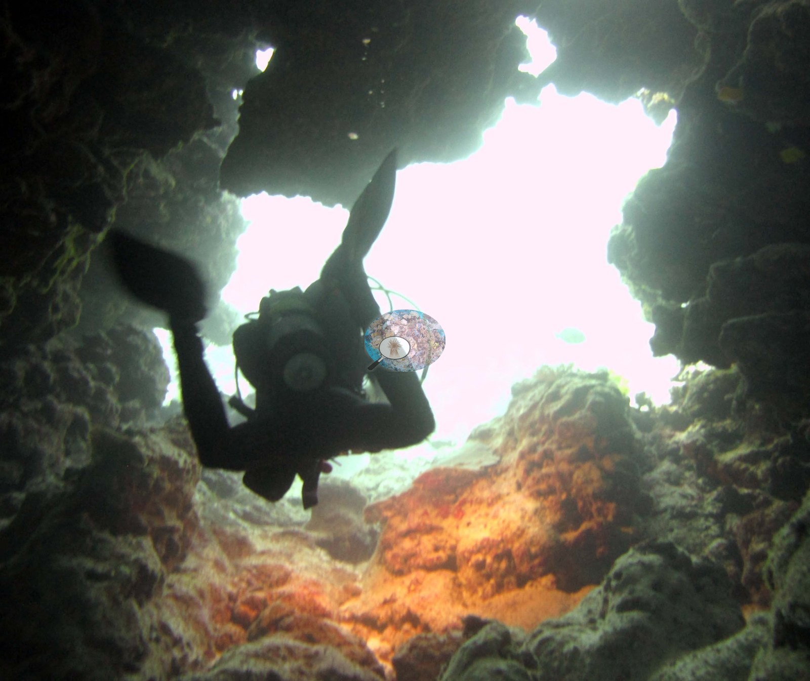
Image 23 is a genuine sea cave, a diver silhouetted in the entrance against bright surface light with orange encrusting growth on the walls, so there is real cavernous relief on this reef.
General benthic features
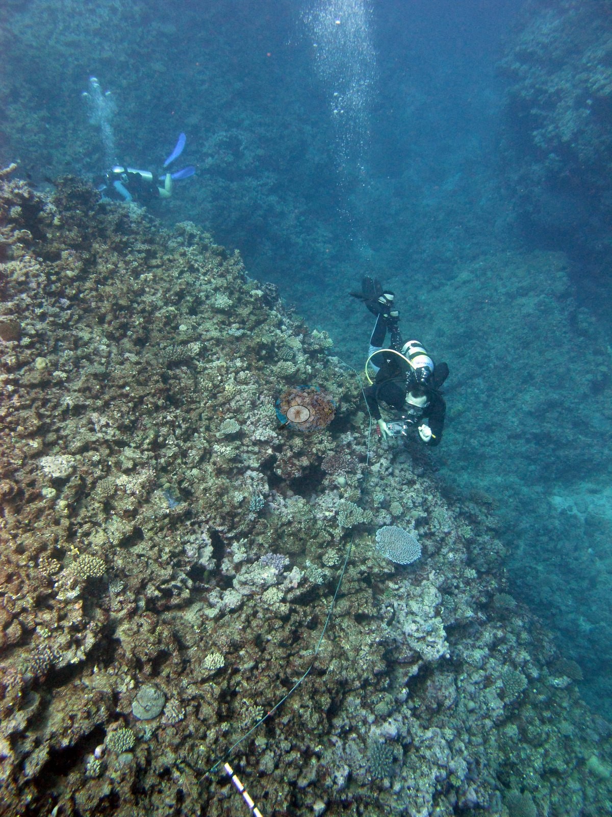
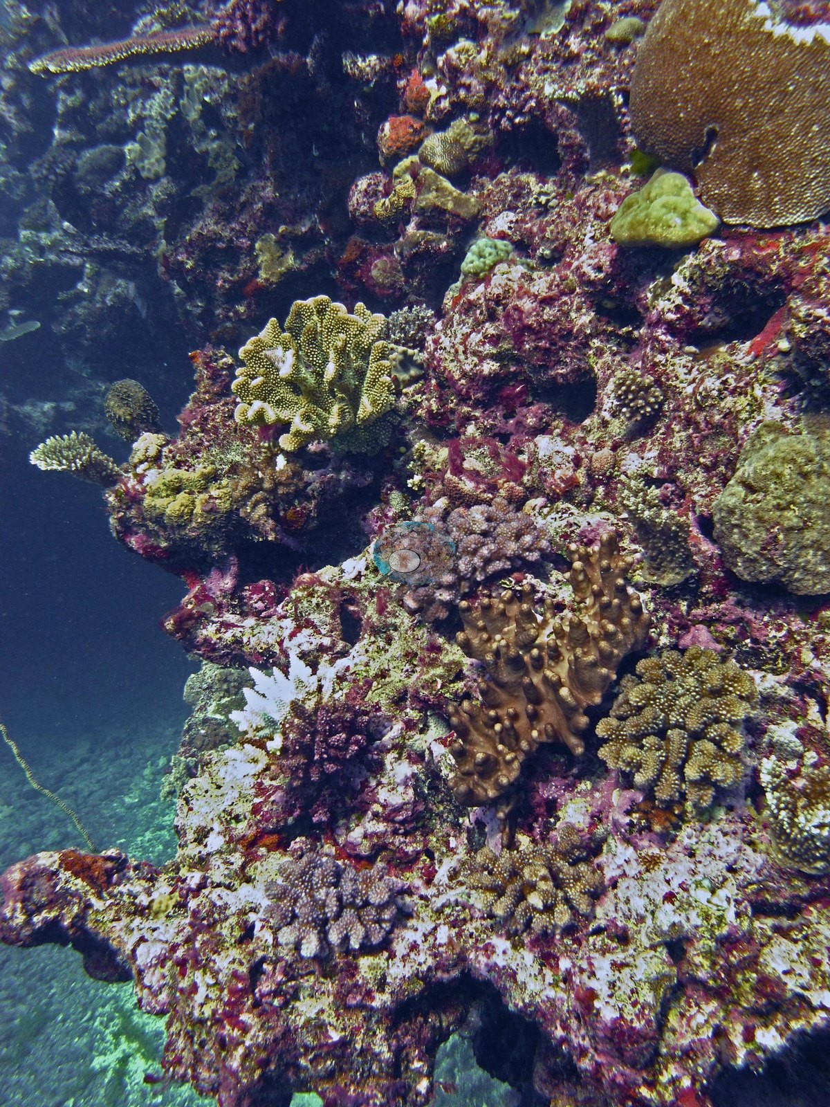
The reef-scale frames are the minority but they are consistent. Image 22 is the fullest: a densely occupied slope of small and medium colonies packed over pink and magenta coralline-crusted framework, with one broad rose-pink encrusting sheet in the foreground and dark blue water beyond. Image 25, from the safety stop, looks down over a much flatter reef top — broad pavement and rubble, coralline-pink and gray, with colonies scattered rather than joined. Image 24 shows the same shallow character near the surface: crusted pavement carrying small tan digitate and branching colonies with wide gaps of bare rock between them. Image 19 and Image 21 are hazy and color-shifted wide shots (the latter with a strong red cast) of mixed low-relief reef with small fish and are of limited use for anything but general structure.
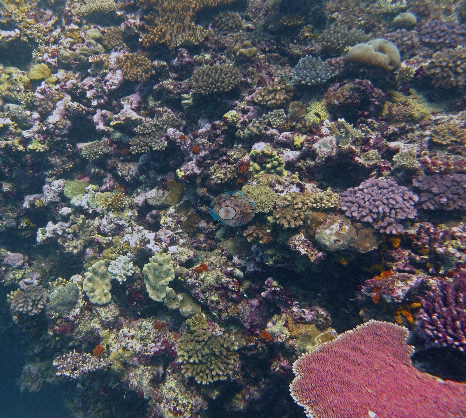
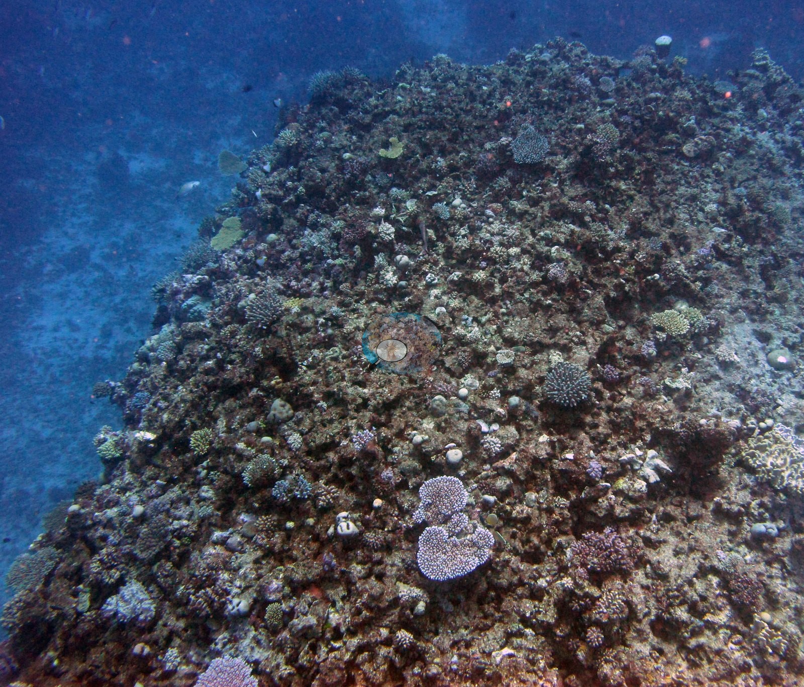
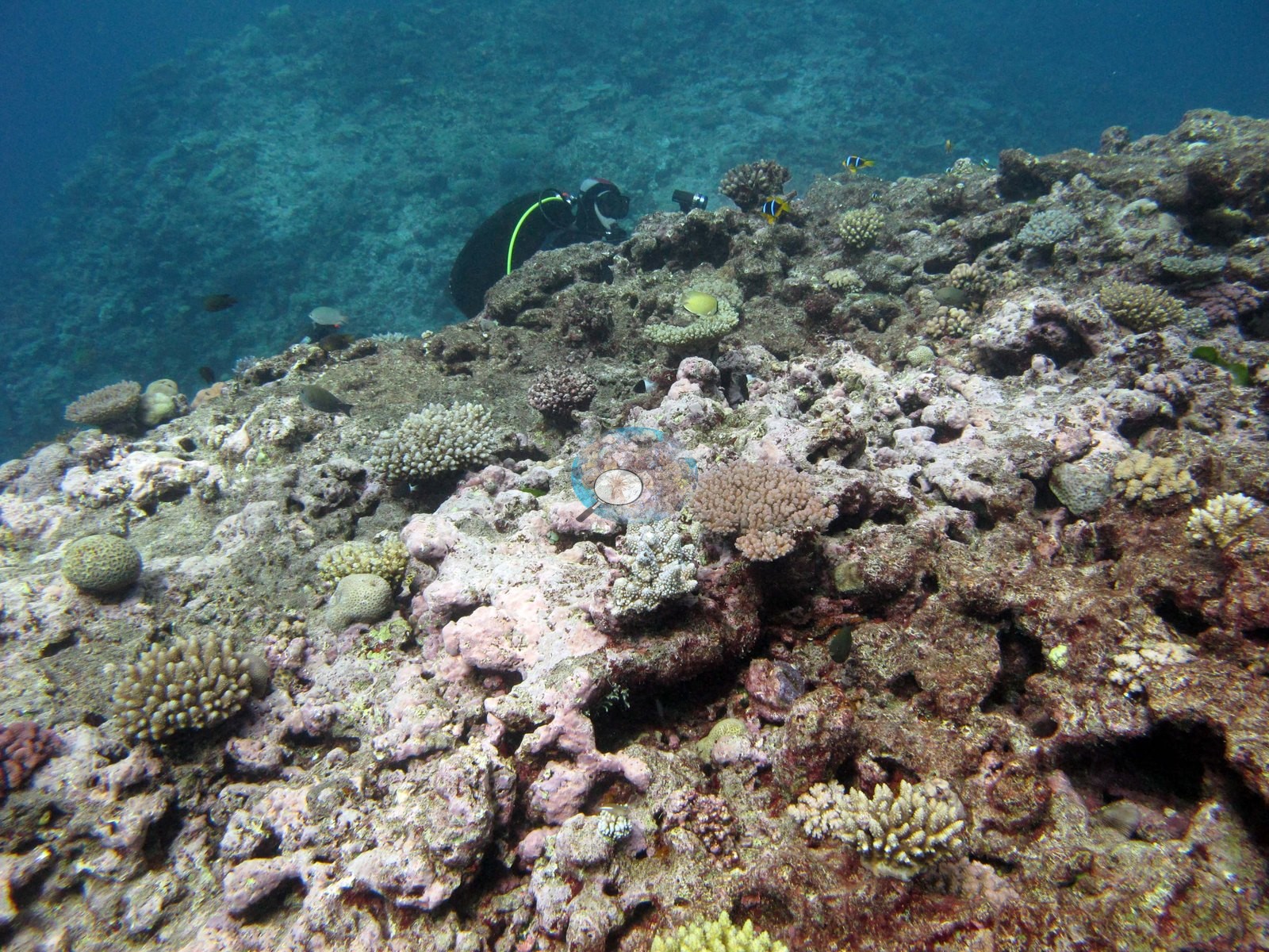
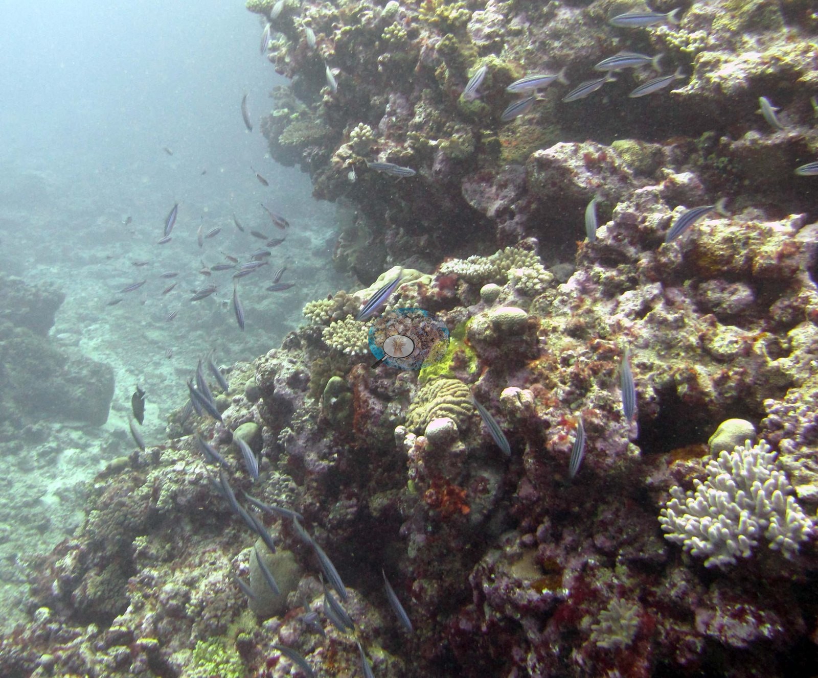
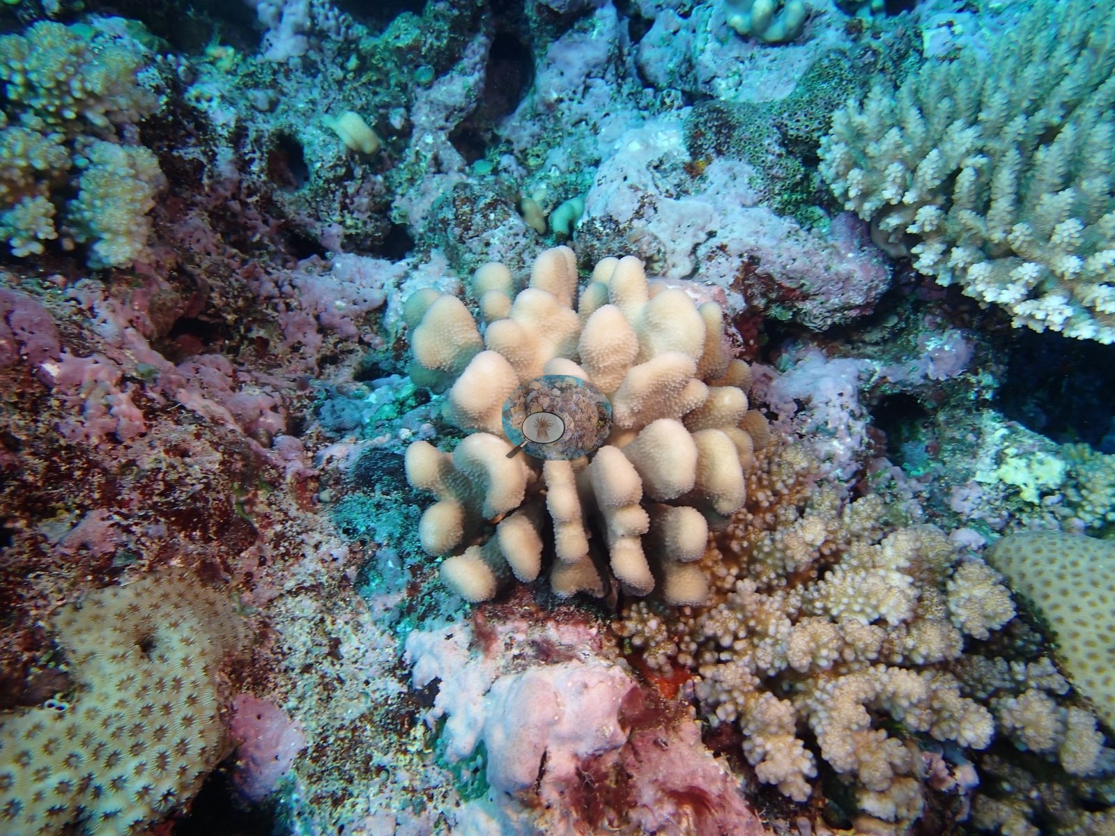
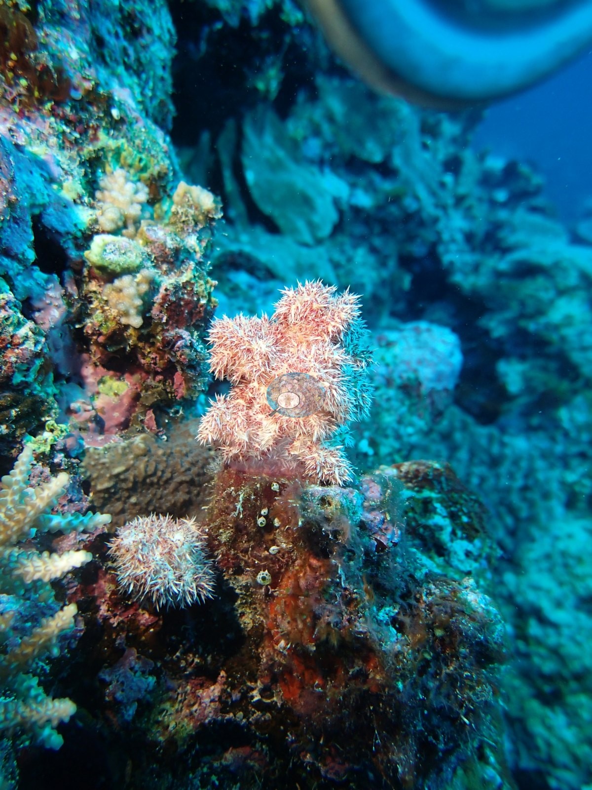
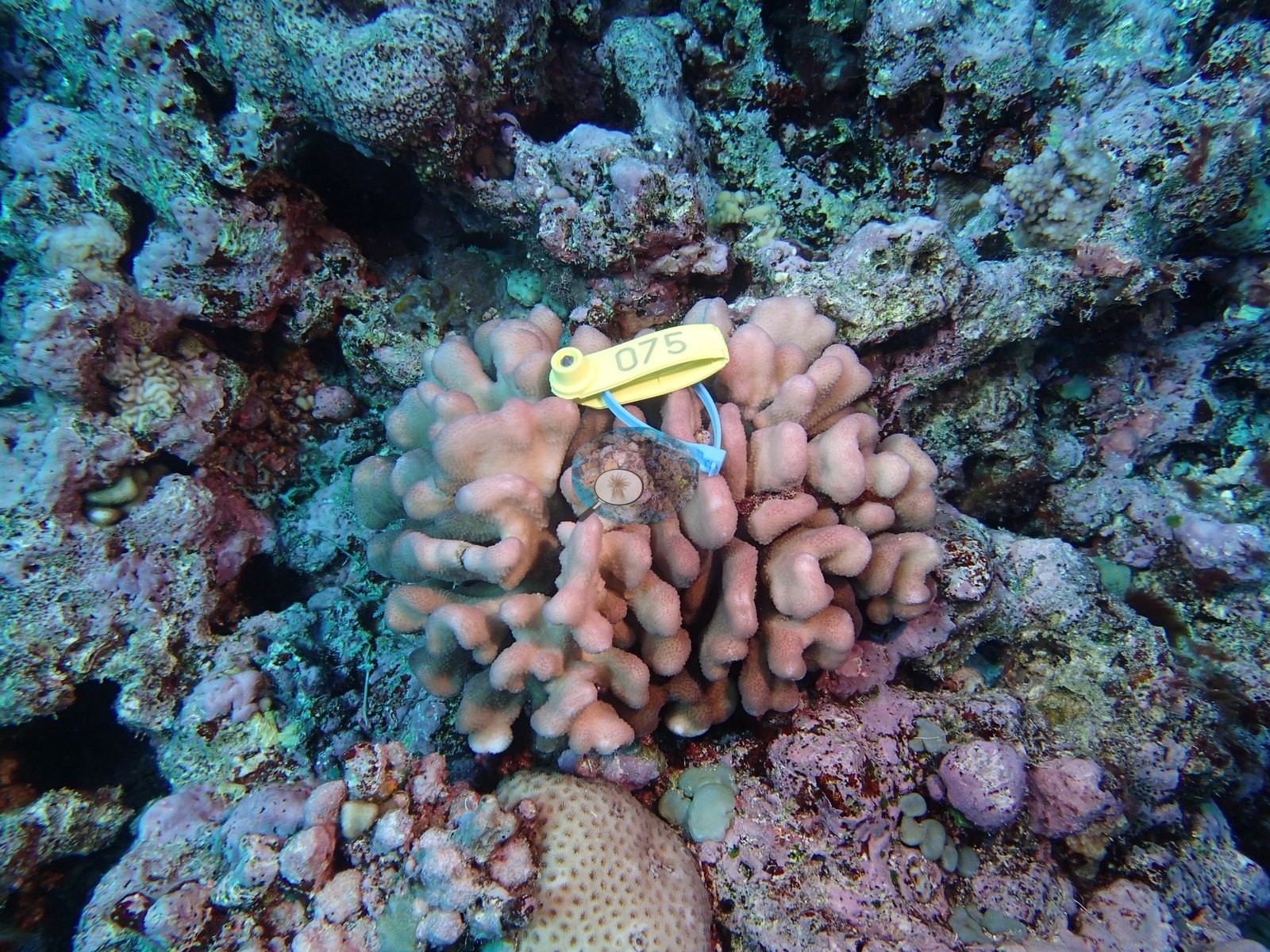
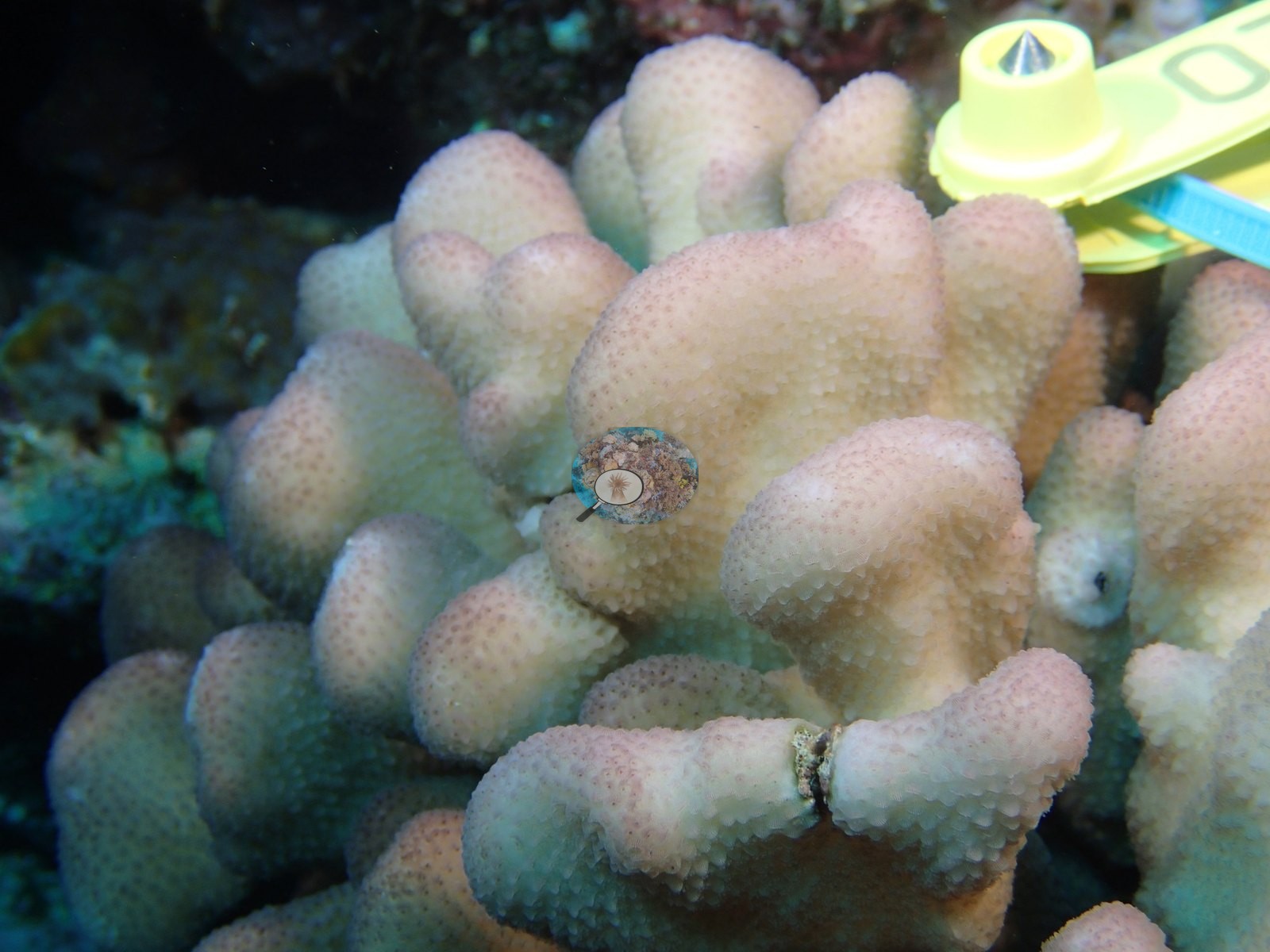
The colony close-ups give the assemblage more texture than the published subset could. Recurring through the set are pale pink and salmon knobby, lobed, and columnar colonies (e.g., Image 5, Image 7, Image 8, Image 9, Image 16) — a Porites-like growth form dominating the macro frames; a cream-tan colony with coarse, widely spaced corallites on crusted rock (e.g., Image 2); a thick-lobed tan colony (e.g., Image 3); a yellow-green massive with deep cerioid honeycomb corallites (e.g., Image 10); and two green colonies, one a smooth plate with pale polyp dots (e.g., Image 11) and one a lumpy surface studded with bright yellow-green polyps (e.g., Image 12).
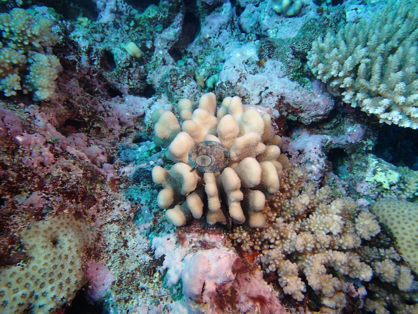
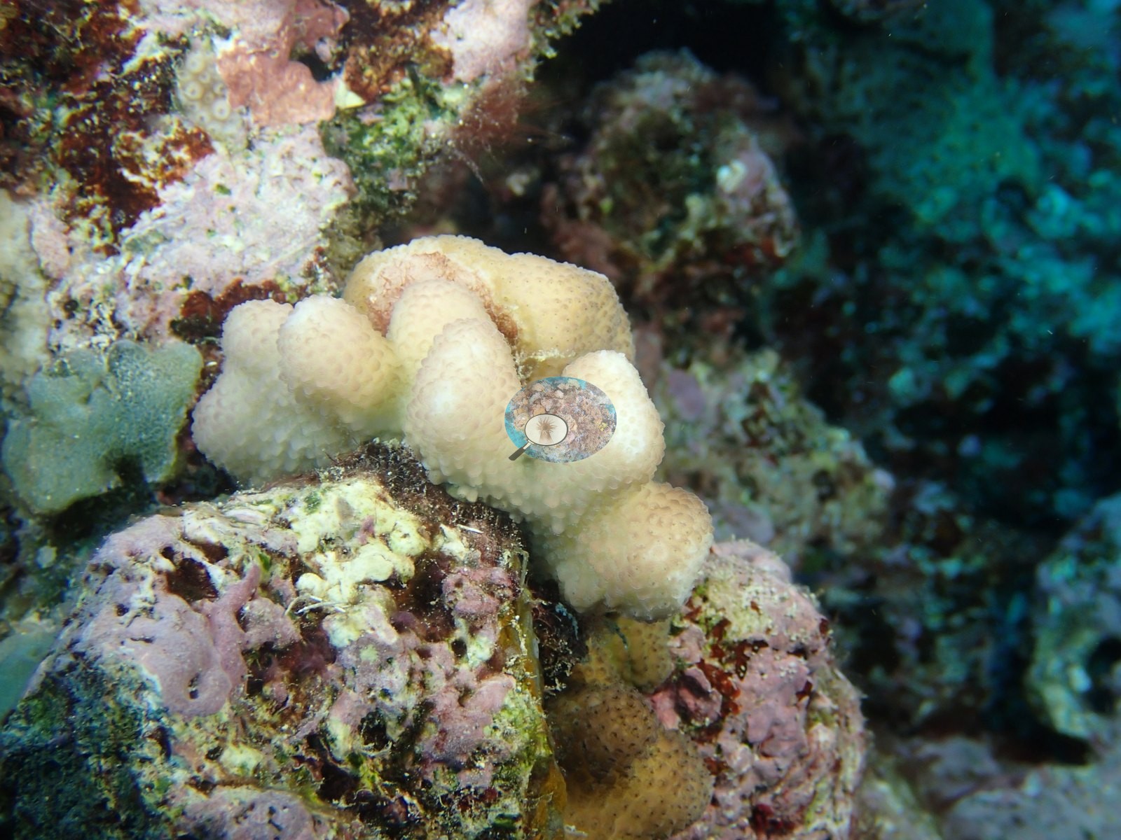
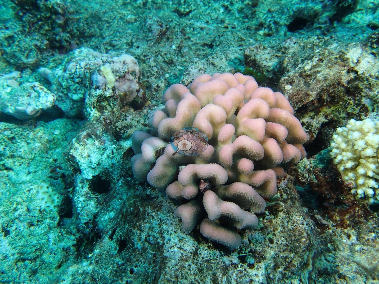
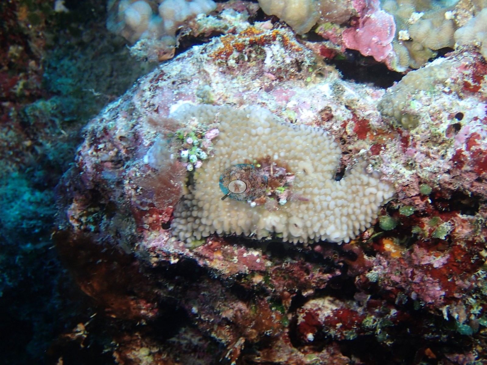
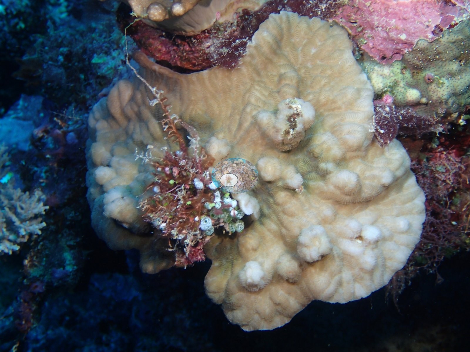
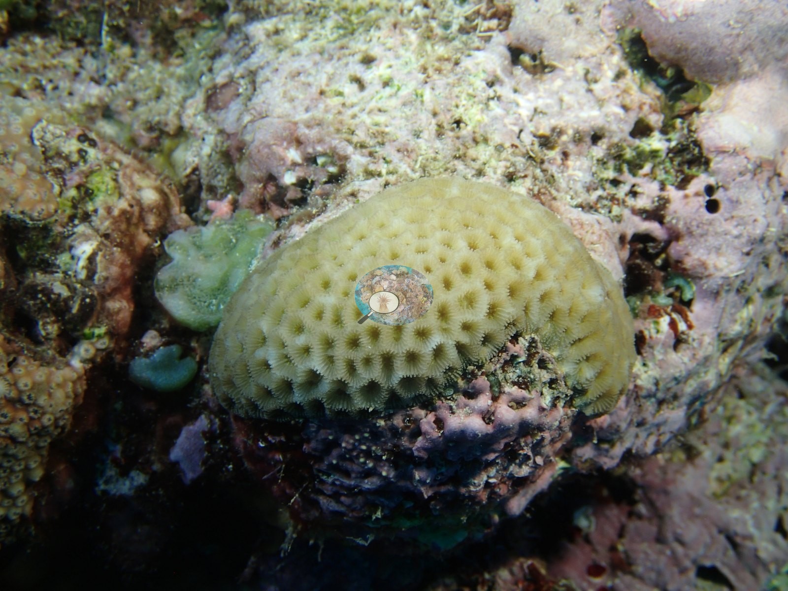
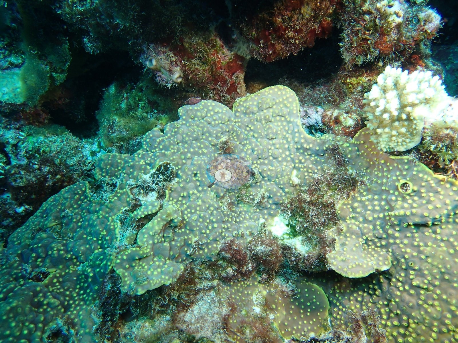
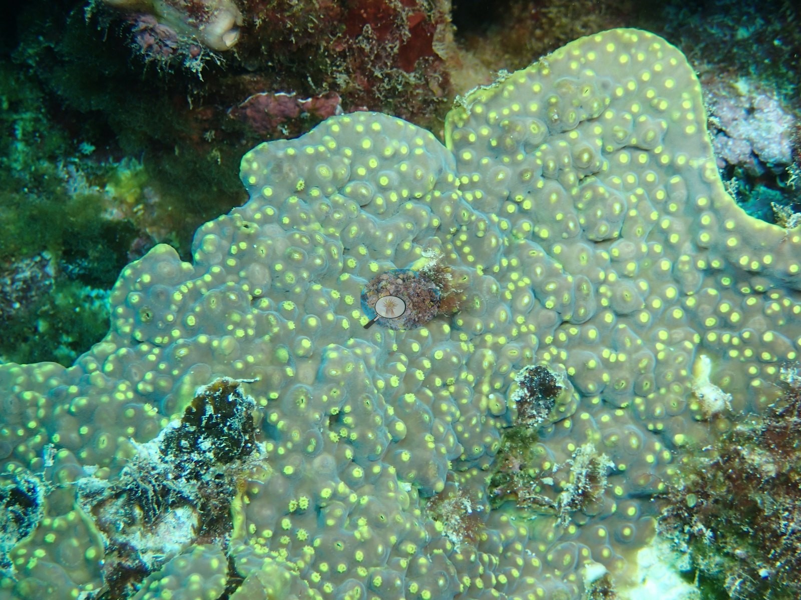
Several of these cannot be separated to genus from a photograph and none is asserted. Image 20, which Anderson left unidentified, shows a pale green-yellow colony of thick blunt columns.
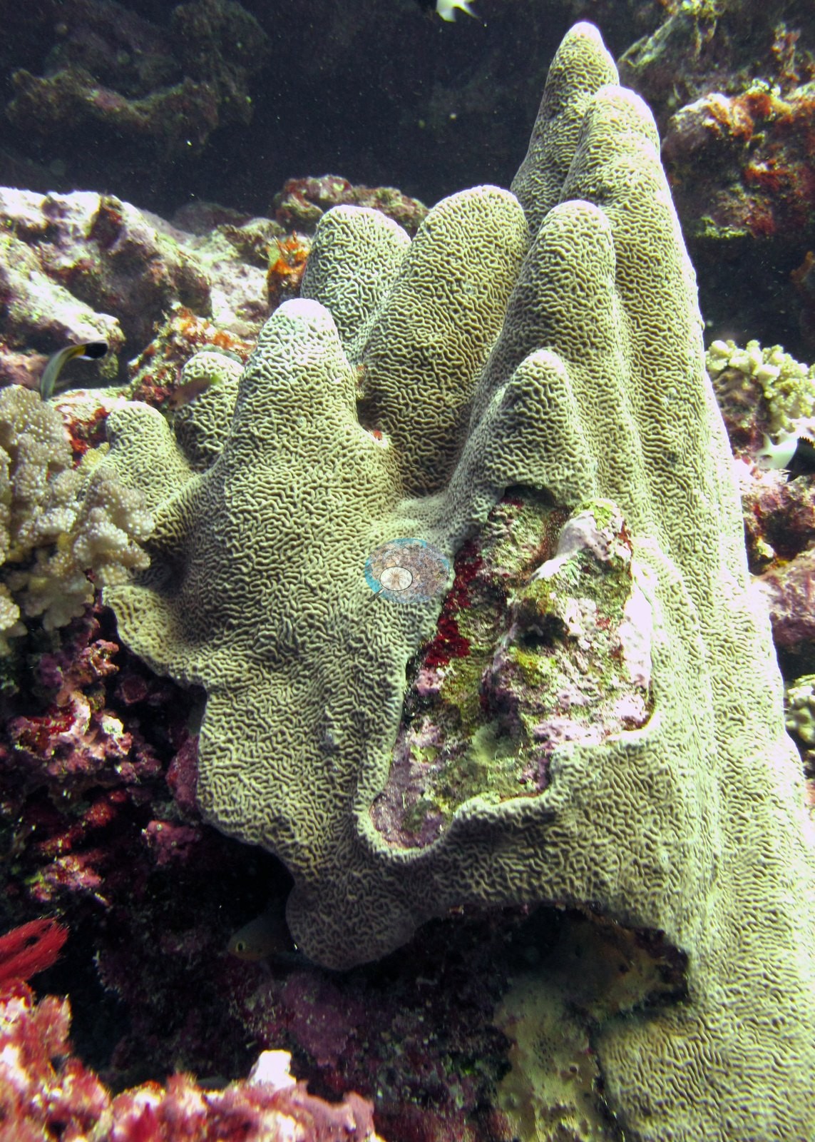
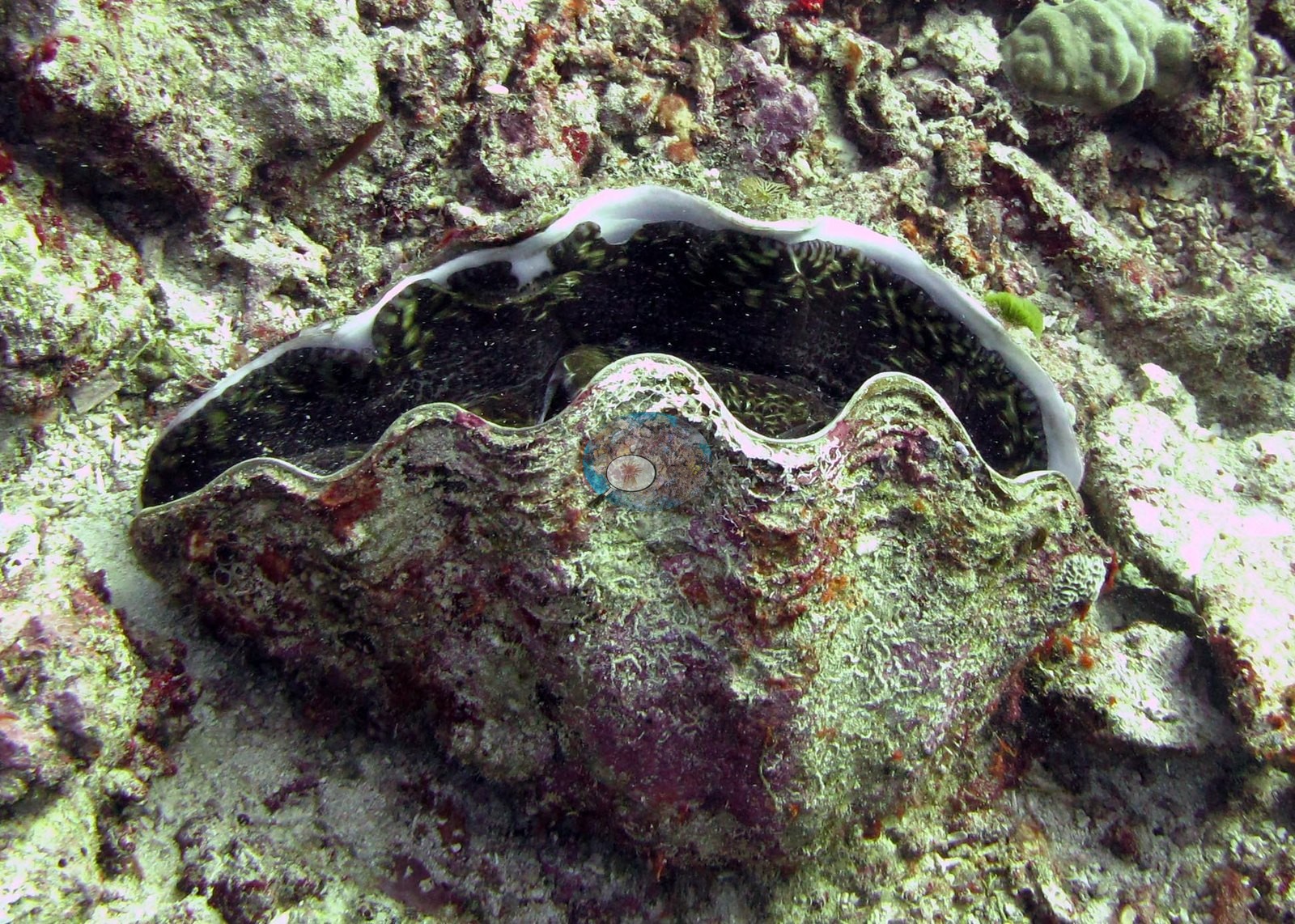
A giant clam sits embedded in the framework with its mantle open in Image 17.
Coral cover
- Reference: Khaled bin Sultan Living Oceans Foundation, Global Reef Expedition (in situ transect)
- Coral cover 25%, from the LOF-GRE in situ benthic transect (the mean of all of this station's own transects in the expedition's per-transect benthic data) — a diver transect, and the authoritative figure here; the frames are consistent with it.
- Transect mean 25 ± 3.3% SE (n = 5 transects; Living Oceans Foundation per-transect benthic data); the headline above is this all-transect mean.
Stressors
One small colony at the edge of one frame shows possible bleaching (one frame, Image 11); no disease lesions or crown-of-thorns feeding scars are visible in any frame, though the green and magenta color casts on several wide shots limit how firmly that can be stated for those. Anderson's sampling sheet records the one colony sampled here as fairly pale but healthy. The Living Oceans Foundation transects here recorded 7.2% cyanobacteria and no recently dead coral.
Fish and other fauna
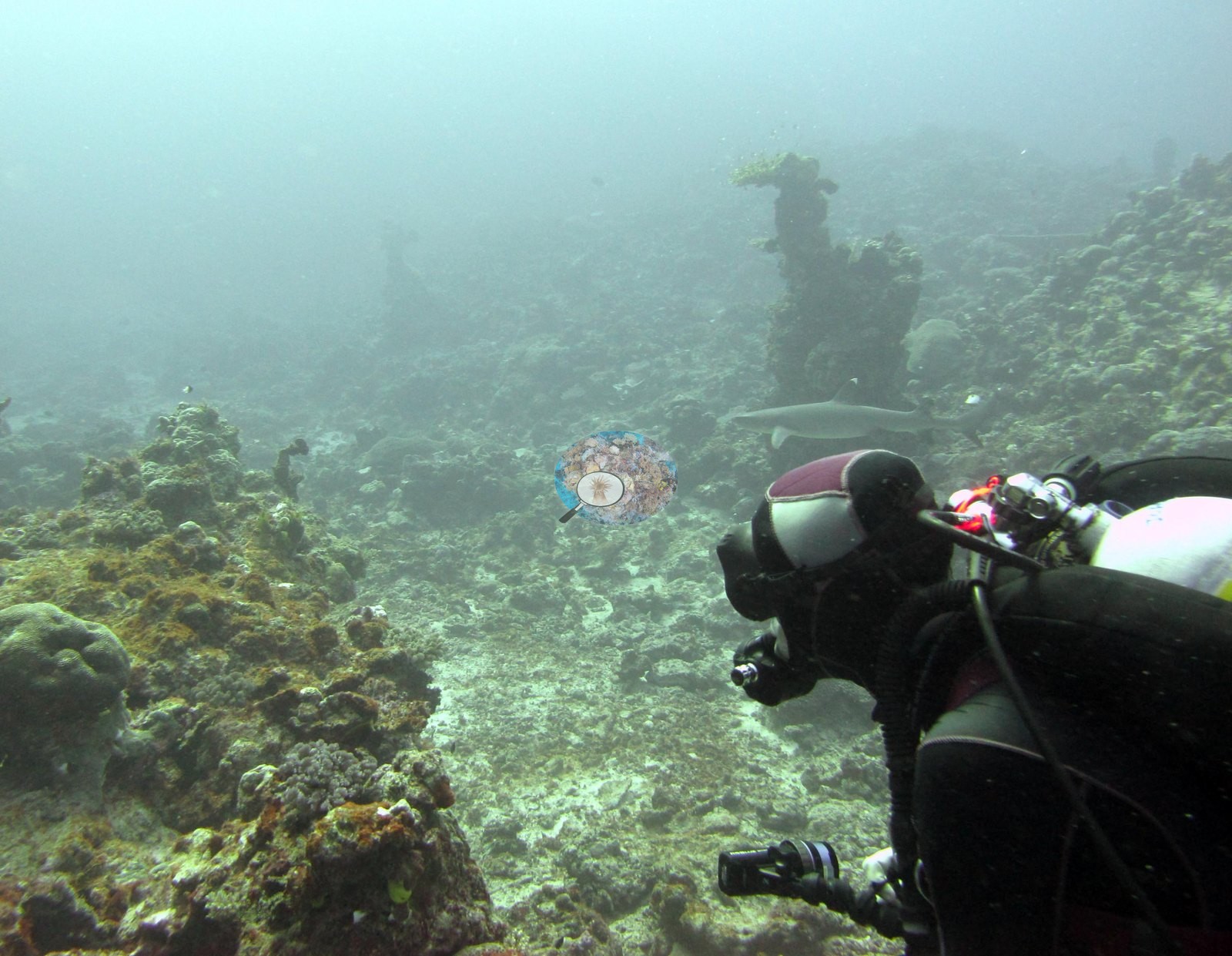
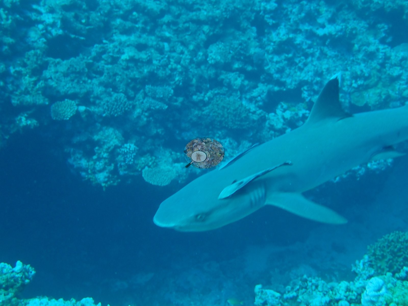
A whitetip reef shark appears in Image 18 and Image 13.
Comparisons with other data
Apart from the Living Oceans Foundation survey shown here, we found no other surveys at this site as of 2026. If you would like to share yours, please email us.
Other notes
The station's photo set holds 26 frames, and roughly half of them are not survey photography at all but the physiological sampling workflow. Four frames show that work directly: a gloved hand reaching in with tagging pliers, dive computer on the wrist (e.g., Image 4); and three colonies each carrying a plastic tag on a colored tie fixed to the substrate beside them — a salmon-pink knobby colony (e.g., Image 7), a pale pink lobed colony photographed close (e.g., Image 8), and a pale colony with its polyps fully extended (e.g., Image 14).
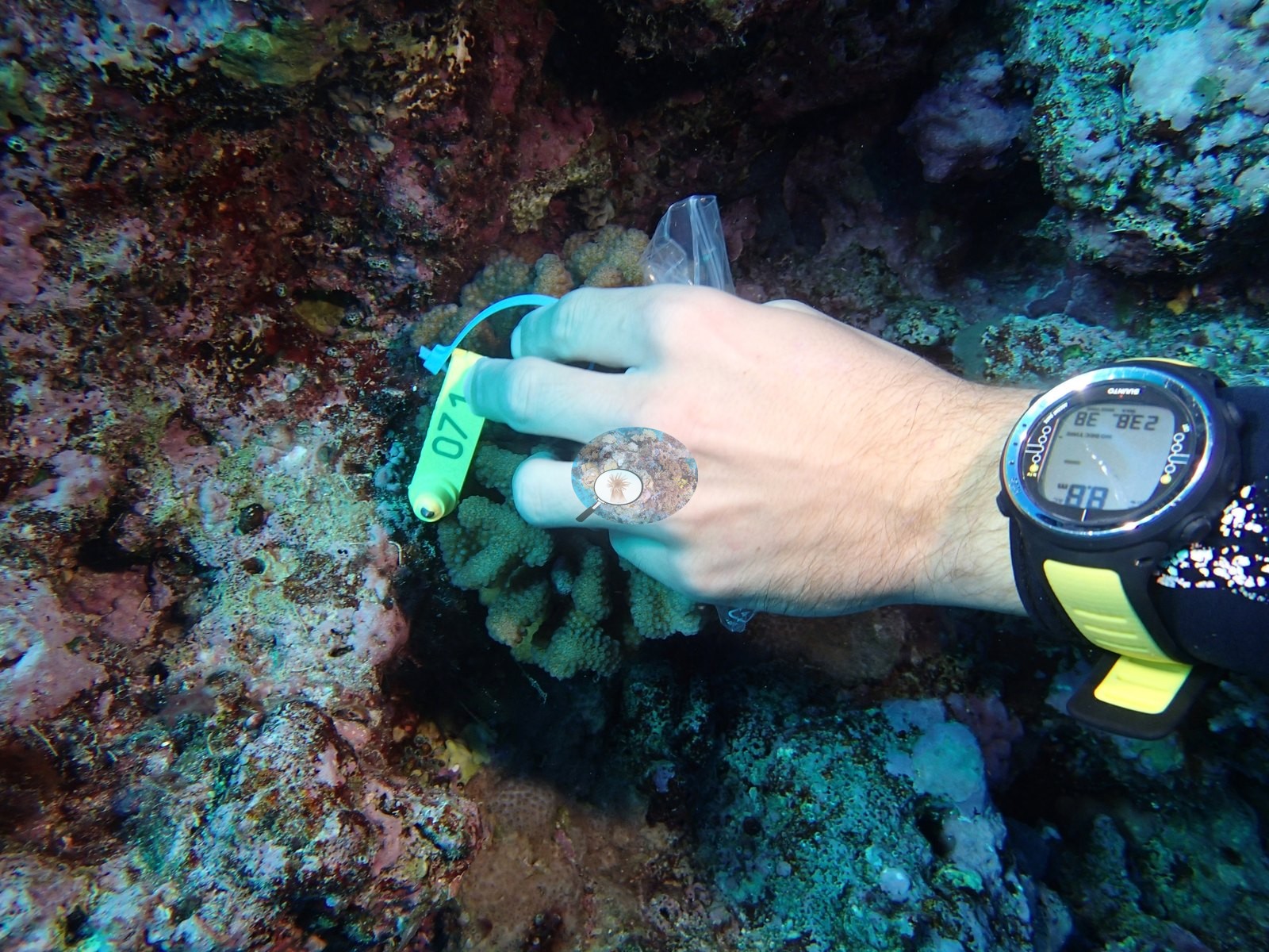
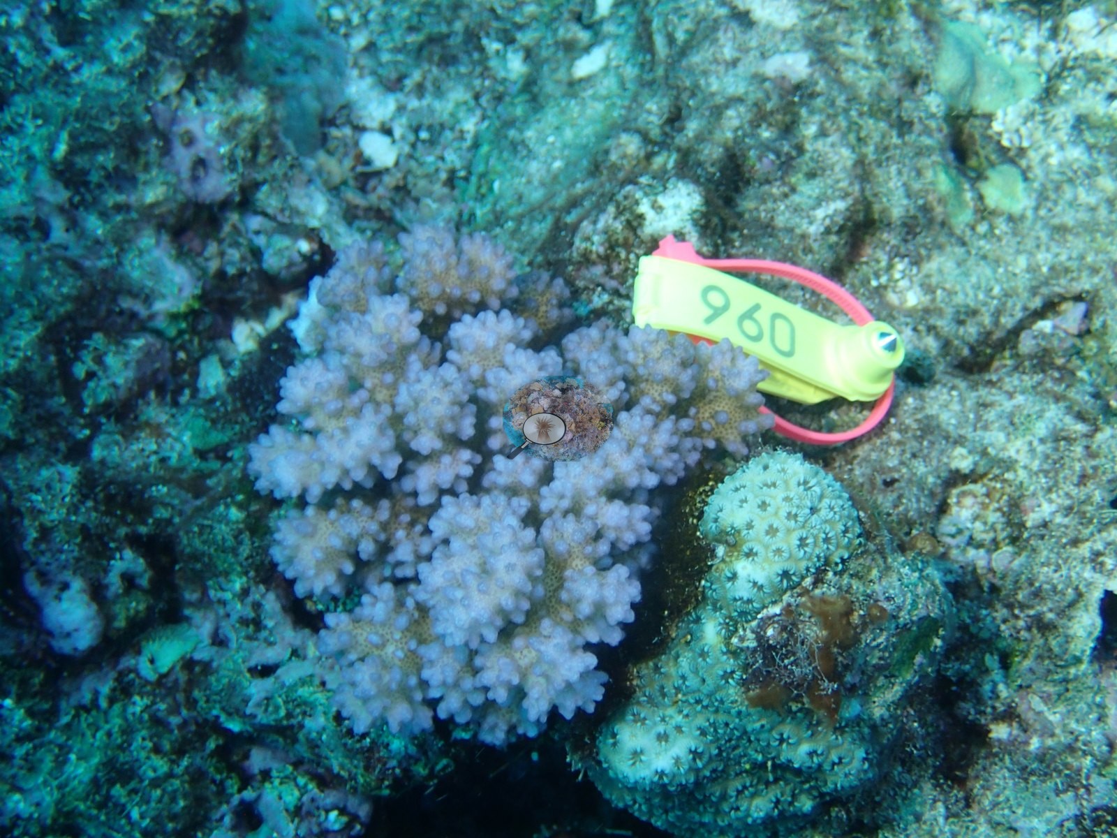
These are tagged colonies, not cover frames; the tags identify nothing, and Anderson's sampling sheet records one colony sampled here (P. damicornis).
Data & links
- Download benthic survey data (CSV) ↗
- Download all LOF-GRE data (933 sites) ↗
- Photo portfolio ↗
- Coral Reef Diagnostics region page ↗
All images from Coral Reef Diagnostics. Data are from the Khaled bin Sultan Living Oceans Foundation Global Reef Expedition and Coral Reef Diagnostics.
 © Coral Reef Diagnostics.