
Part of the regional summaries: Fiji - Moala, Fiji - Moala Group (Moala / Totoya / Matuku), and Fiji - Lau
More info
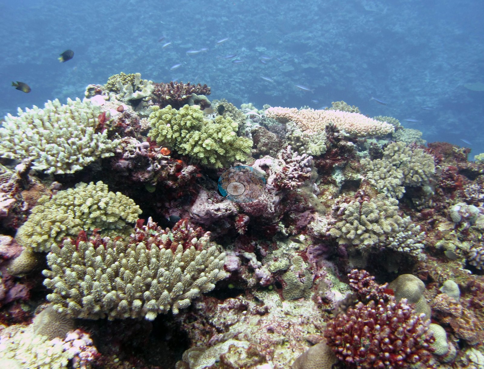
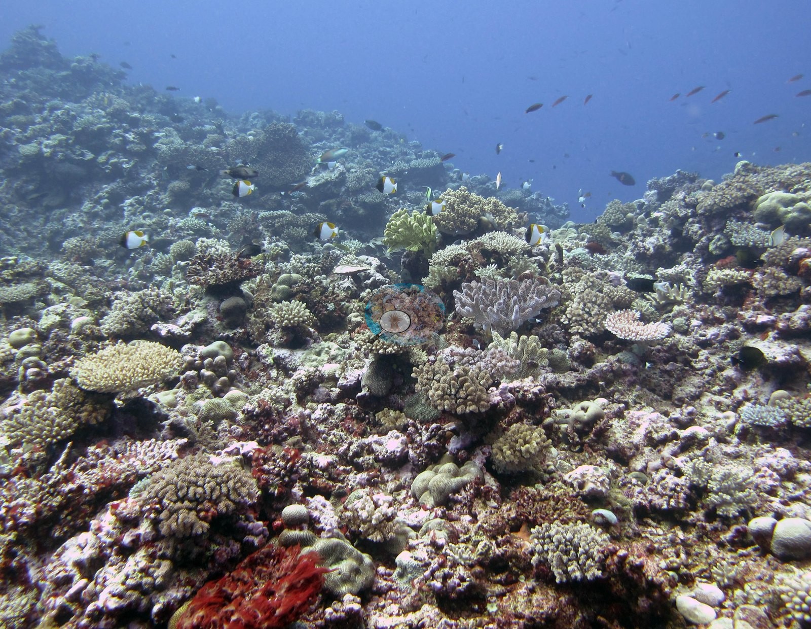
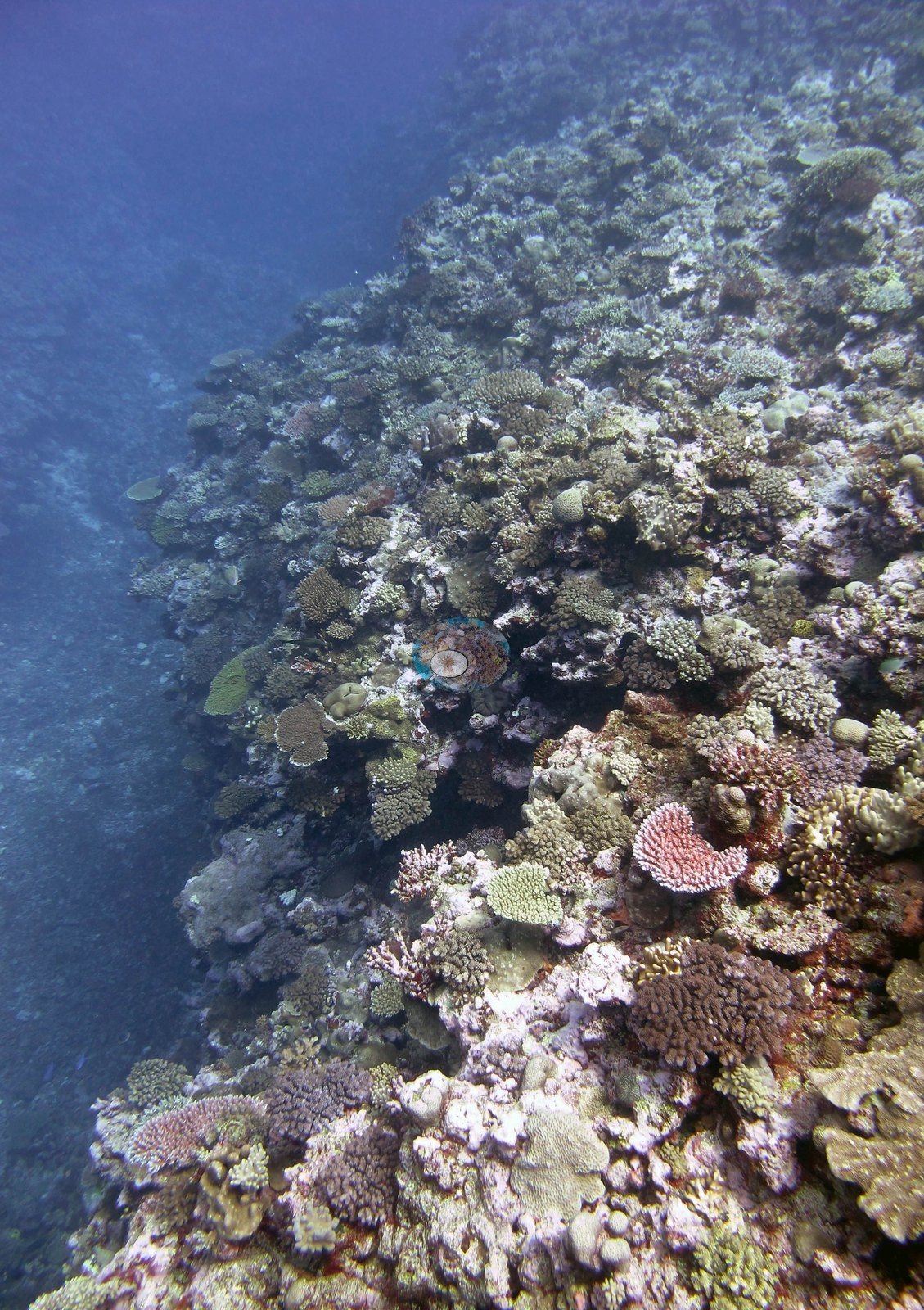
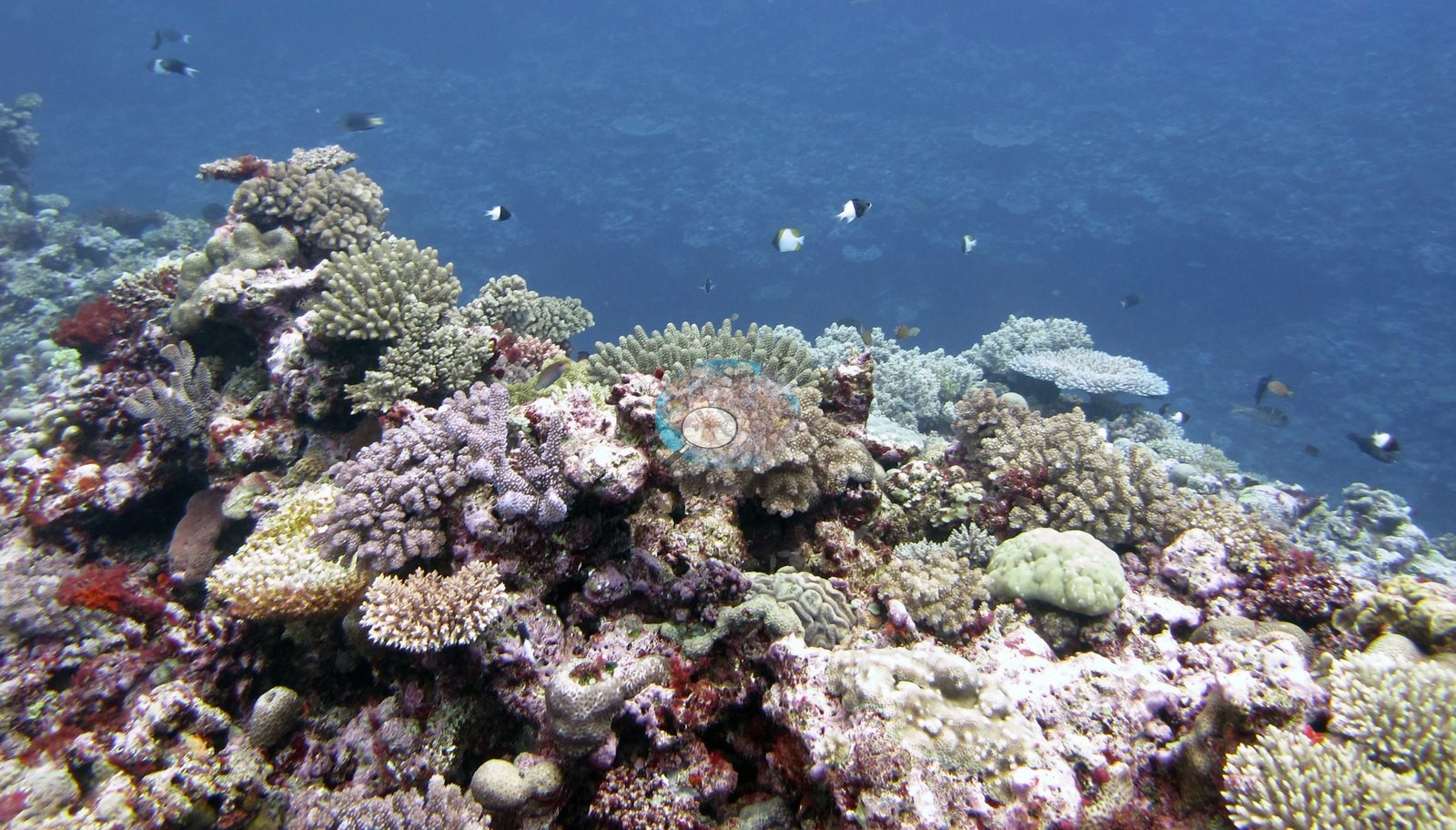
Fiji (NOAA CRW virtual station)A high-relief Moala site shot from atop coral pillars and bommies, with an extremely mixed and colorful community of branching, table, foliose, and brain corals in mint green, maroon, and cream, and very little bare rock. Many frames document coral tagging and fluorometer measurements, and no disease or crown-of-thorns was seen, though the one colony Anderson sampled was pale and about half bleached. Surveyed on the Living Oceans Foundation's Global Reef Expedition.
E dua na vanua cecere e Moala a taba mai na dakuna ni duru ni lase kei na vatu ni lase (bommies), e dua na isoqosoqo ni lase e veiwaki sara ka roka vakaidina, na lase vakatabataba, vakatepeli, vakadrau ni kau (foliose), kei na vakamona (brain), e roka (mint green, maroon, cream), ka lailai sara na vatu lala. E levu na itaba e vakaraitaka na kena vakatakilakilataki (tagging) na lase kei na kena vakatagedegedetaki ena fluorometer, ka sega ni raici e dua na mate se bula (crown-of-thorns), ia na dua ga na lase a taura kina na ivakaraitaki ko Anderson e vulavula vakalailai (pale) ka rauta ni veimama e vulavula (bleached).
AI translation of this summary; the English is authoritative. (low confidence)
CTD-cast midpoint; the cast read 27.2°C throughout
Two other CRD dive sites with their own pages within 5 km shown in orange — click a marker to open its page.
🗺 Detailed map of the Global Reef Expedition dive sites, Moala, Fiji (Khaled bin Sultan Living Oceans Foundation; opens in a new window)
Site description
Setting
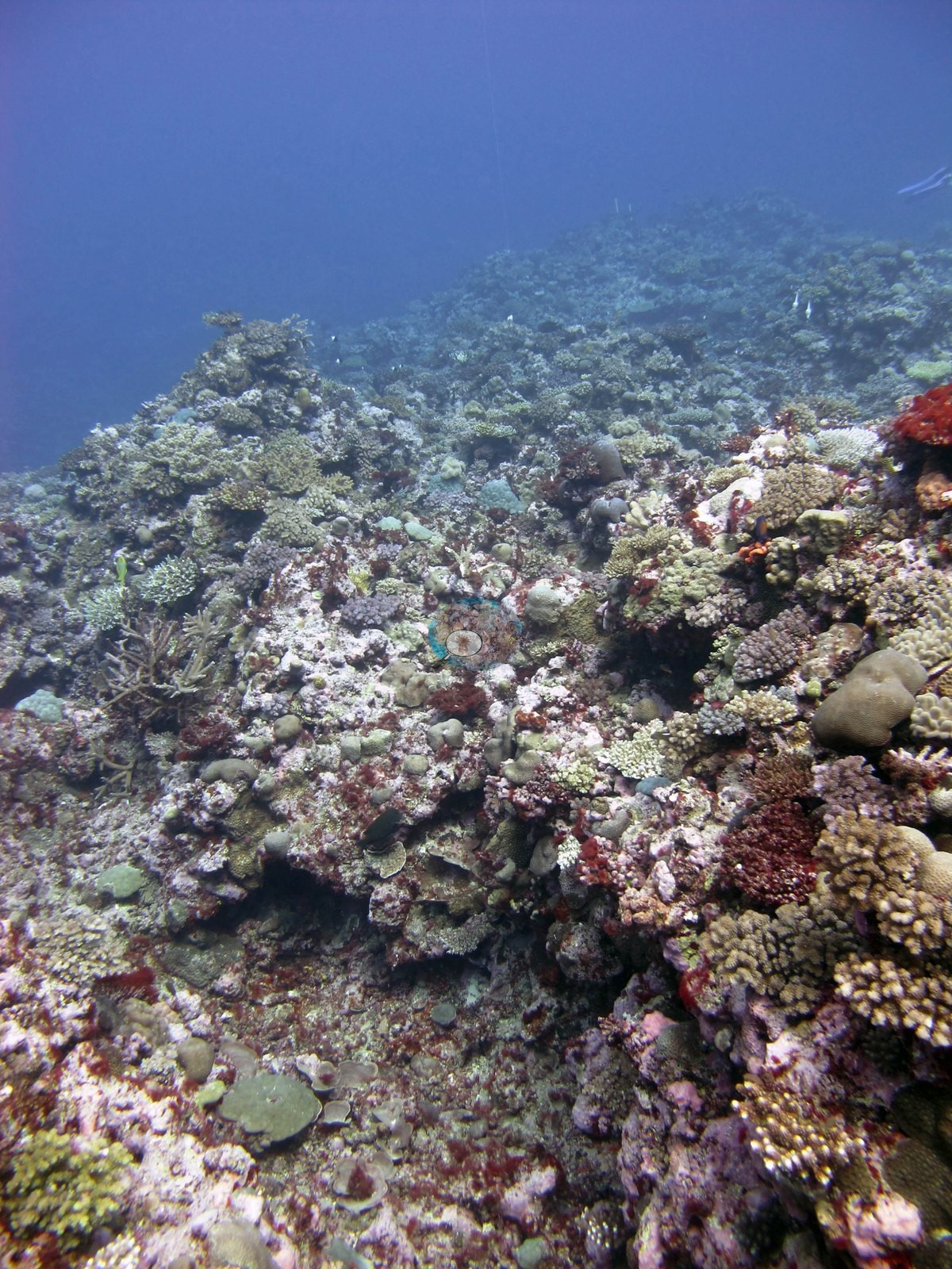
This is not a flat terrace: several frames are shot from the top of a coral pillar or bommie looking out and down, as Anderson's notes on them also record (e.g., Image 15, Image 22).
General benthic features
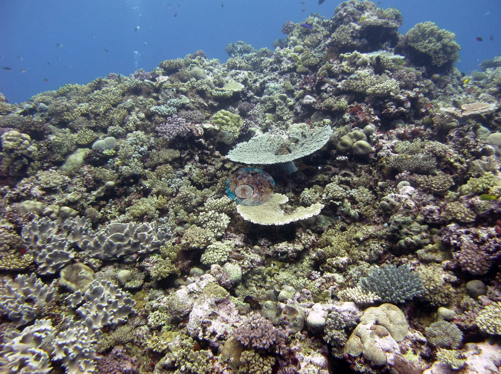
The habitat frames show a high-relief, extremely mixed community with very little bare rock in view — small corymbose and digitate Acropora in cream, mustard, lilac, and purple-brown, knobby Pocillopora-type clumps, low tables, smooth to knobbly massives, upright leafy bifacial fronds, scrolled foliose plates, and small brain-textured heads, all interlocking with pink and mauve coralline crust filling the gaps (e.g., Image 2, Image 3, Image 12, Image 14, Image 21, Image 22, Image 23, Image 24, Image 27, Image 28, Image 29).
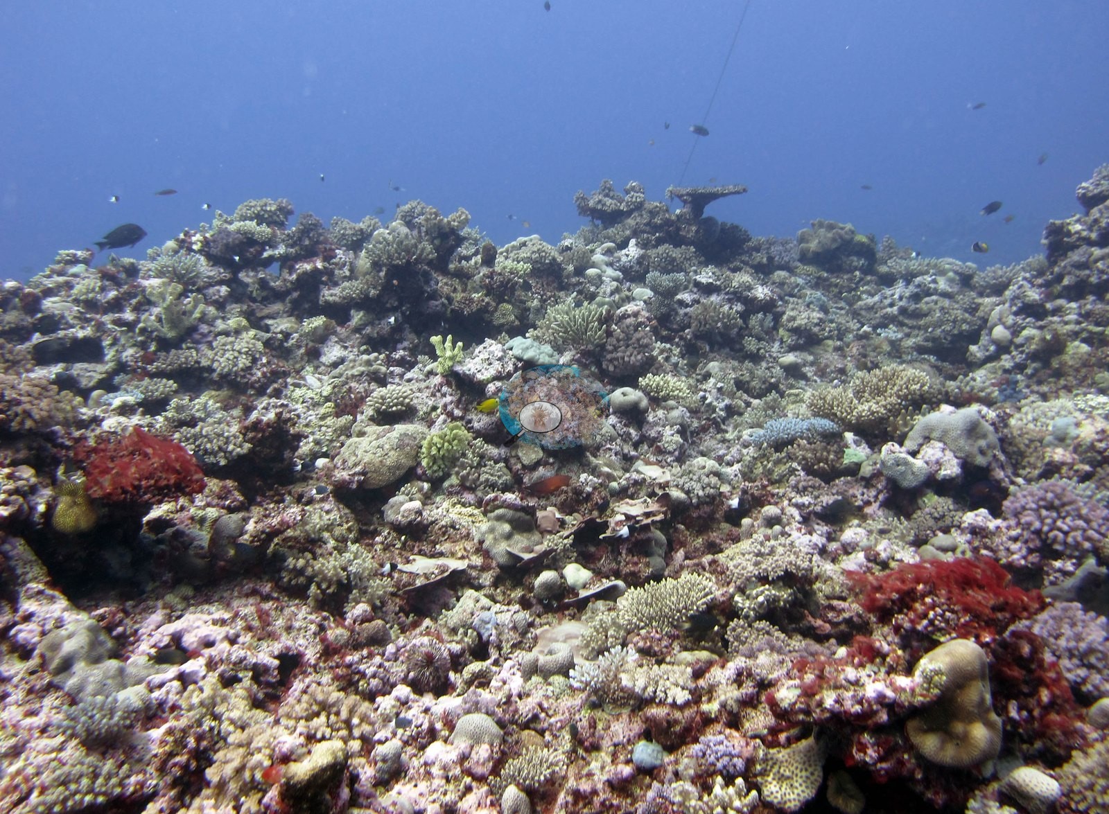
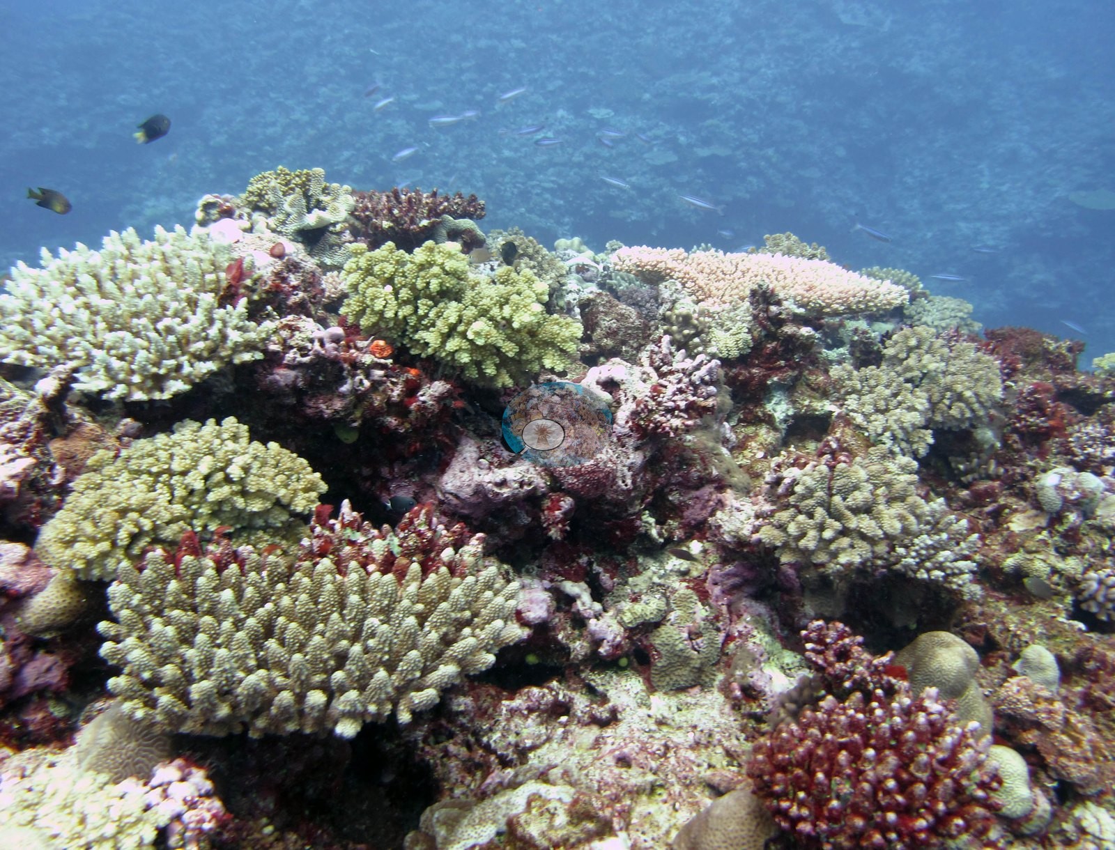
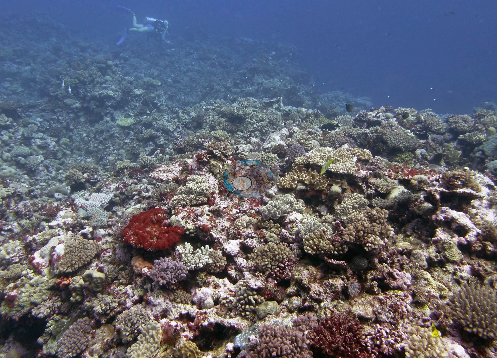
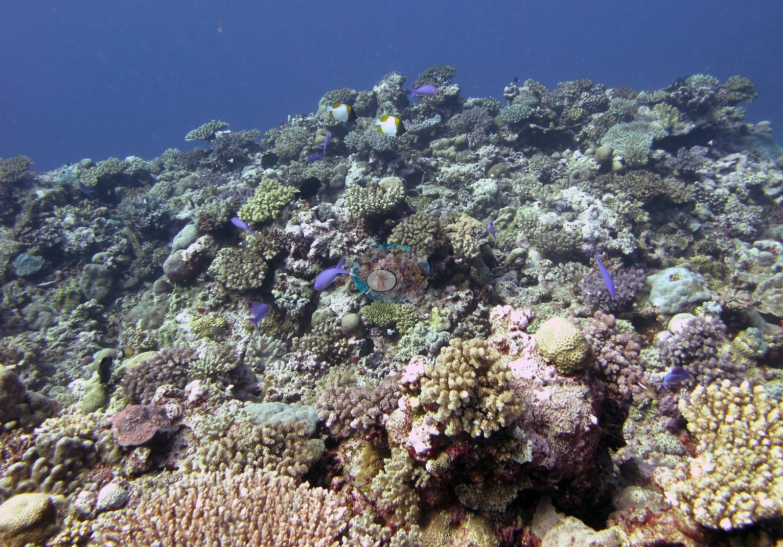
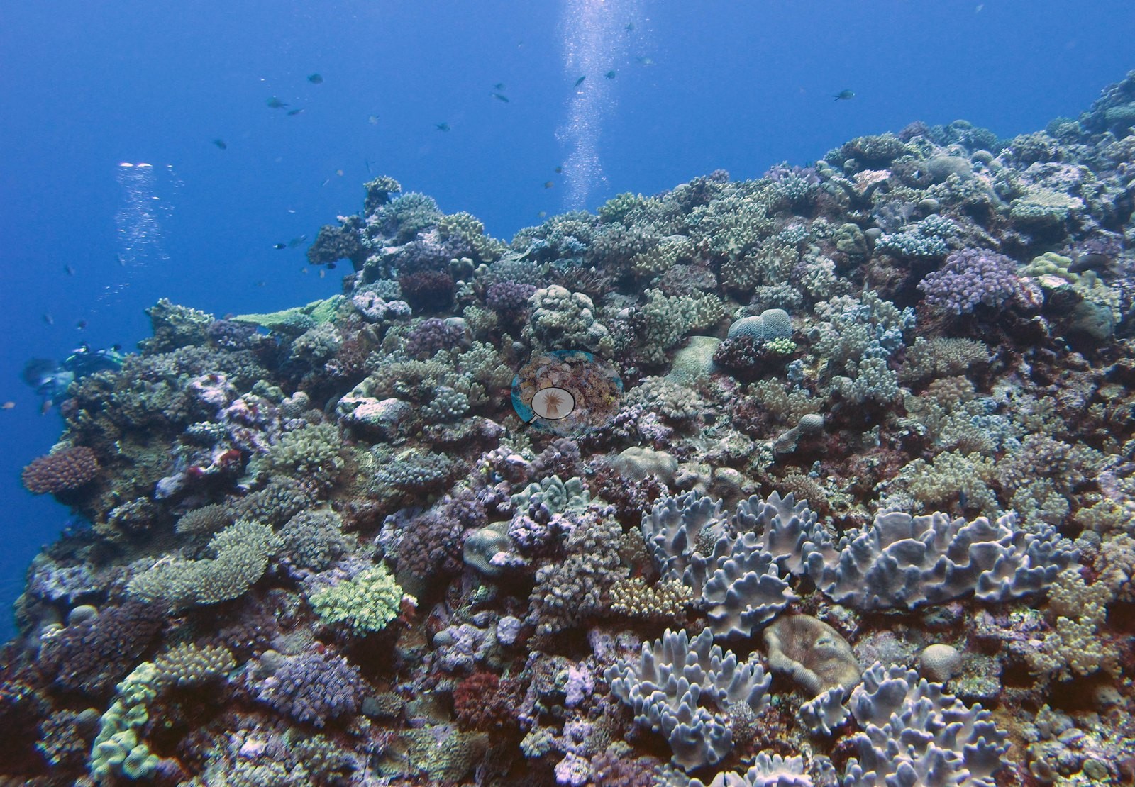
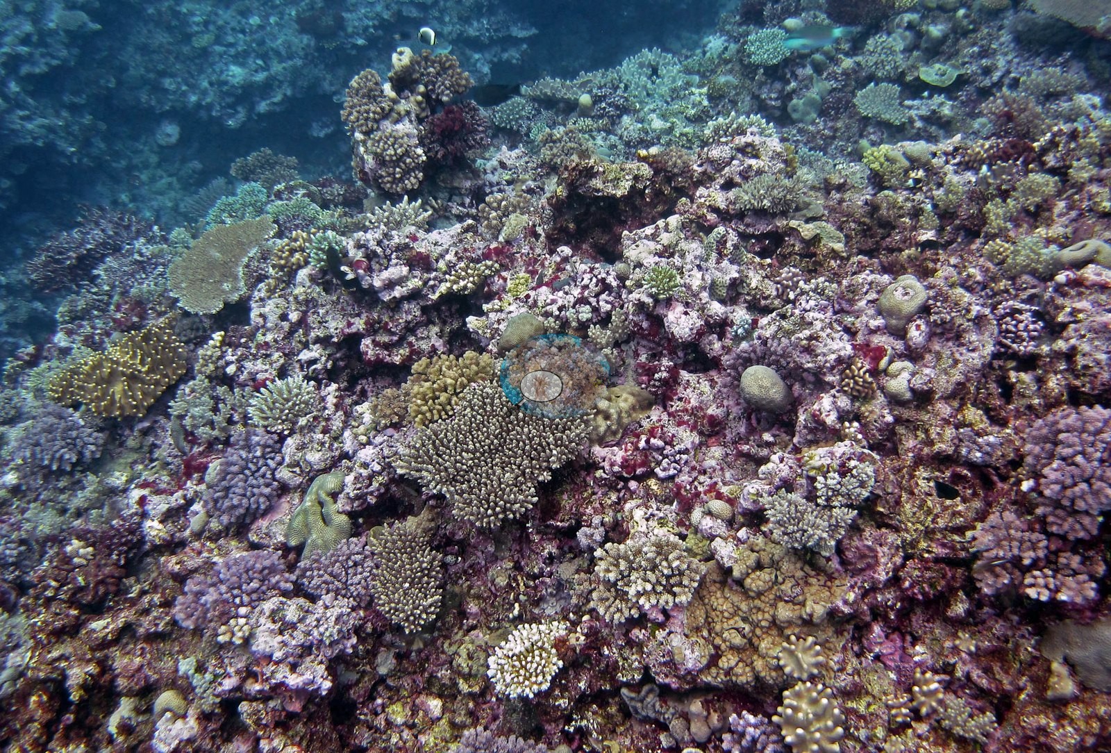
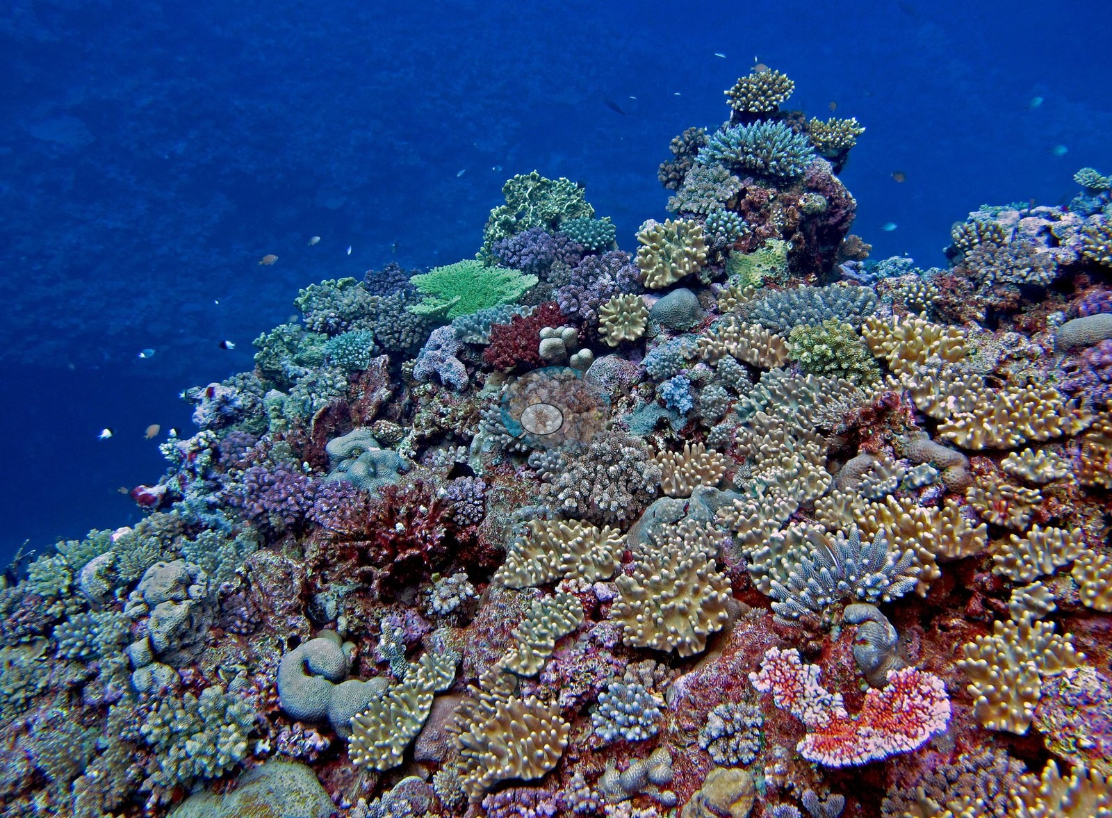
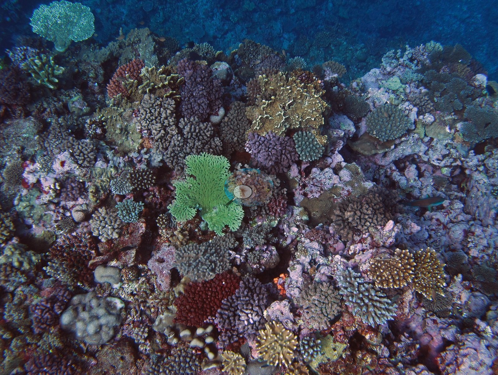
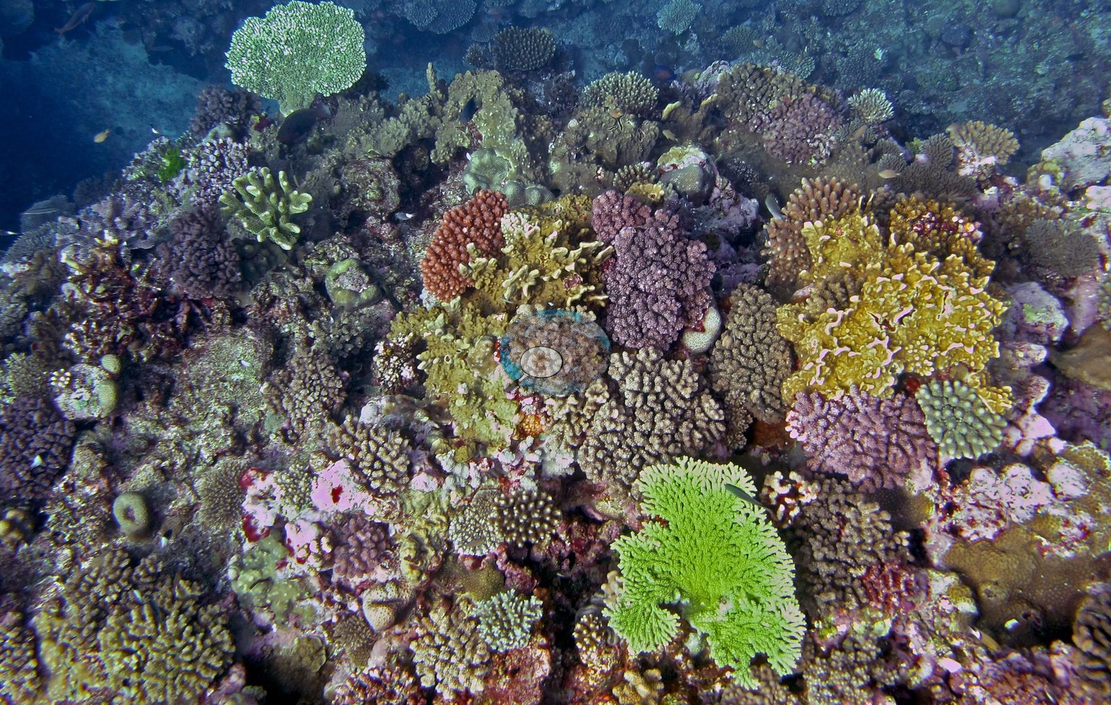
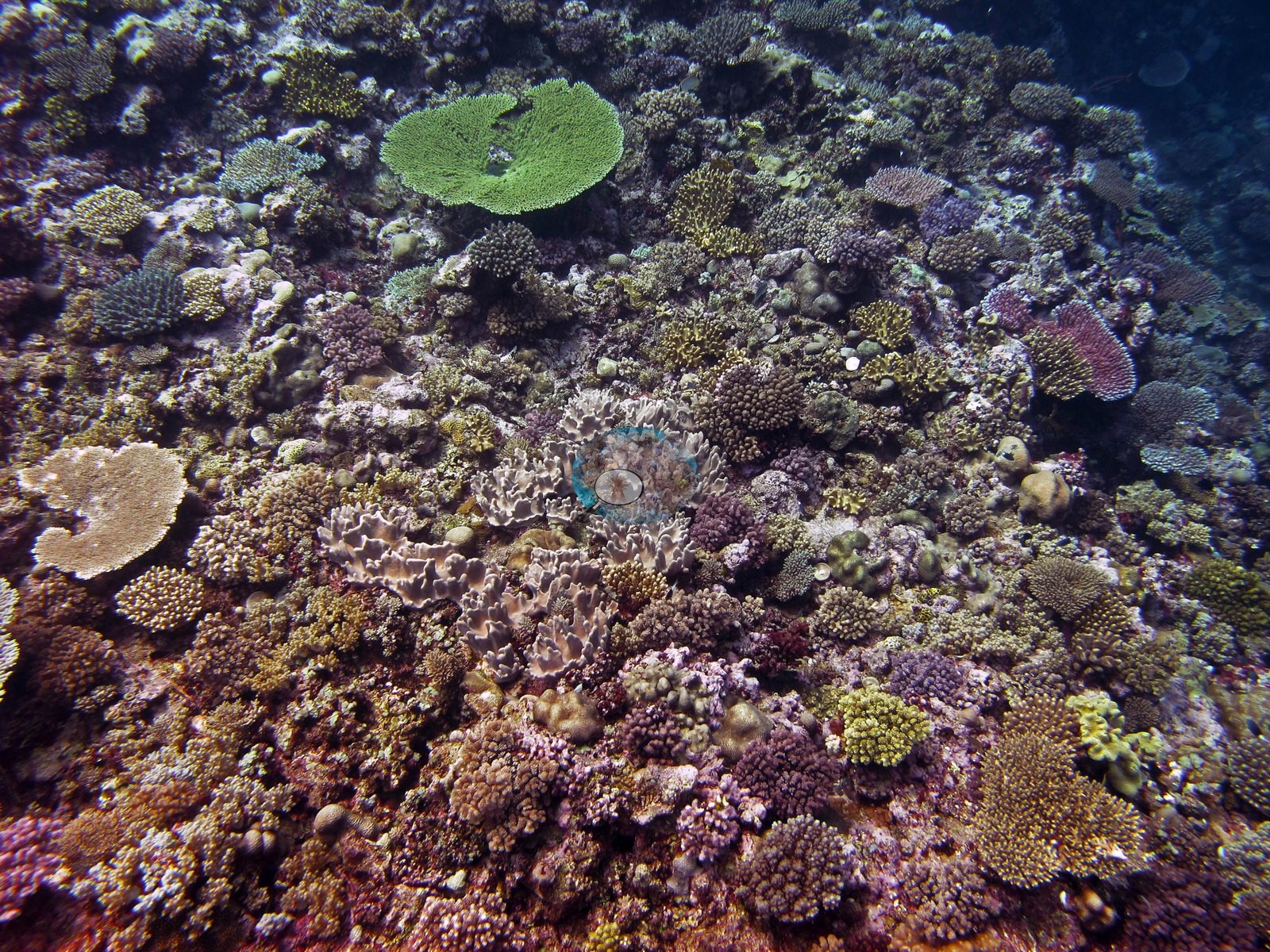
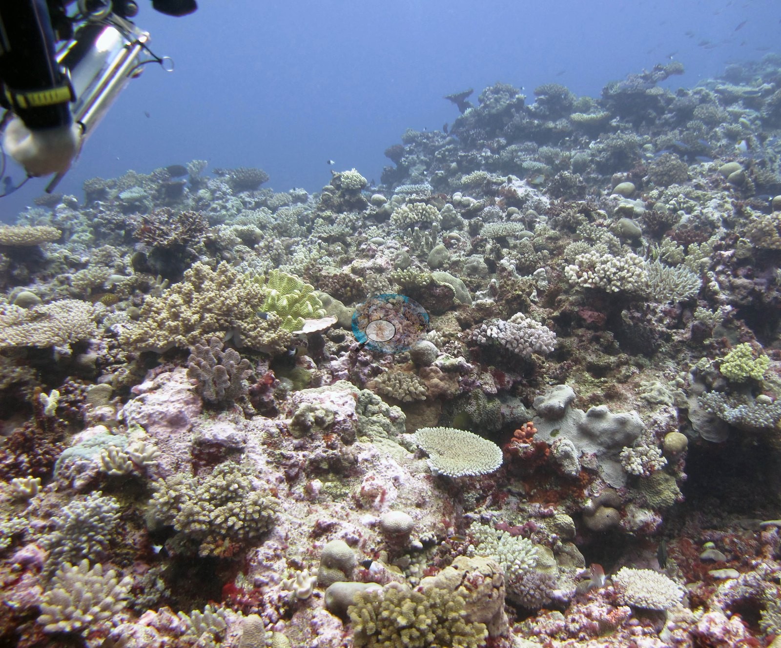
Color is the striking feature in the well-lit frames: bright mint-green and lime tabulate and foliose colonies sit next to deep maroon-red and burgundy encrusting colonies, blue-gray digitate clumps, and pale cream corymbose cushions in the same field of view (e.g., Image 24, Image 27, Image 28, Image 29). Two frames are shot up the slope into open blue with the reef crest as a silhouette (e.g., Image 1, Image 19), one is looking down a rubble-and-pavement channel (e.g., Image 5), and two show the same reef in flatter light on Anderson's second dive of the day (e.g., Image 2, Image 12).
Coral cover
- Reference: Khaled bin Sultan Living Oceans Foundation, Global Reef Expedition (in situ transect)
- Coral cover 33%, from the LOF-GRE in situ benthic transect (the mean of all of this station's own transects in the expedition's per-transect benthic data) — a diver transect and the authoritative figure; no cover estimate is made here from the photographs.
- Transect mean 33 ± 2.7% SE (n = 5 transects; Living Oceans Foundation per-transect benthic data); the headline above is this all-transect mean.
Stressors
No disease lesions, crown-of-thorns feeding scars, or fresh mortality are visible in any of the frames reviewed; Anderson's sample record for the colony tagged and sampled (F31, a P. damicornis) notes it as pale, about 50% bleached, and partly overgrown with algae, as the frame of it (Image 25) shows.
The Living Oceans Foundation transects here recorded 2.6% cyanobacteria and no recently dead coral.
Comparisons with other data
Apart from the Living Oceans Foundation survey shown here, we found no other surveys at this site as of 2026. If you would like to share yours, please email us.
Other notes
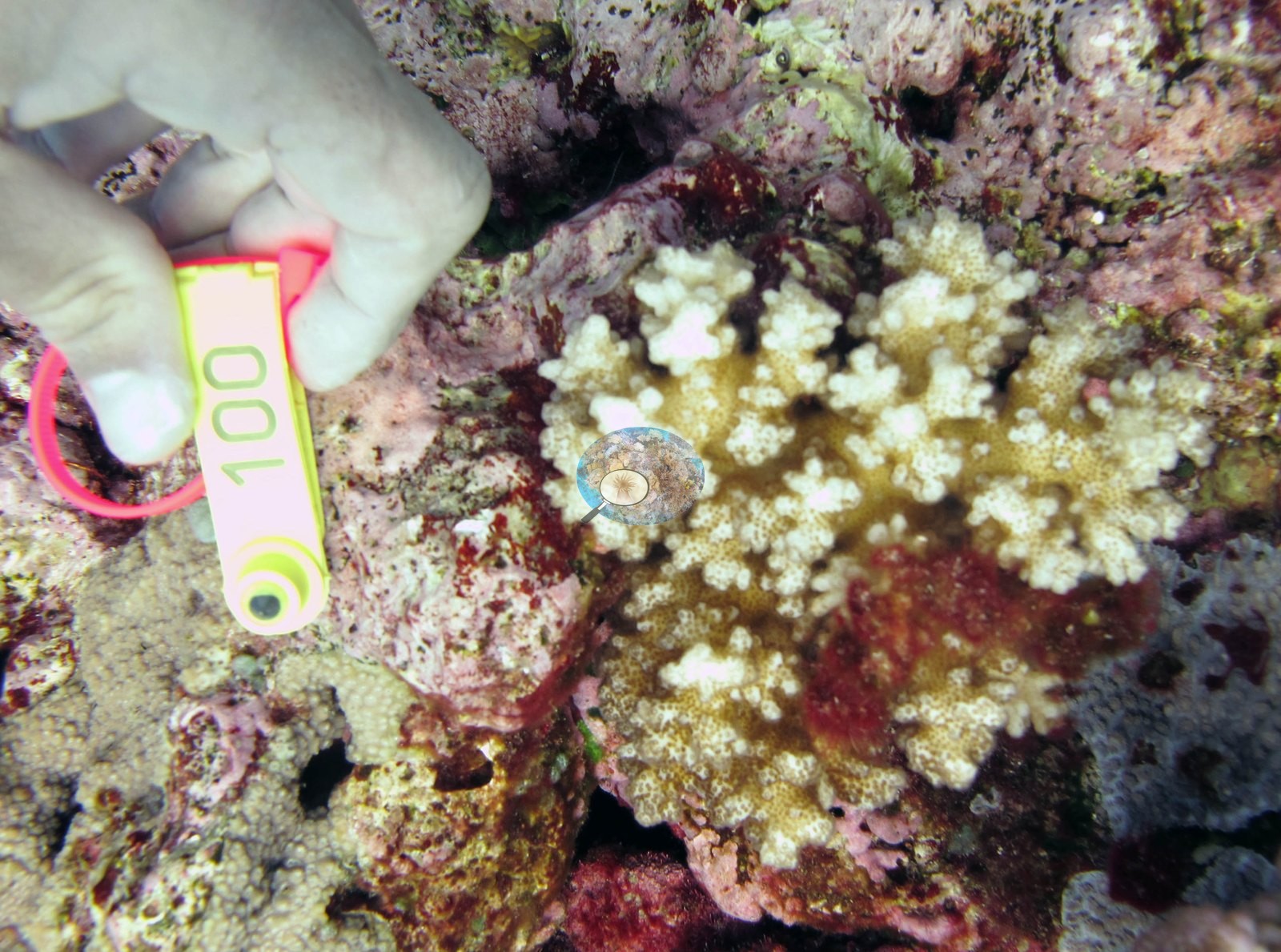
Nine frames are close-in sampling macros taken over the shoulder of the collector: a gloved hand steadying a small branching or corymbose colony while a plastic tag is fixed to it and, in several, the fiber-optic tip of a Diving-PAM fluorometer is held against the colony surface (e.g., Image 6, Image 7, Image 8, Image 9, Image 10, Image 13, Image 16, Image 17, Image 18).
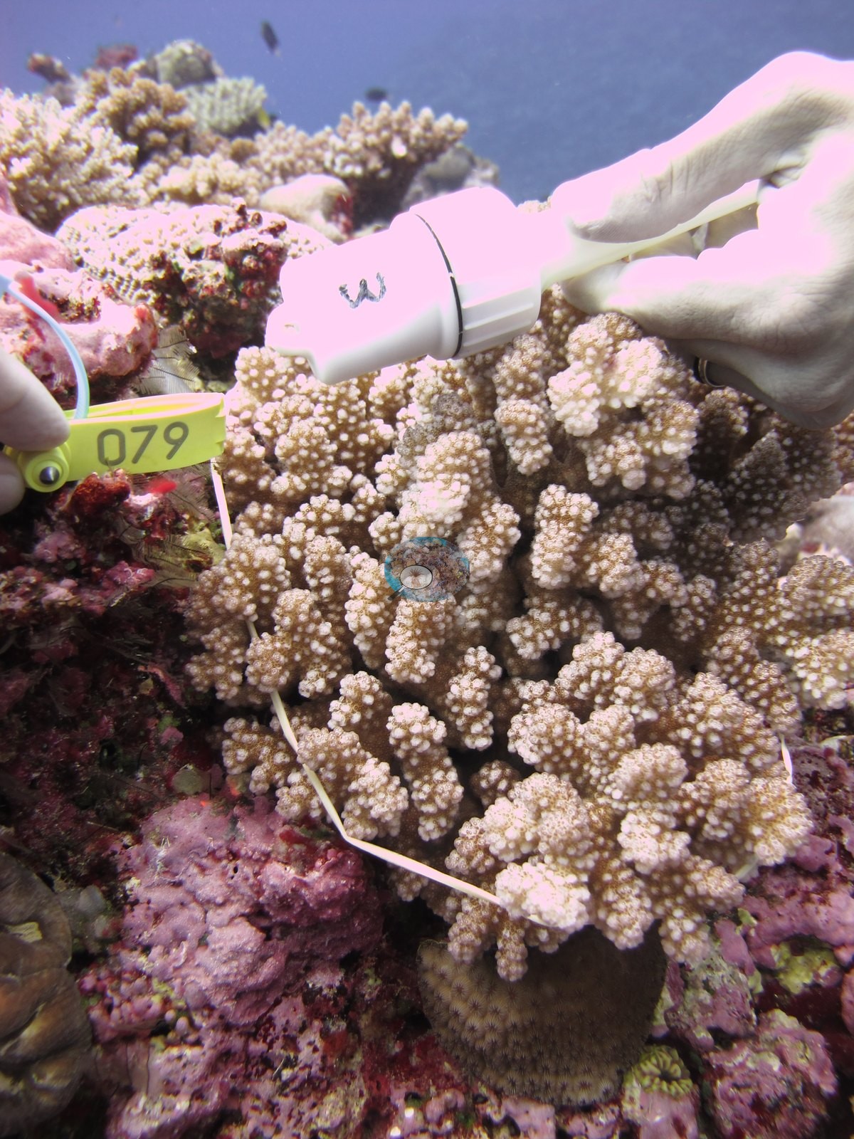
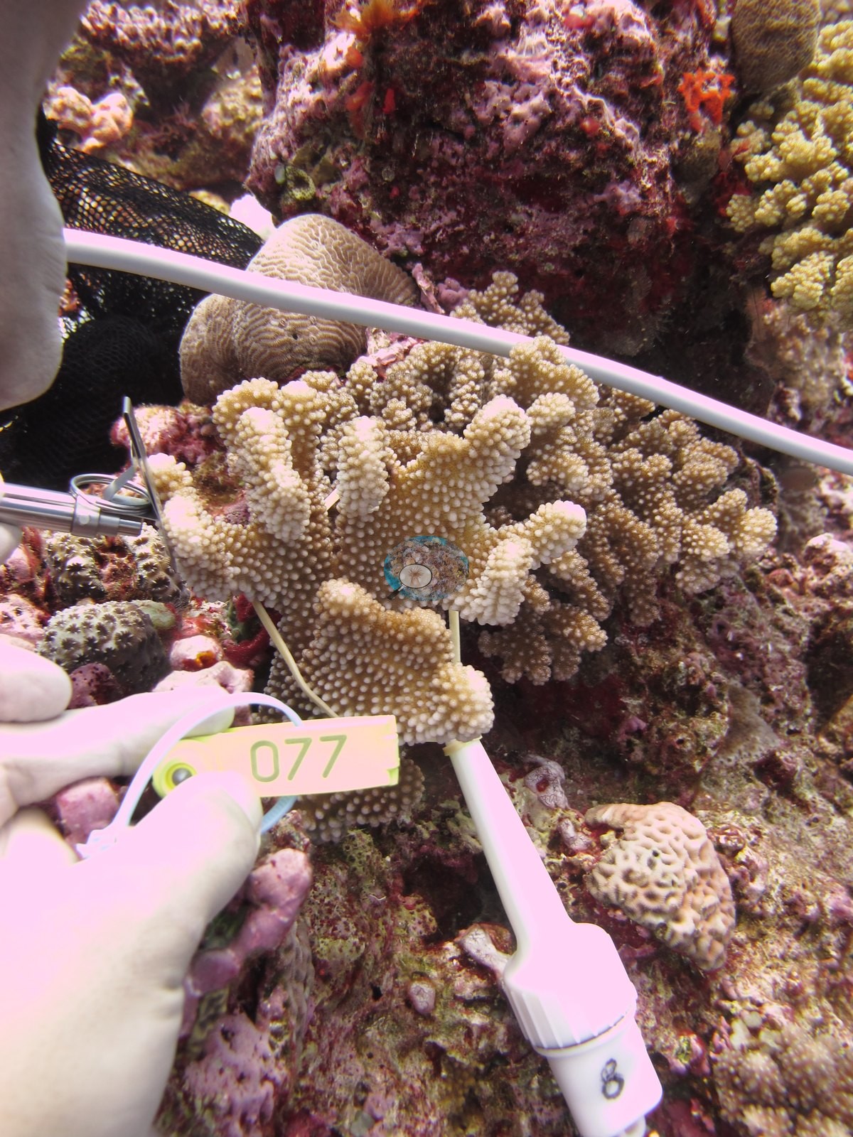
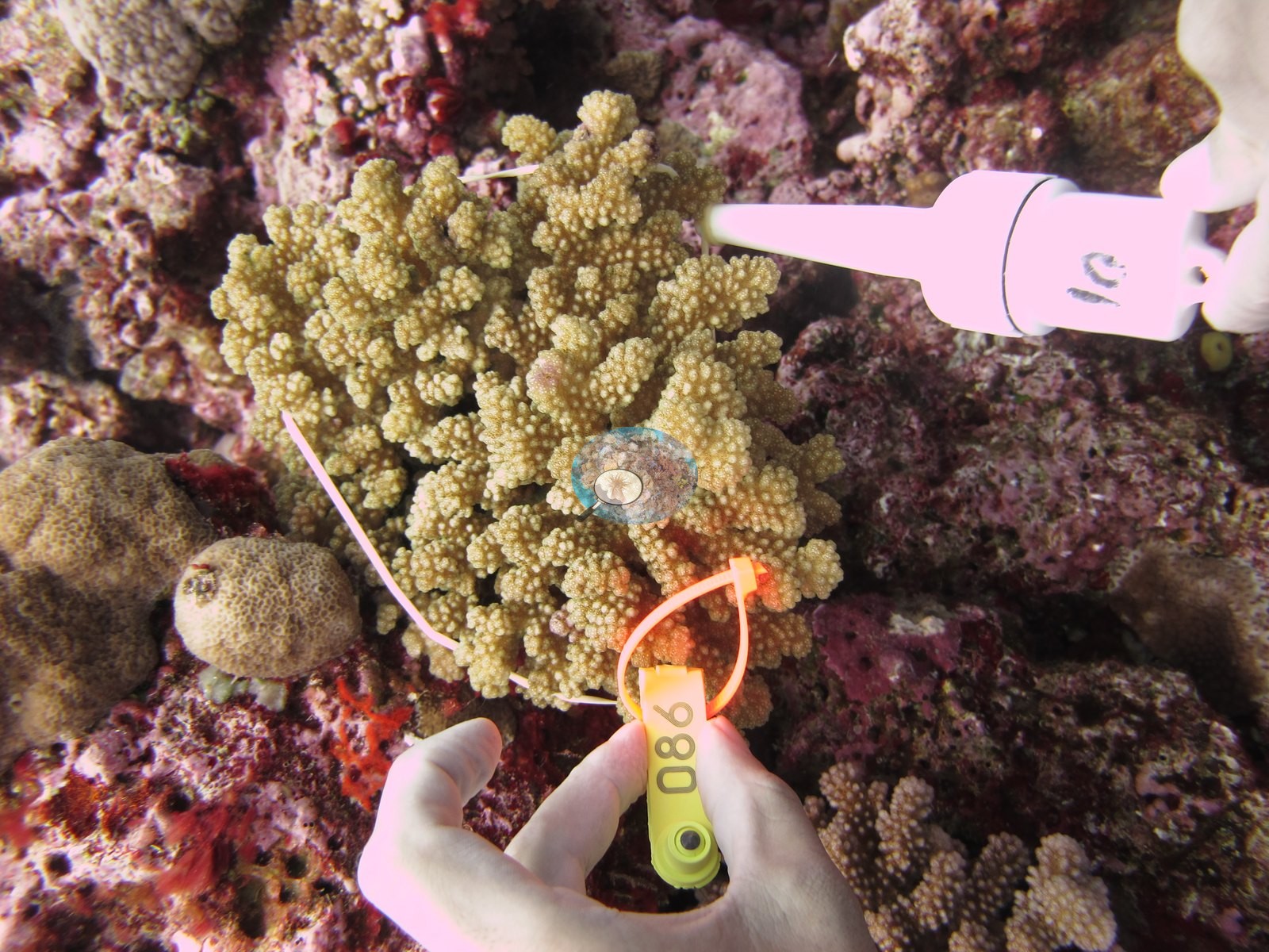
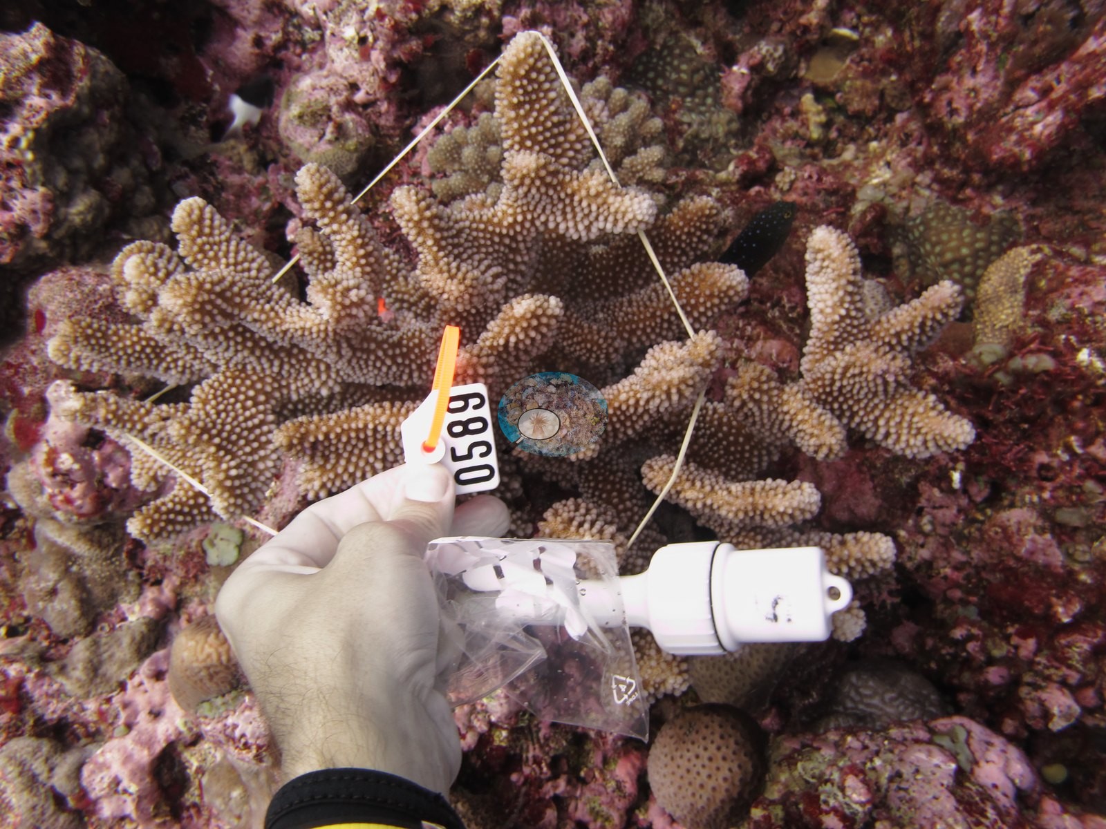
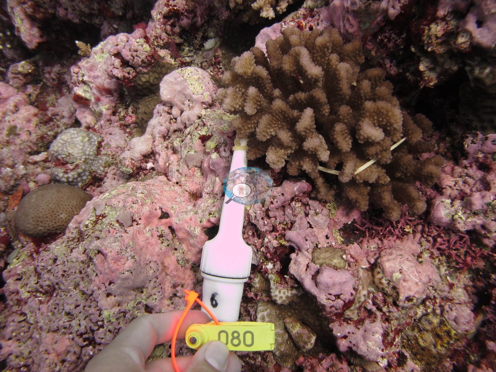
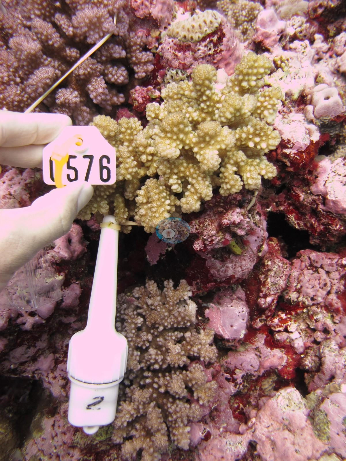
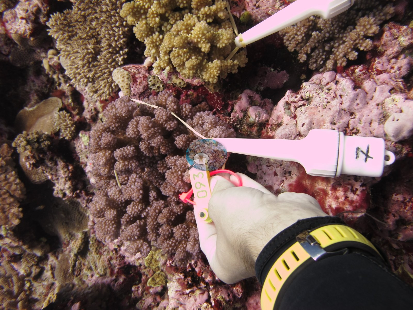
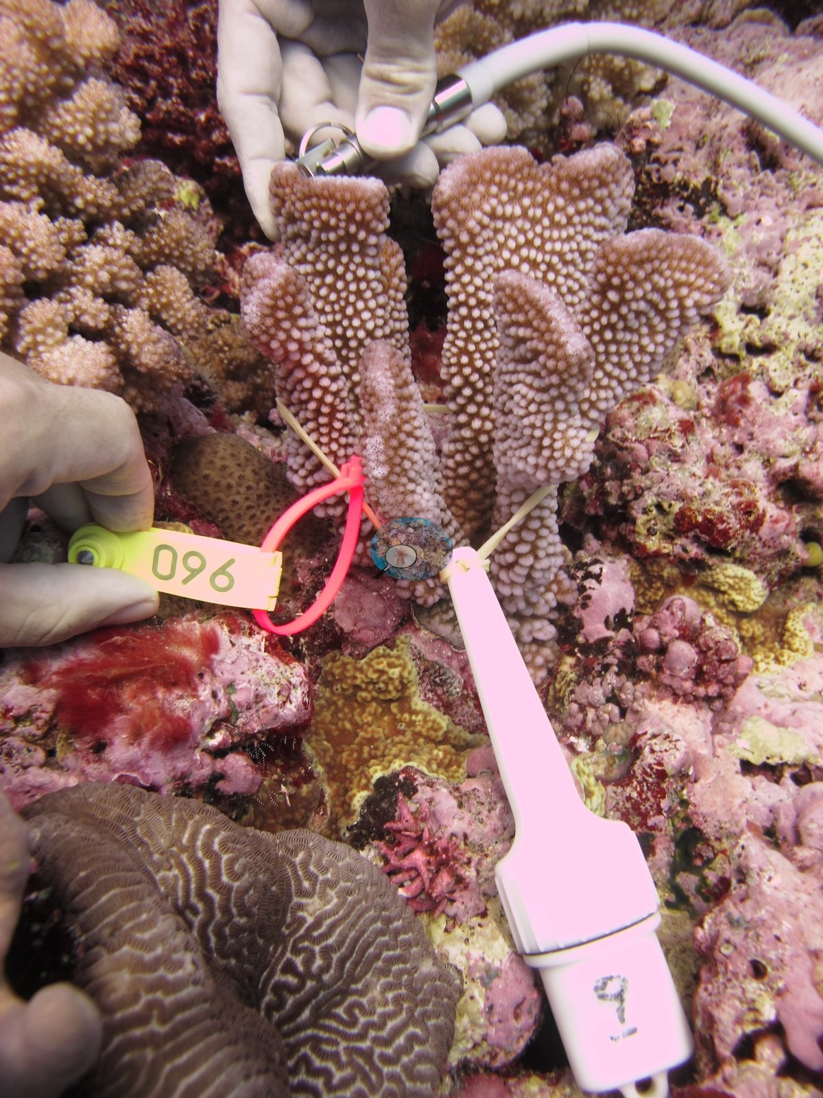
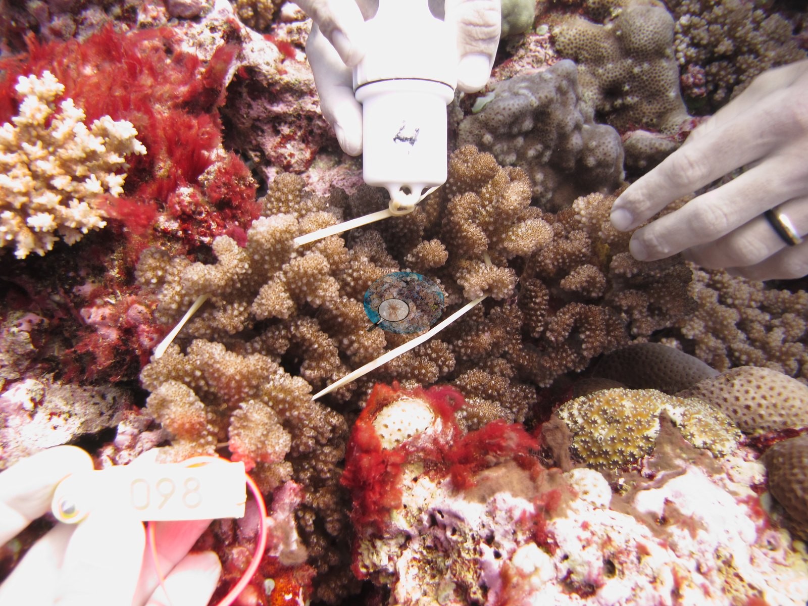
These are method frames — they show single tagged colonies at arm's length, not the reef — and must not be read as habitat or cover. A tenth frame shows the PAM instrument itself being carried over the reef (e.g., Image 4).
One further frame is a fluorescence exposure: a nodular, coarse-polyped colony glowing orange-red with individual polyps picked out against a dark maroon background (e.g., Image 20); fluorescence frames carry no cover information either.
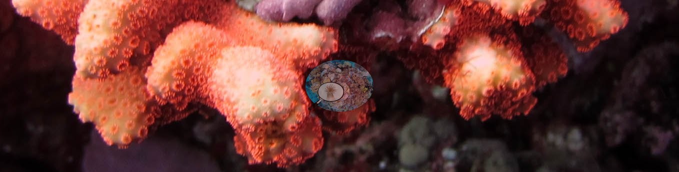
The frames cannot settle species-level calls anywhere — the growth forms recur across several genera at this size — and they cannot be used to revise the transect.
Survey team's site notes: On the east side of the island (FJML23, FJML15, FJML22, FJML16), sites were on the fore reef slope. Coming up the slope from 32m, not very high coral cover, but CCA was common. Small colonies of Platygyra, Lobophyllia, and Echinopora were common.
CV model comparison
More info
The photo row below is a methods comparison only: it is never averaged with the transect and never used as the headline. It compares site means only: the photos come from the same survey dive as the transect but not from the same points.| Method | Hard coral cover (%) | Photos (n) ± SE |
|---|---|---|
| Transect mean (all transects) | 33.0 | |
| CoralSCOP edited | 41.0* | n = 19 · SE 2.0 |
| Claude (edited photos) | 34.8* | n = 19 · SE 2.9 |
The two computer-vision methods agree within 6.2 pp (34.8–41.0%) and read 1.8–8.0 pp above the in-water transect (33.0%); the headline is the in-water transect because a diver transect is independent of the photographs.
*Based on only 19 reef-habitat photos; this estimate is less certain.
See a more detailed, stand-alone version of this table.
Data & links
- Download benthic survey data (CSV) ↗
- Download all LOF-GRE data (933 sites) ↗
- Photo portfolio ↗
- Coral Reef Diagnostics page for this area ↗
All images from Coral Reef Diagnostics. Data are from the Khaled bin Sultan Living Oceans Foundation Global Reef Expedition and Coral Reef Diagnostics.
 © Coral Reef Diagnostics.