
Part of the regional summaries: Fiji - Cicia, Fiji - Northern Lau (Vanua Balavu / Cicia / Nayau), and Fiji - Lau
More info
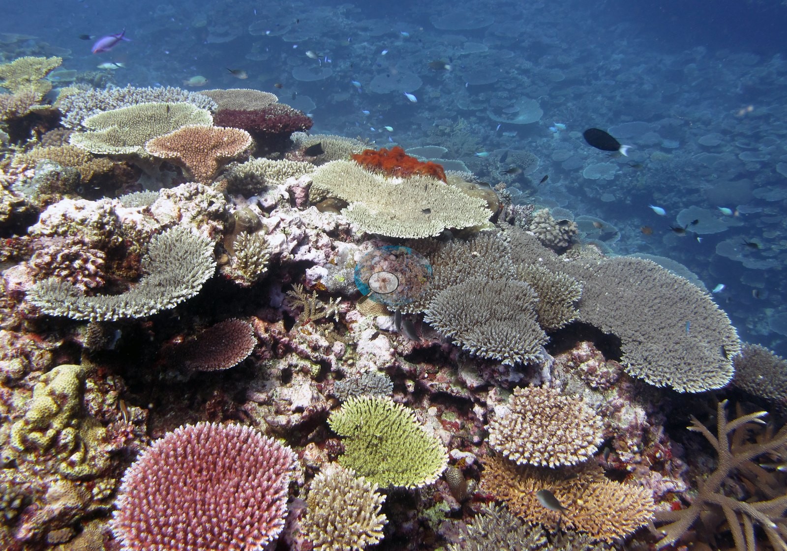
Fiji (NOAA CRW virtual station)A flat-topped coral mound off Cicia whose entire upper surface is paved with table Acropora stacked like shingles, in cream, sea-green, olive, mauve, and salmon, under clouds of anthias and fusiliers. Coral cover is about 50%, with no bleaching, disease, or crown-of-thorns seen. Surveyed on the Living Oceans Foundation's Global Reef Expedition.
E dua na ulu ni lase (coral mound) e savasava na dakuna mai Cicia, ka na kena yasana i cake taucoko e vakaubitaki ena Acropora vakatepeli e veitabaki vaka na kau ni vale, e roka (cream, sea-green, olive, mauve, kei na salmon), e ra ni ilawalawa levu ni ika anthias kei na fusilier. Na ubi ni lase (coral cover) e rauta ni 50%, ka sega ni raici e dua na vulavula, mate, se bula (crown-of-thorns).
AI translation of this summary; the English is authoritative. (low confidence)
CTD-cast midpoint; the cast read 27.2°C throughout
Three other CRD dive sites with their own pages within 5 km shown in orange — click a marker to open its page.
🗺 Detailed map of the Global Reef Expedition dive sites, Cicia, Fiji (Khaled bin Sultan Living Oceans Foundation; opens in a new window)
28 of the 29 photographs on this page are shown beside the text that discusses them under "Site description" below.
Site description
General benthic features
Of the 28 frames in this station's folder, eighteen are reef-scale, and every one of them shows the same thing: a broad, flat-topped carbonate mound whose entire upper surface is paved with tabulate Acropora. Image 9 is the clearest — the whole dome silhouetted against blue with plates stacked shingle-fashion from the base to the summit and no visible break in the framework.
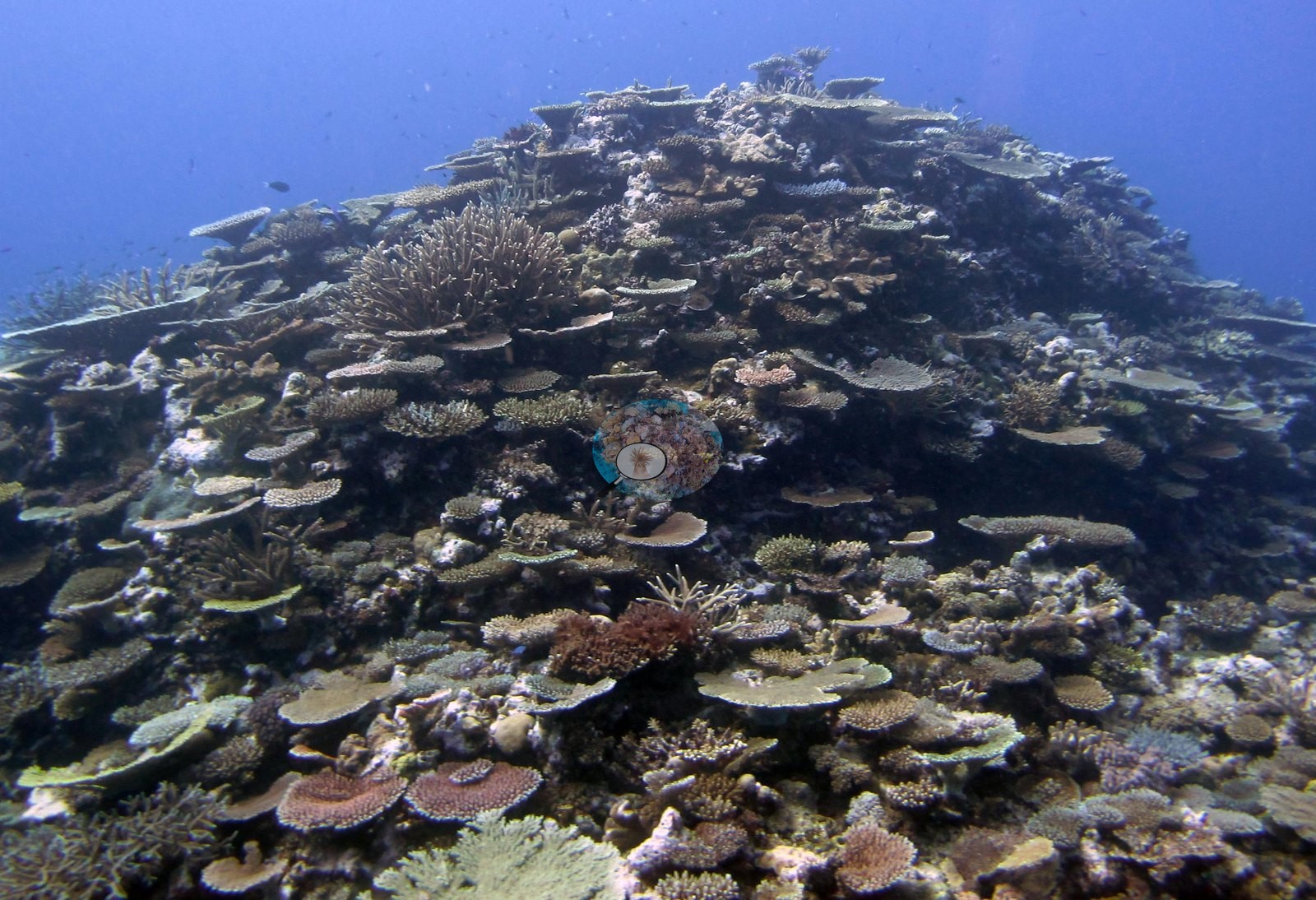
Image 15 and Image 26 show the same structure from closer in, tables layered over lower plating and corymbose colonies.
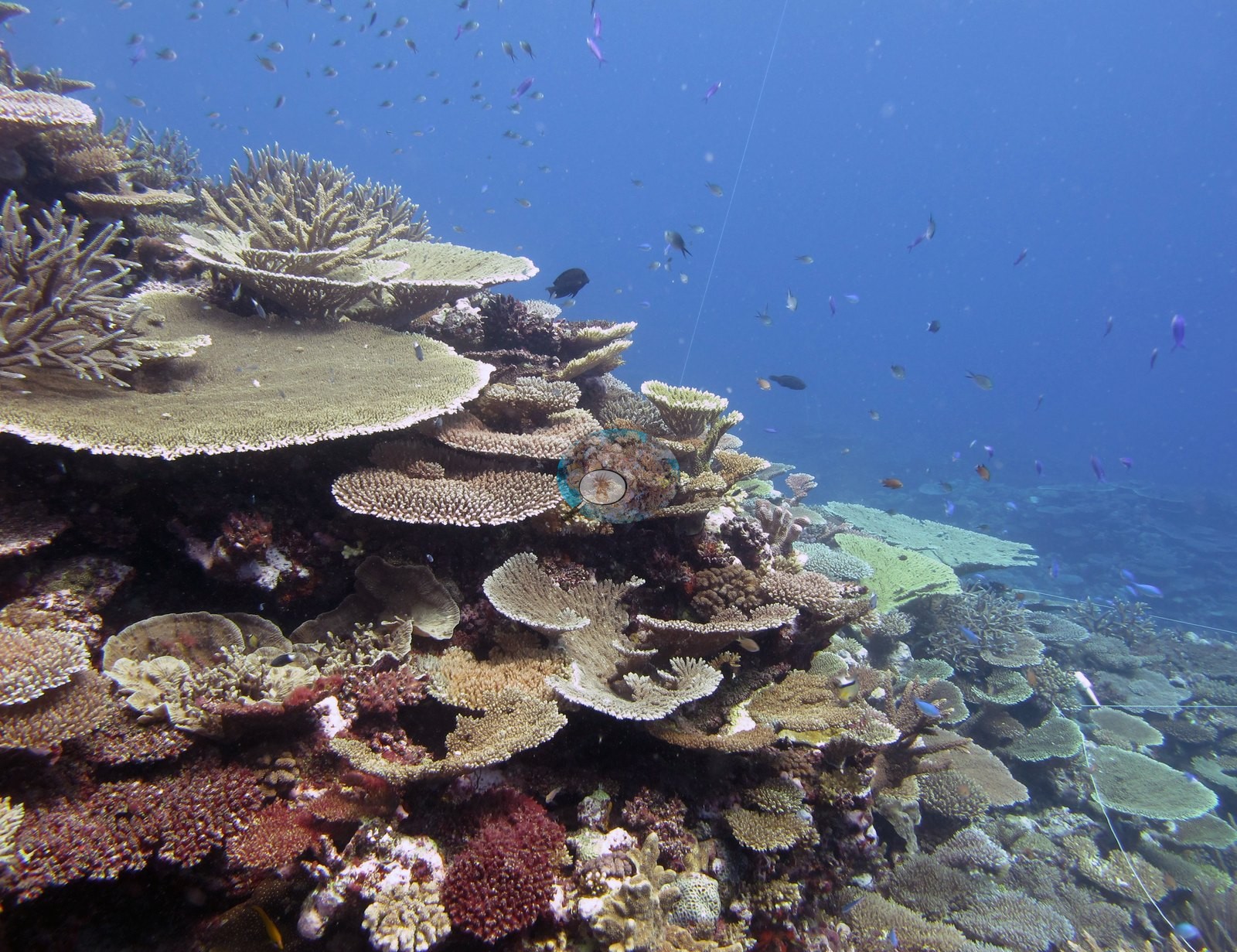
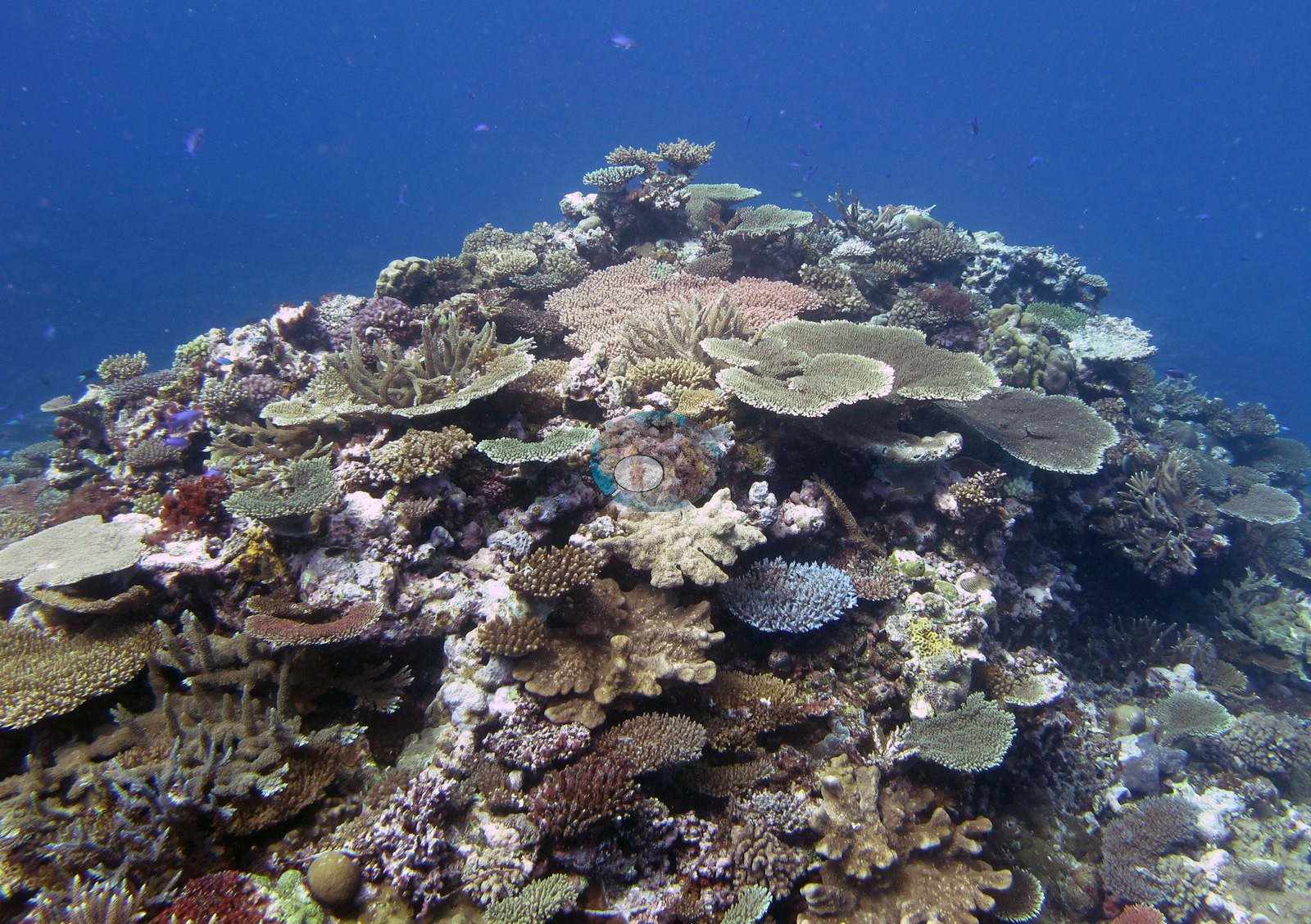
Image 2 shows the texture clearly: a shoulder of reef at Anderson's noted 10 m carrying tables in an unusual spread of colors — cream, pale sea-green, olive, mauve, and salmon — with pink corymbose Acropora, a bright orange encrusting sponge, staghorn thickets at the edges, and a wall of schooling fish behind.
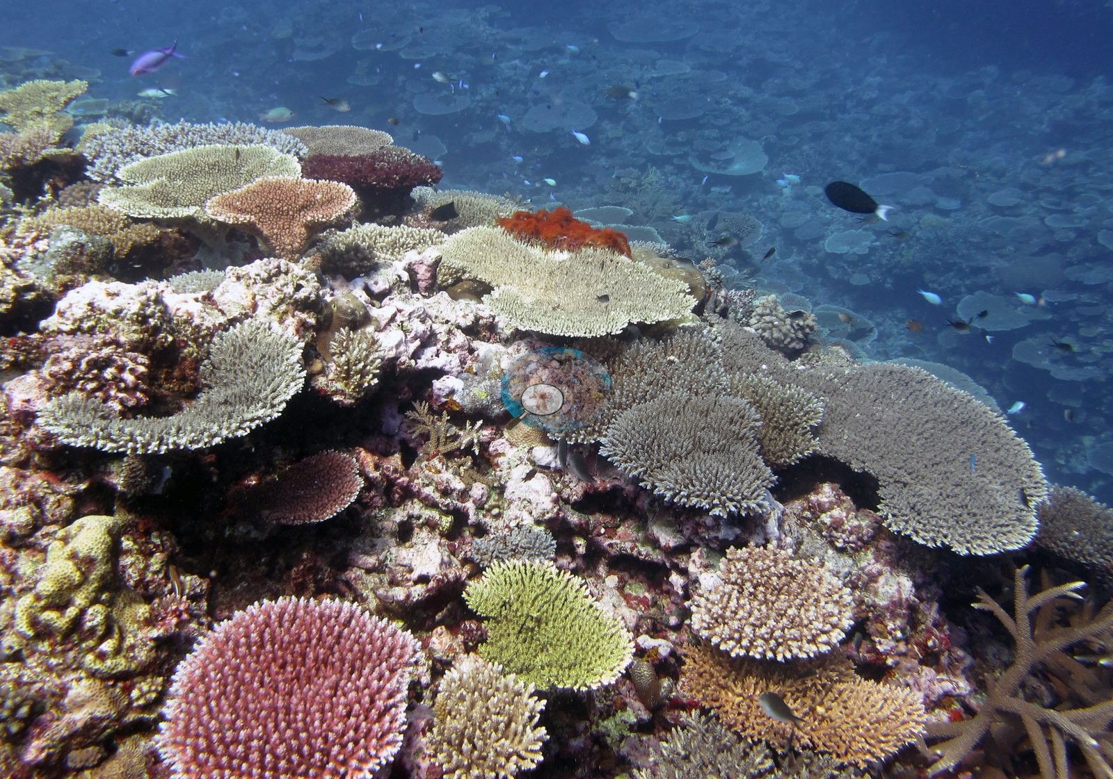
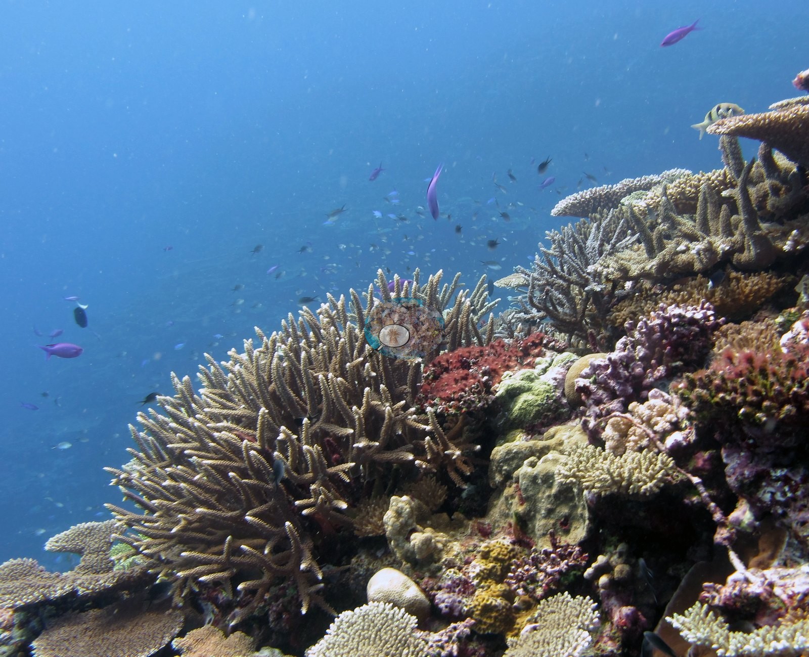
Image 27, Image 8, Image 3, Image 1, Image 10, Image 21, Image 24, Image 25, Image 14, Image 16, Image 23, and Image 11 repeat that assemblage across the site with divers' exhaust bubbles and clouds of anthias, fusiliers, and blue-green damselfish over it; Image 28 and Image 22 are hazier green-cast versions of the same terrain.
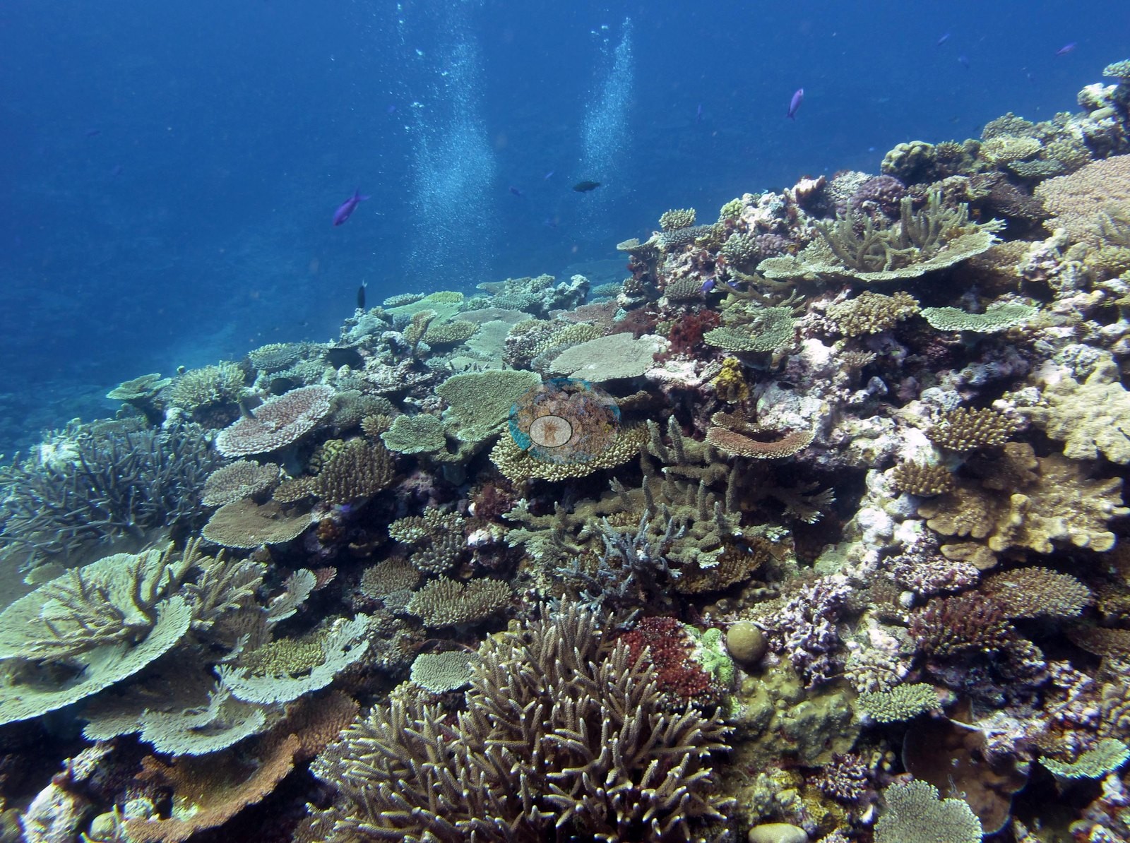
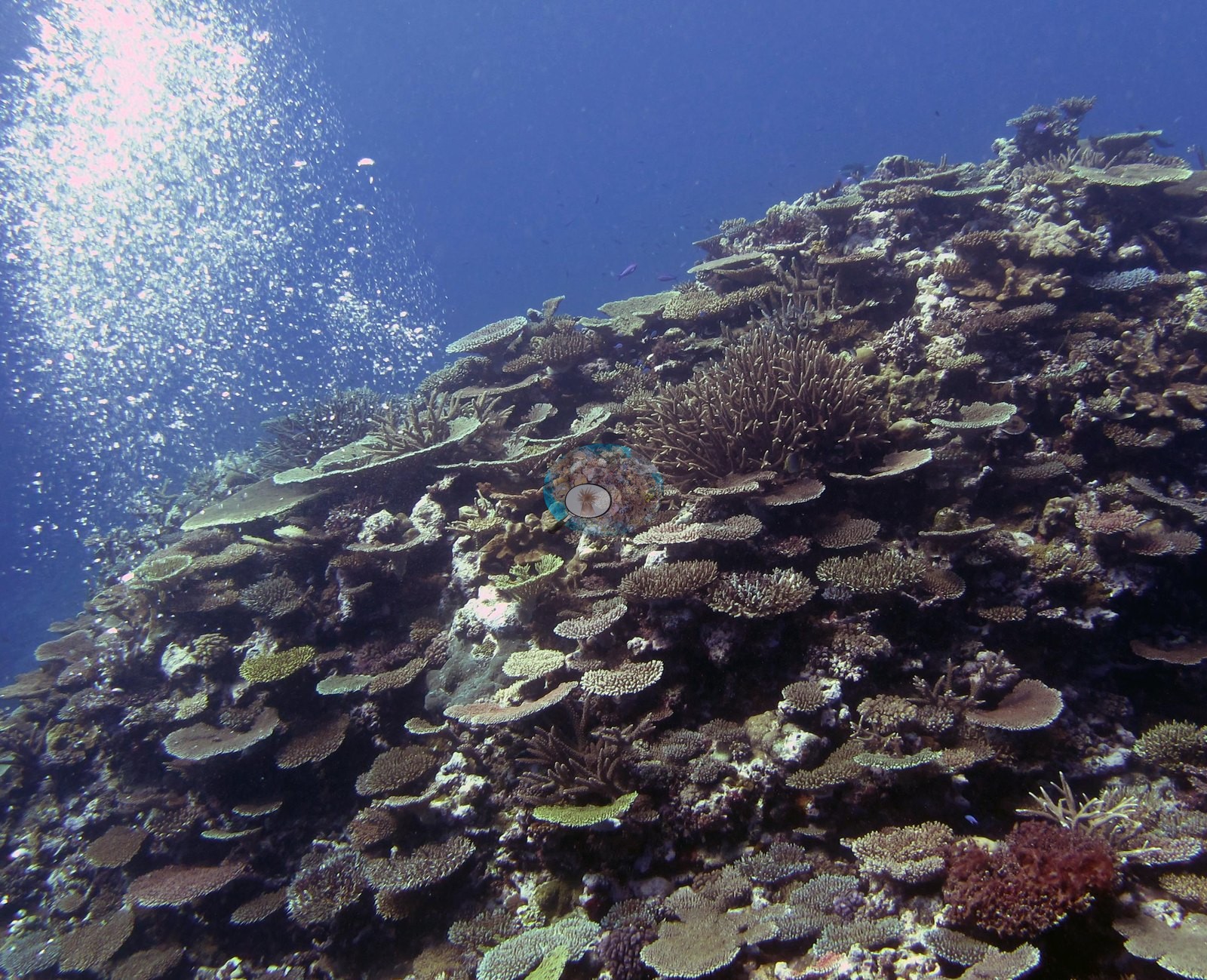
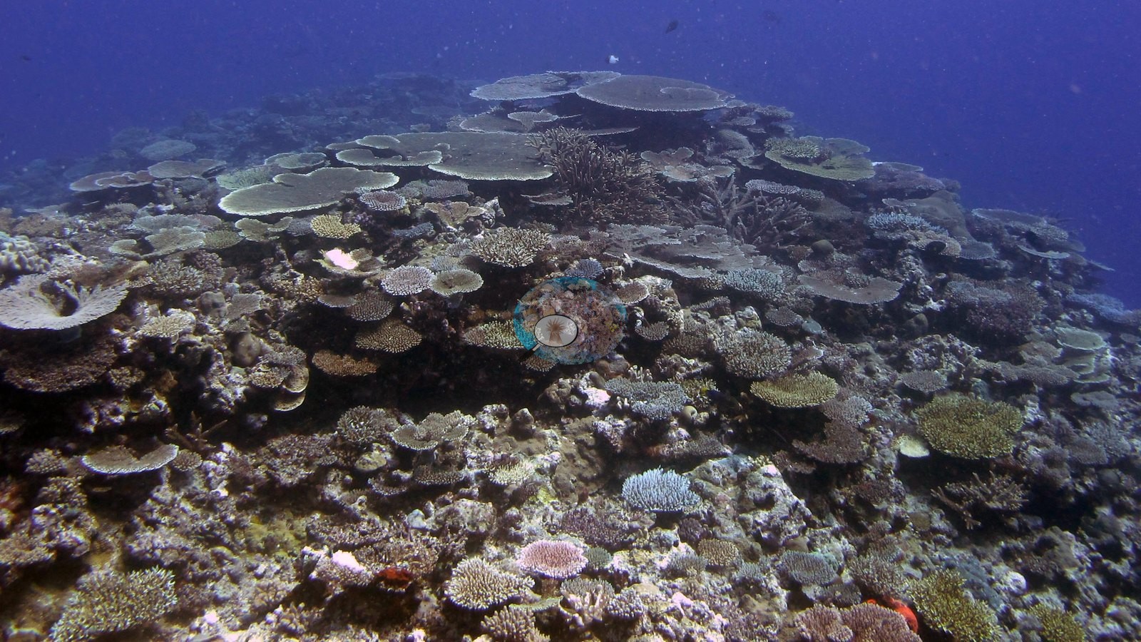
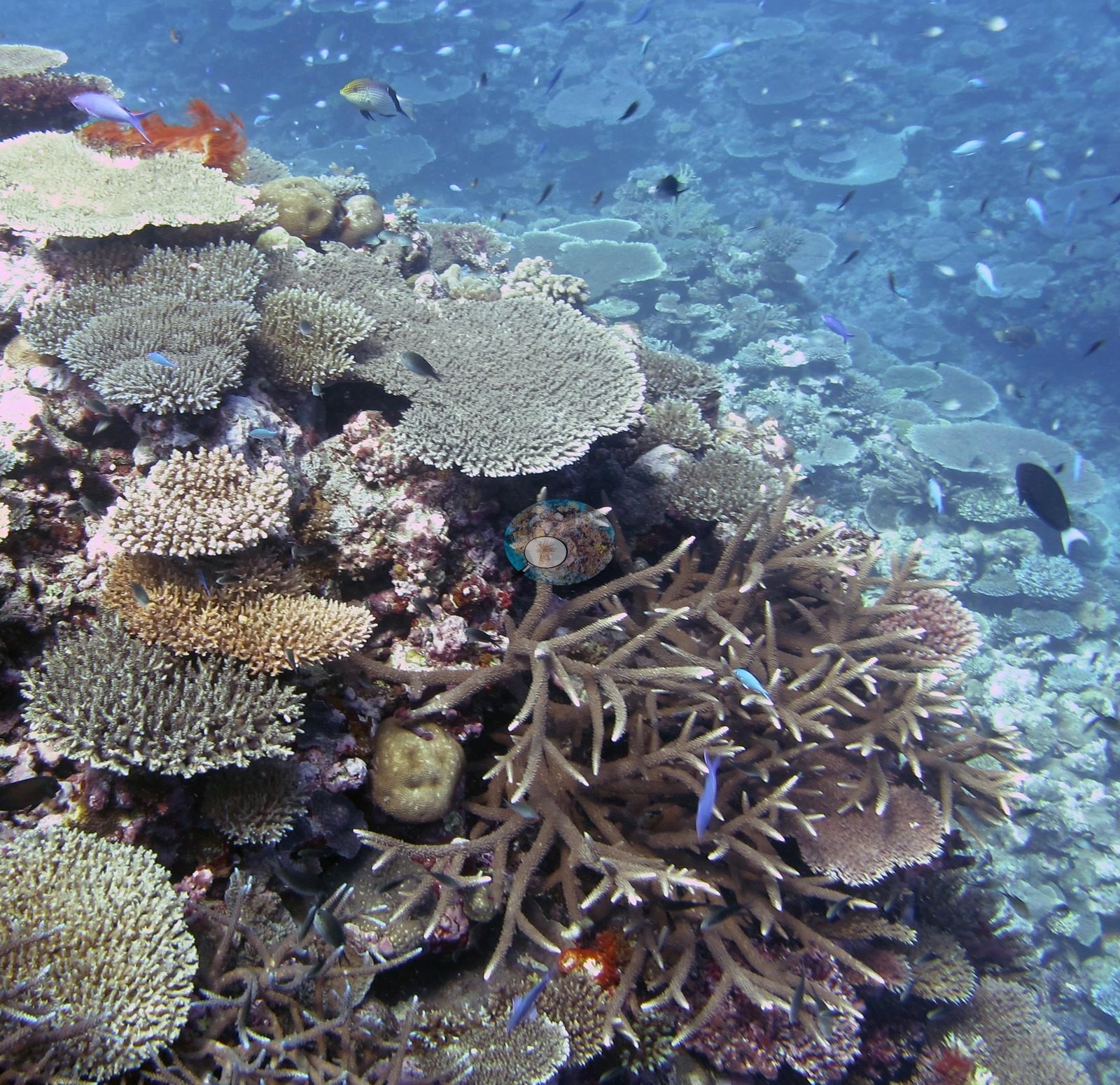
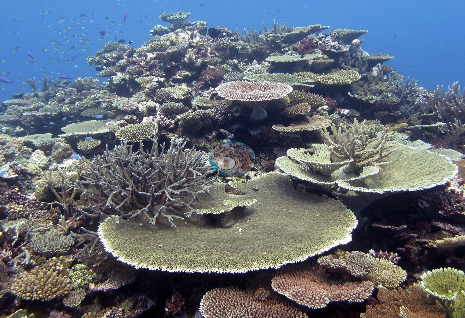
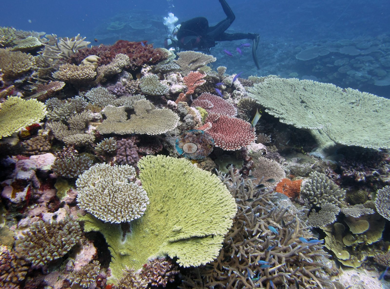
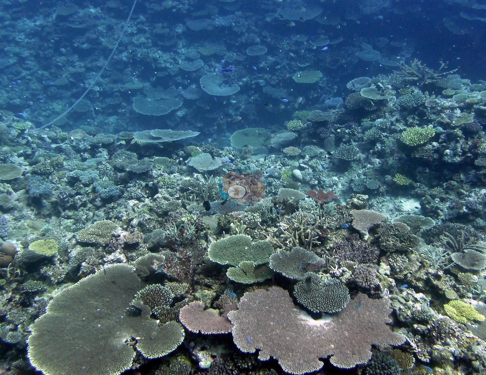
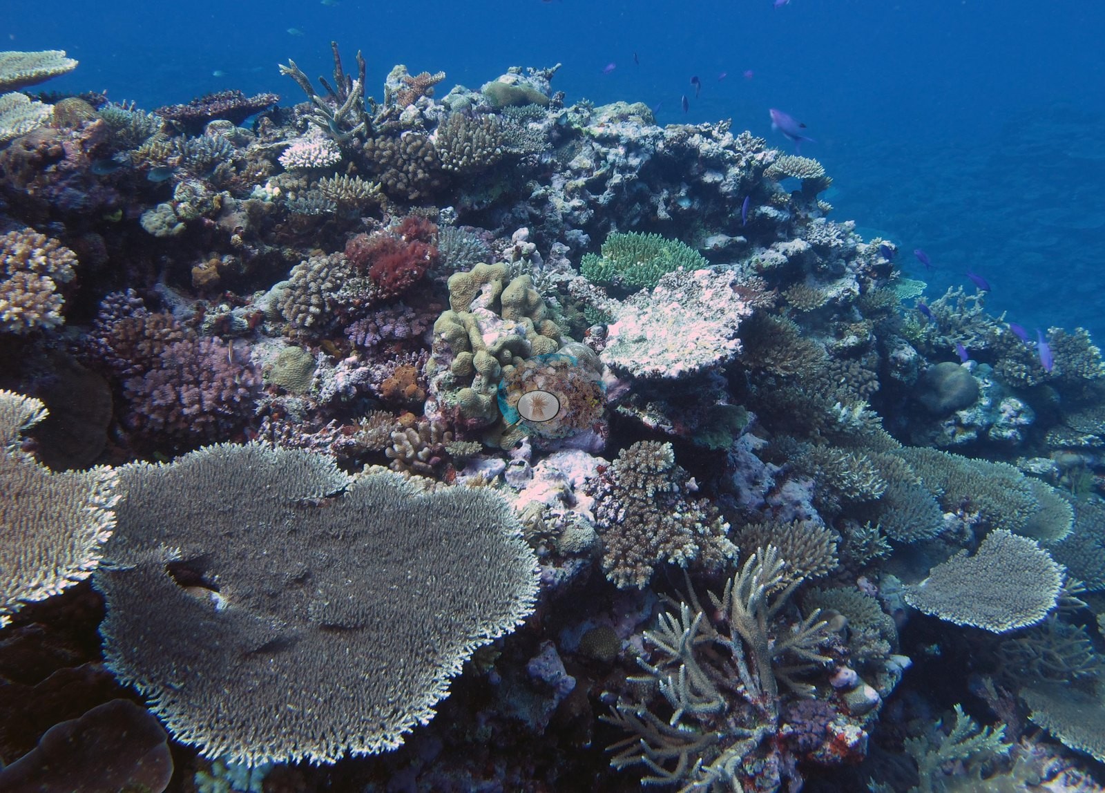
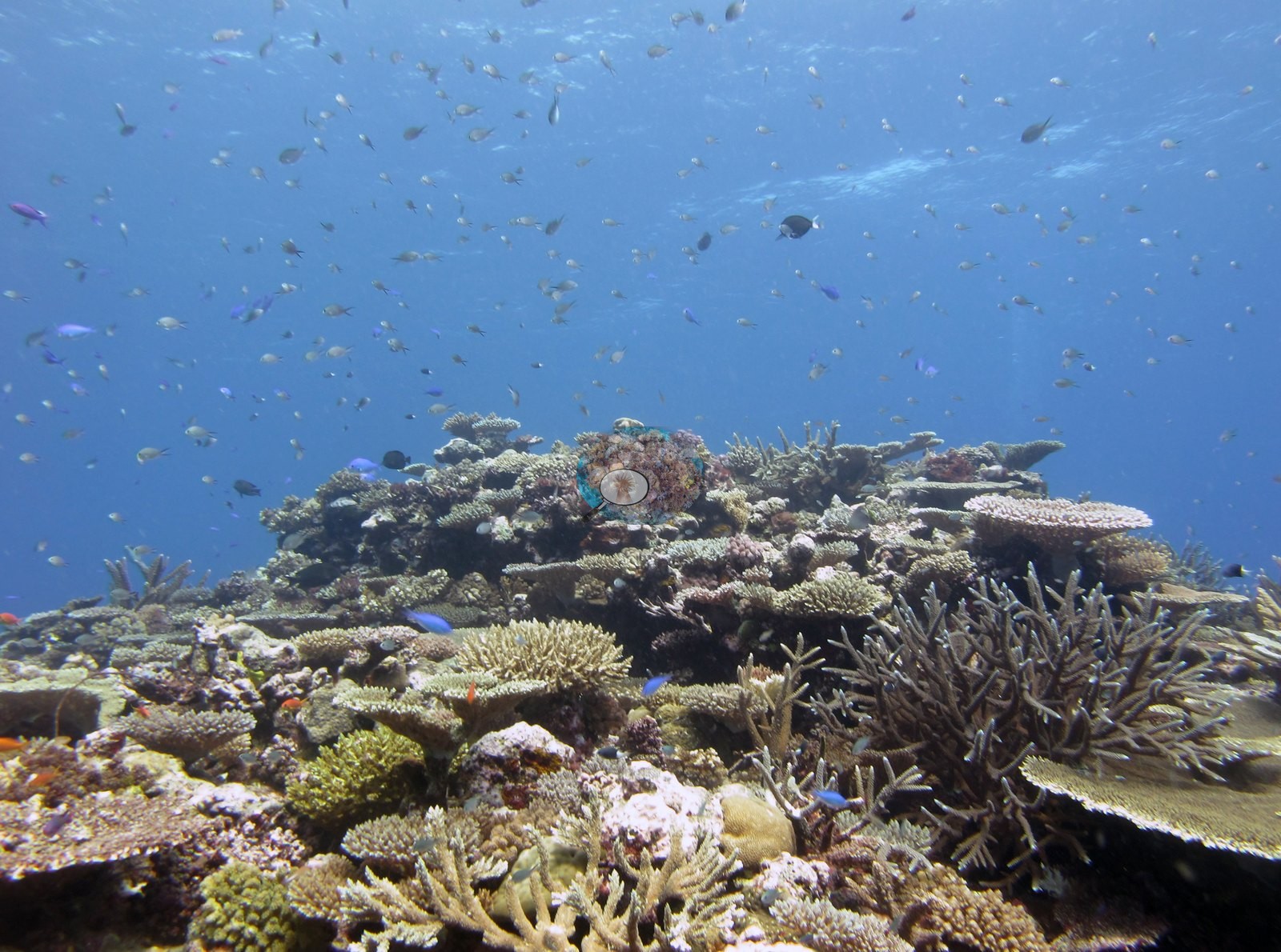
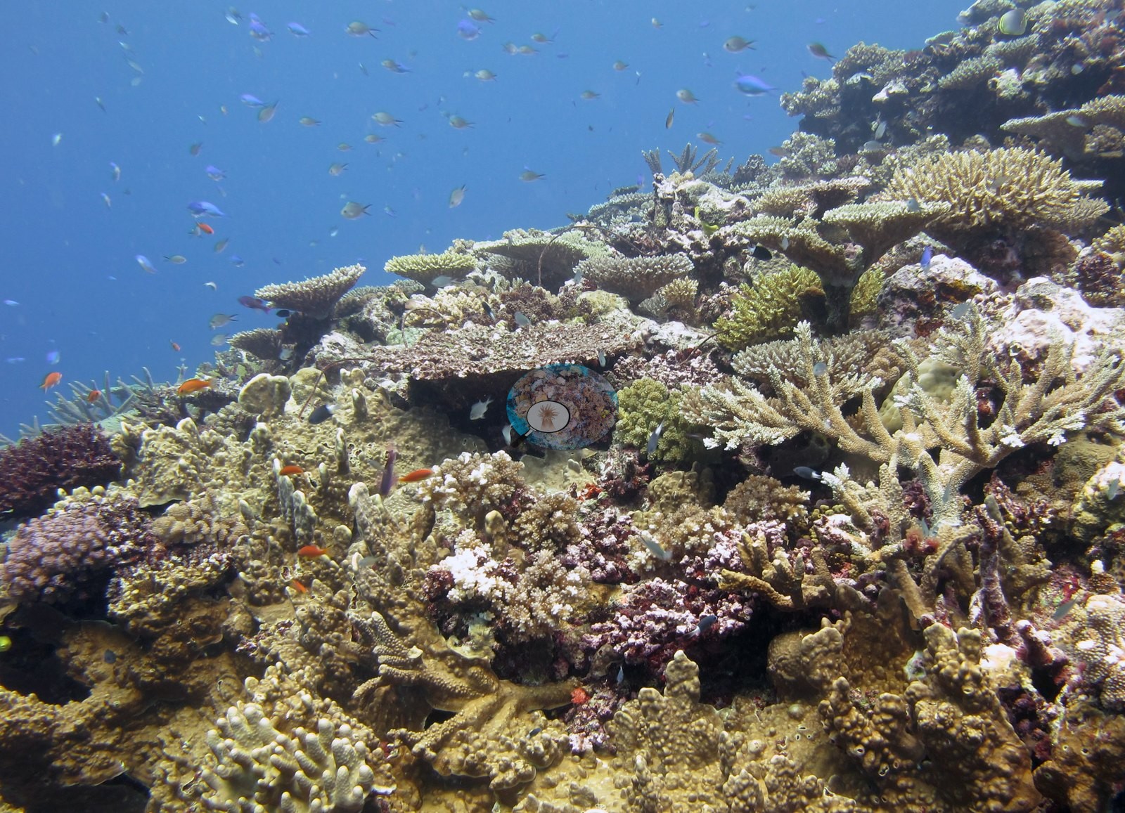
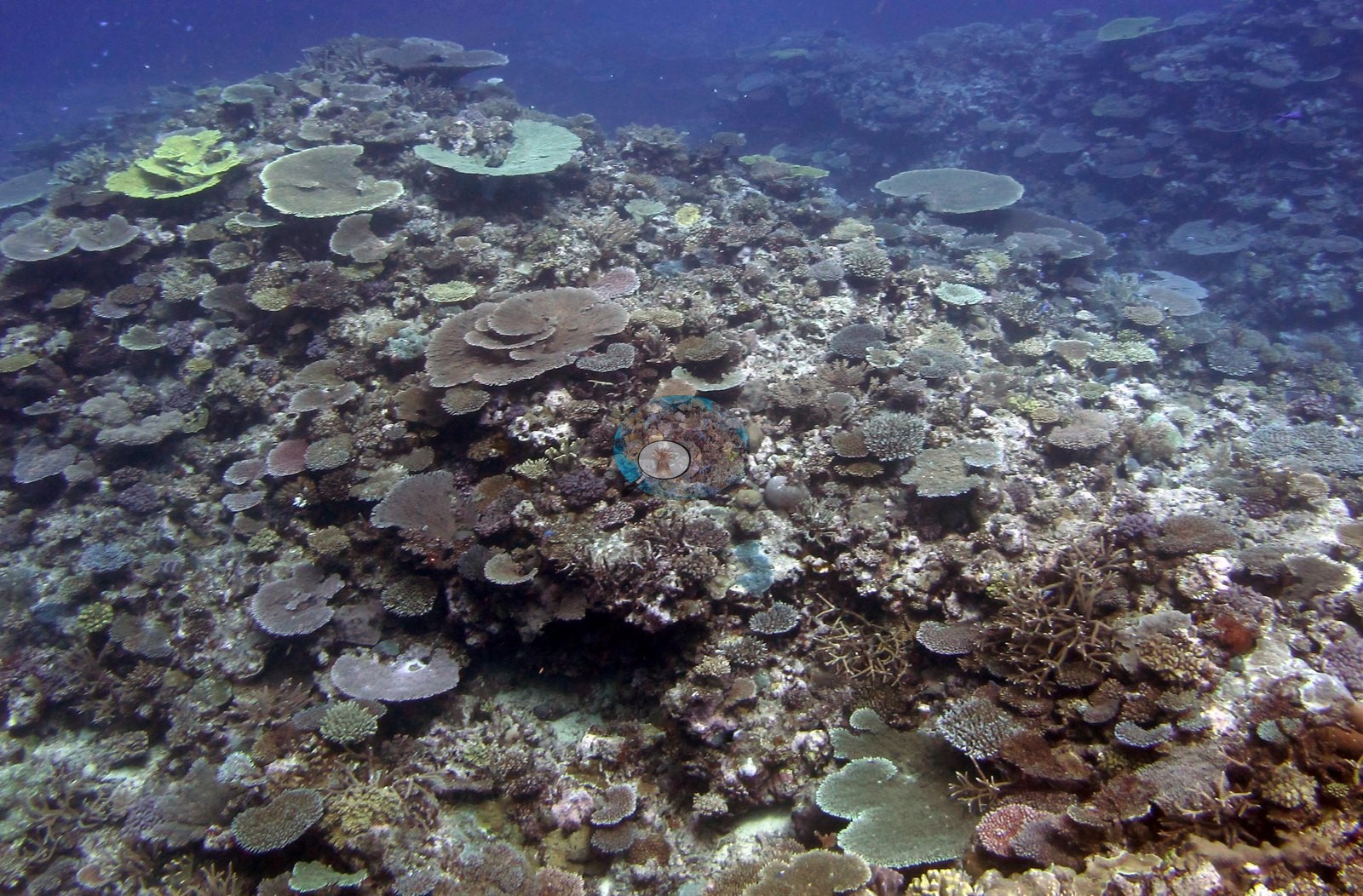
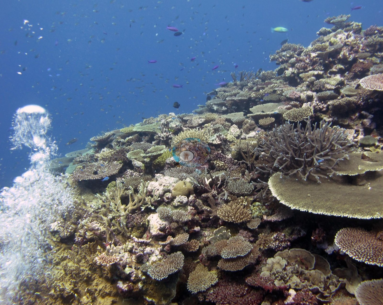
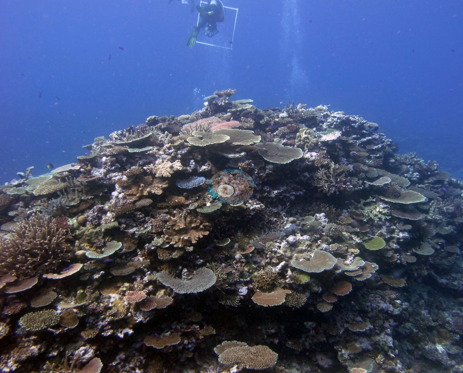
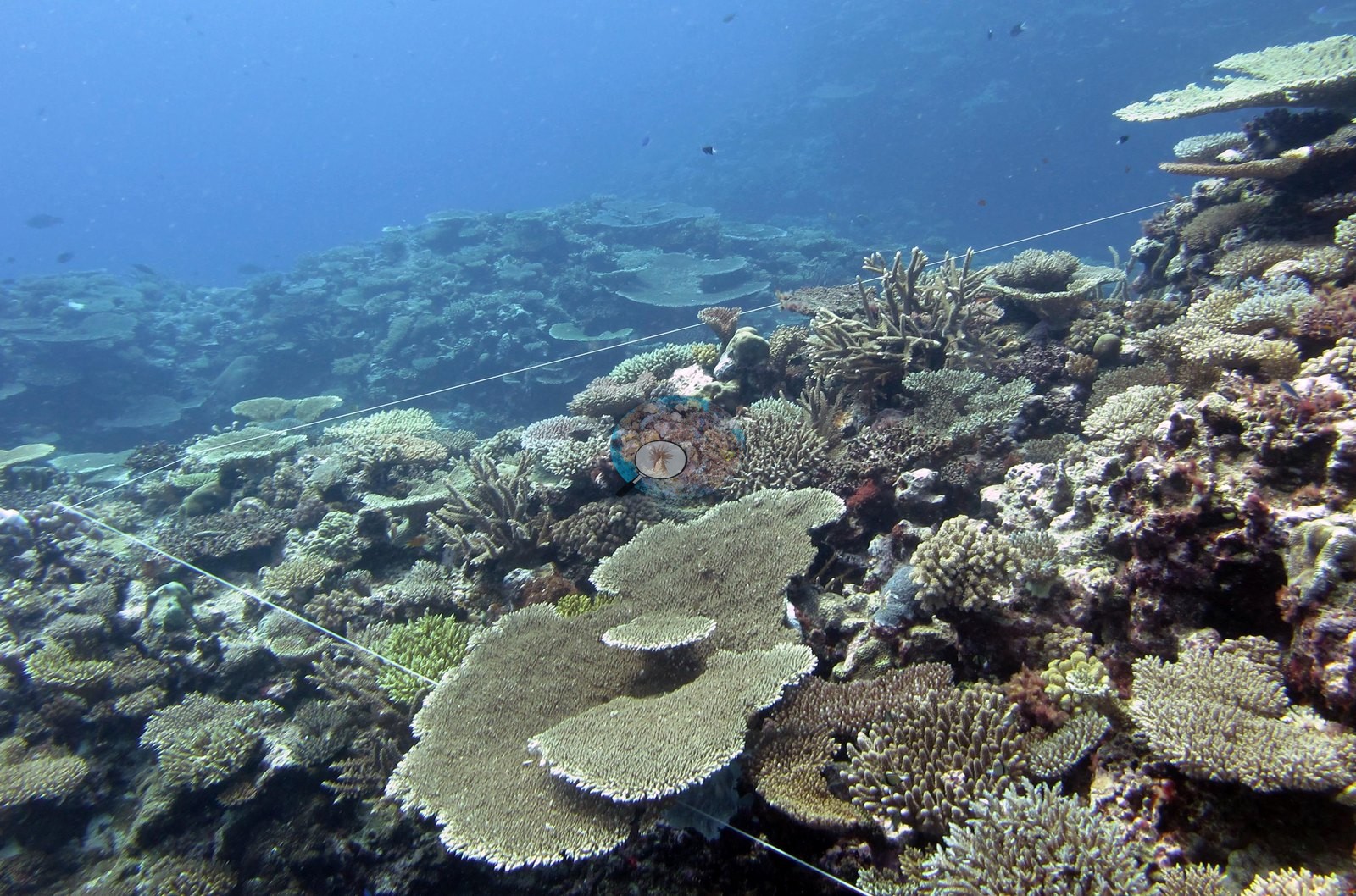
Growth forms are overwhelmingly acroporid — tabulate first, then staghorn and corymbose — with encrusting red algae and pink coralline crust binding the framework between plates and only small patches of turfed dead surface showing through.
Coral cover
- Reference: Khaled bin Sultan Living Oceans Foundation, Global Reef Expedition (in situ transect)
- Coral cover 50%, from the LOF-GRE in situ benthic transect (the mean of all of this station's own transects in the expedition's per-transect benthic data) — a diver transect, not a computer vision index, and the authoritative value for this site.
- Transect mean 50 ± 12% SE (n = 4 transects; Living Oceans Foundation per-transect benthic data); the headline above is this all-transect mean.
Stressors
No bleached tissue, disease lesion, or crown-of-thorns feeding scar resolves anywhere in the reef-scale set. One frame shows an anchor chain lying across the reef, which Anderson noted as a bad place to anchor (e.g., Image 24). The Living Oceans Foundation transects here recorded 5.3% cyanobacteria and no recently dead coral.
Comparisons with other data
FJCC45 (Cicia, Lau Archipelago) was surveyed 18 Jun 2013 during the Khaled bin Sultan Living Oceans Foundation Global Reef Expedition. The benthic community is highly diverse and coral-dominated often with a substantial macroalgae component. Environment and disturbance regime govern benthic community structure. Scleractinian diversity is very high and often dominated by acroporids, pocilloporids, poritids, and faviids. The edge of the shallow slope is often dominated by large Pocillopora eydouxi colonies, thick-branched and elkhorn-like Isopora sp, and unusually large Favia stelligera and/or Pavona maldivensis stands. The expedition's 2013 field report records 212 crown-of-thorns starfish removed from one shallow outbreak reef elsewhere on Cicia, not tied to this station.
Other notes
The remaining ten frames document tagged-colony work rather than the reef: individual knobby Pocillopora-form and corymbose acroporid colonies each carrying a plastic livestock-style tag on a colored cable-tie tether, with a gloved hand holding a white cylindrical instrument head against the colony surface in most of them (e.g., Image 4, Image 5, Image 6, Image 7, Image 12, Image 13, Image 17, Image 19, Image 20), and one frame of a second diver working a large housed camera over a tagged colony (e.g., Image 18).
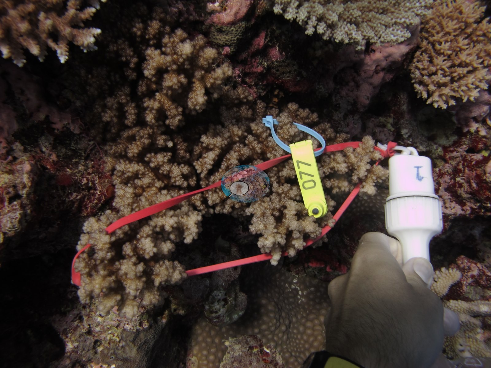
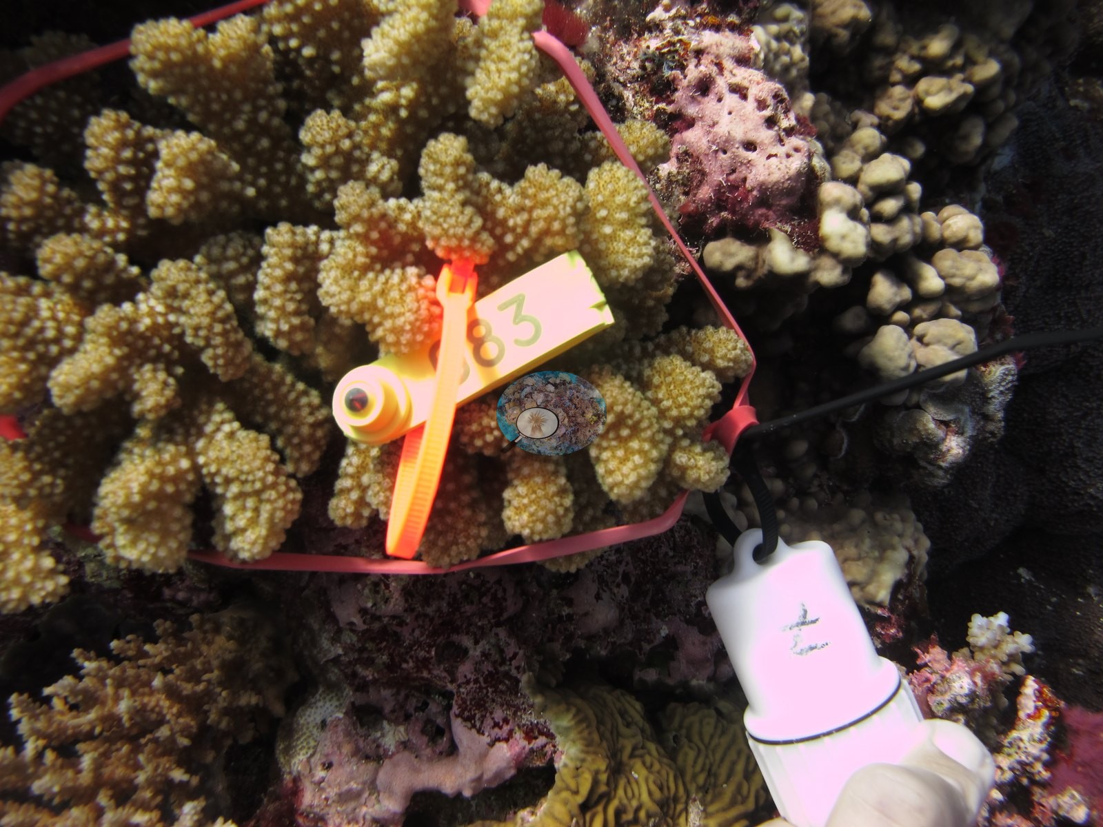
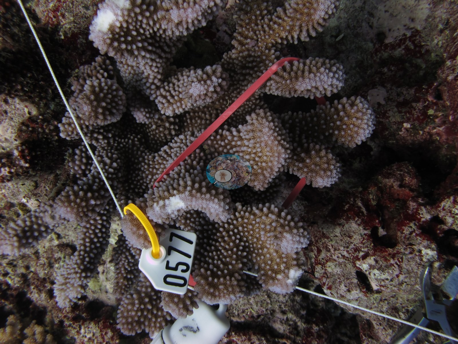
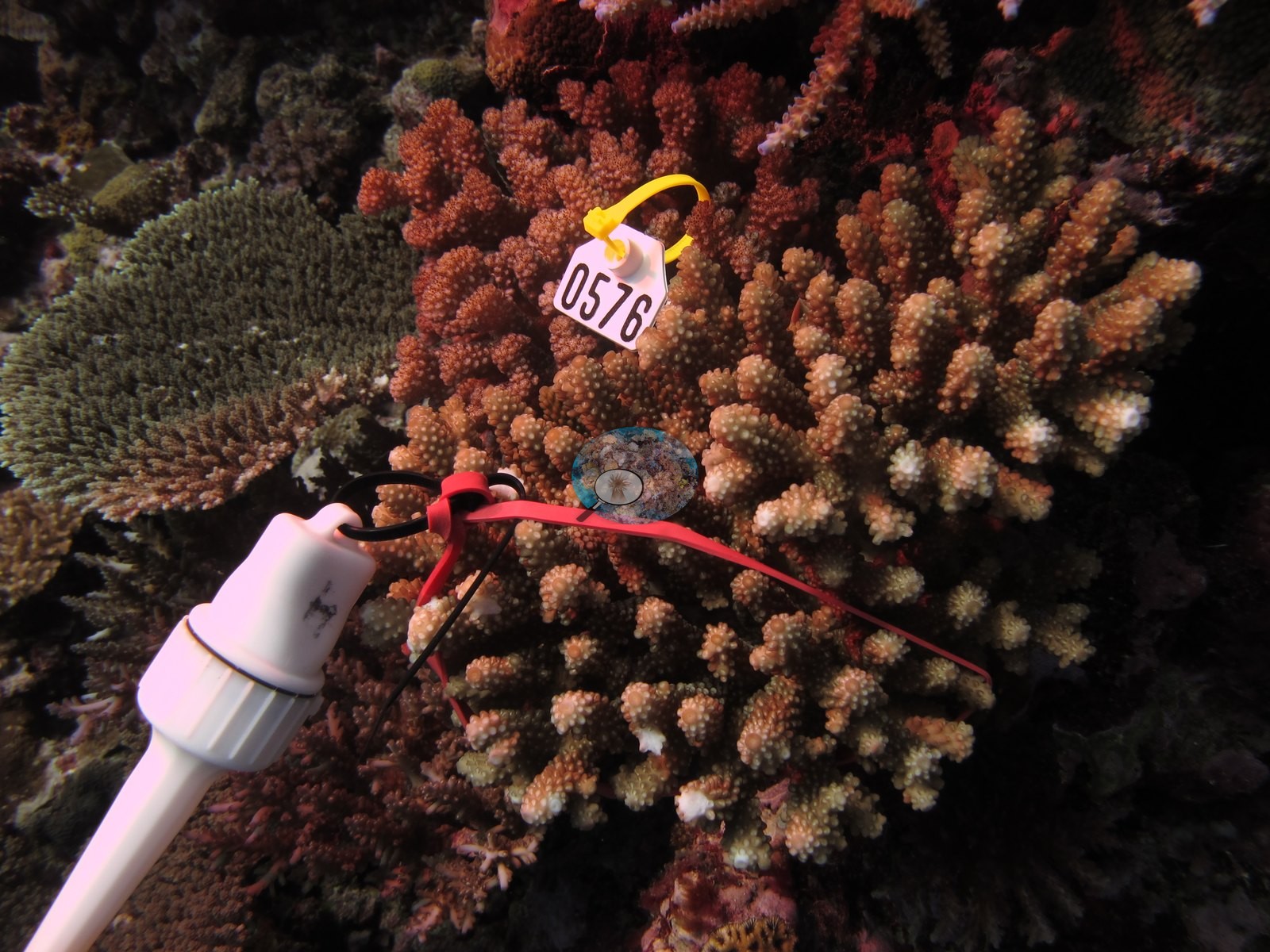
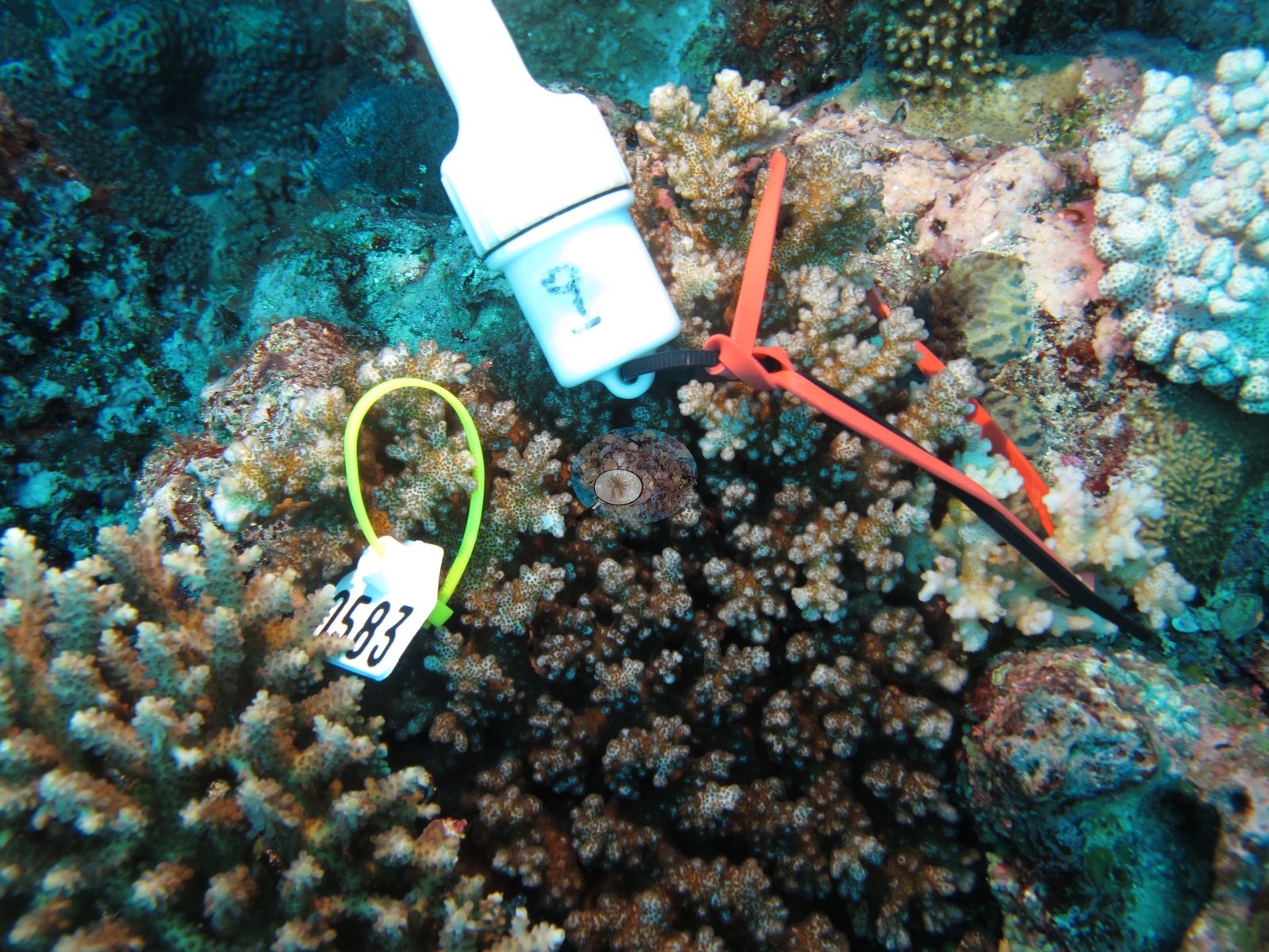
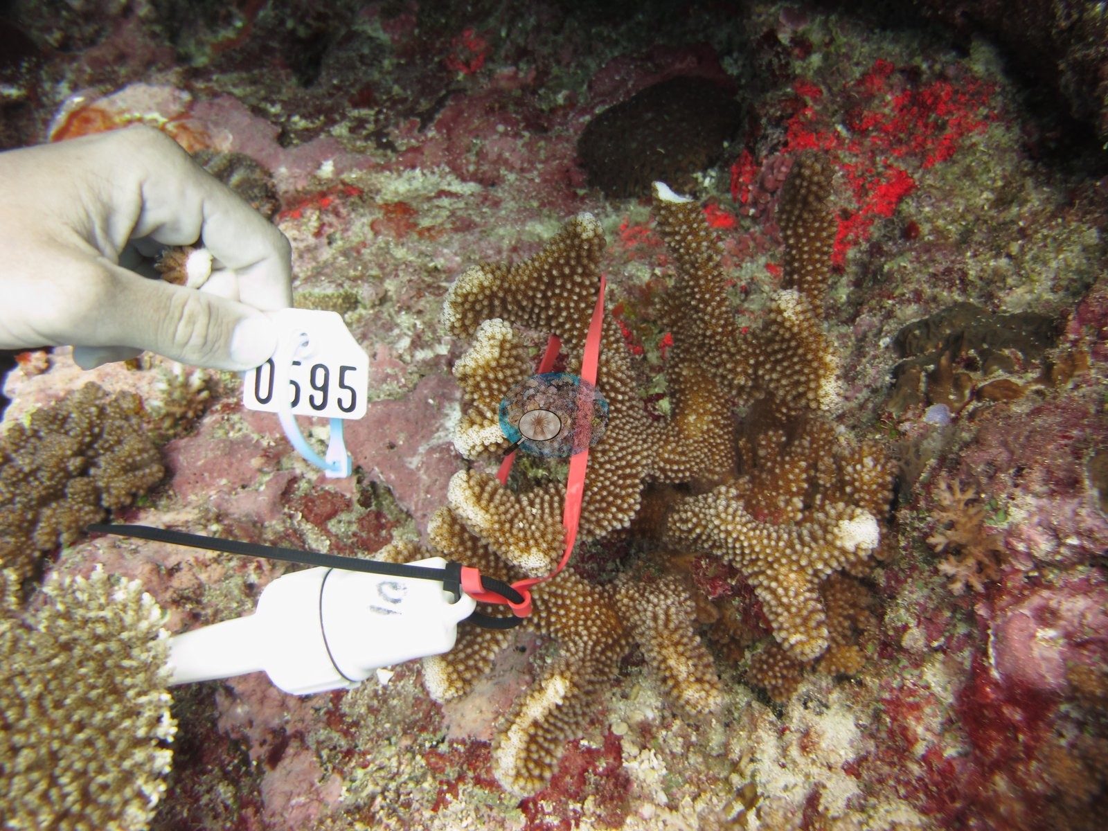
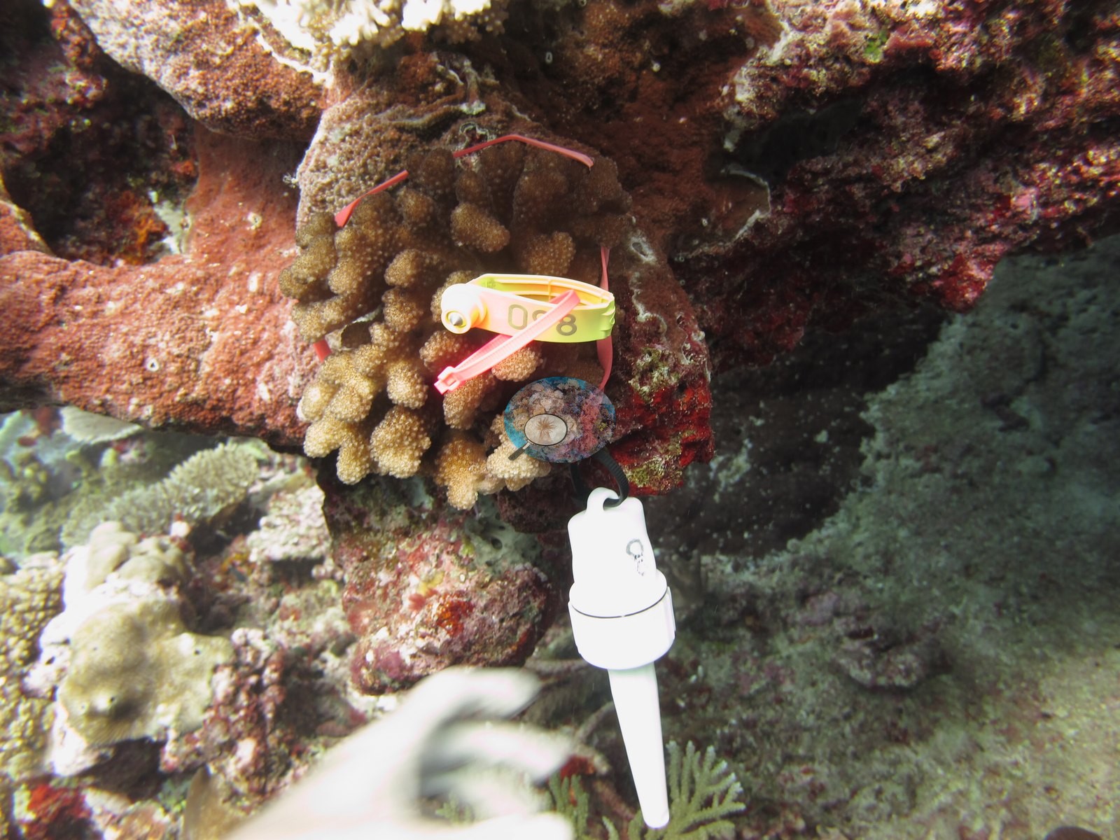
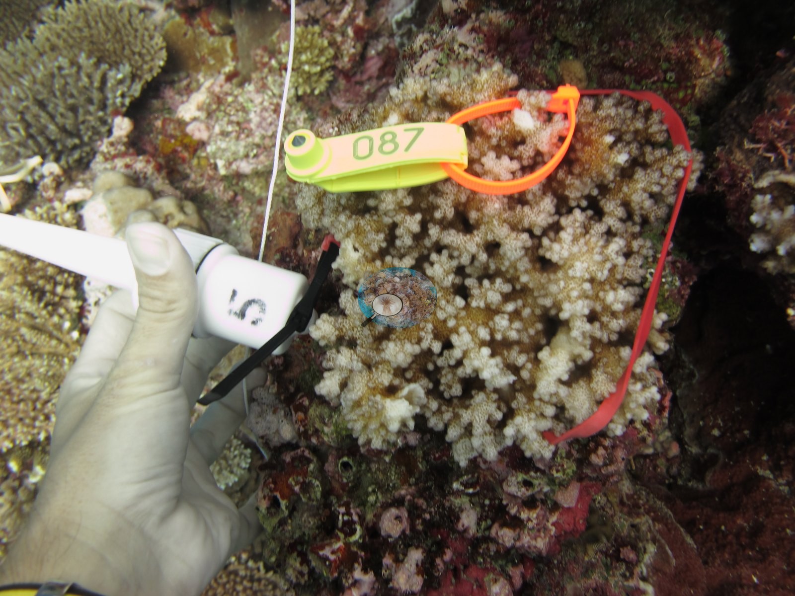
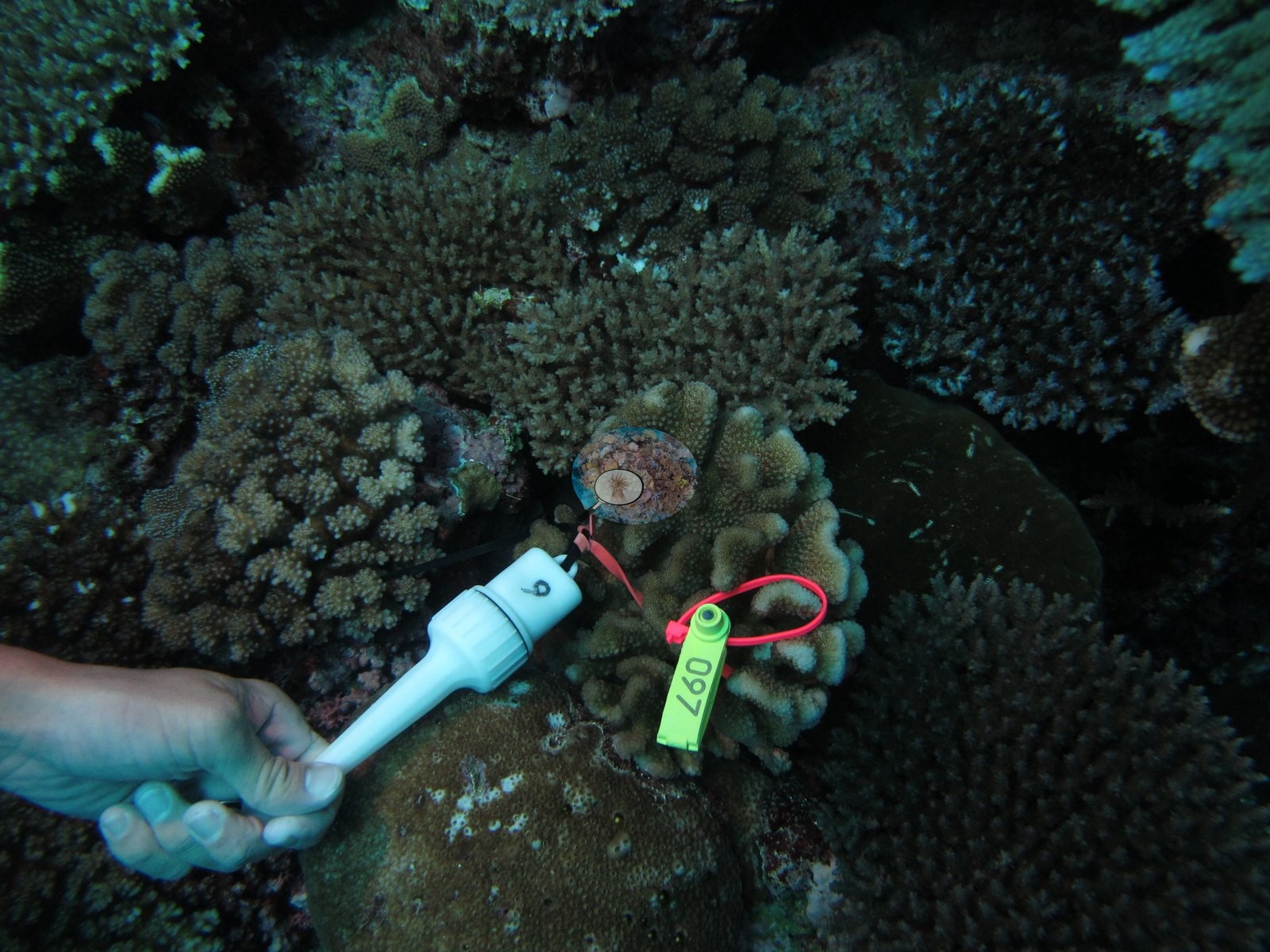
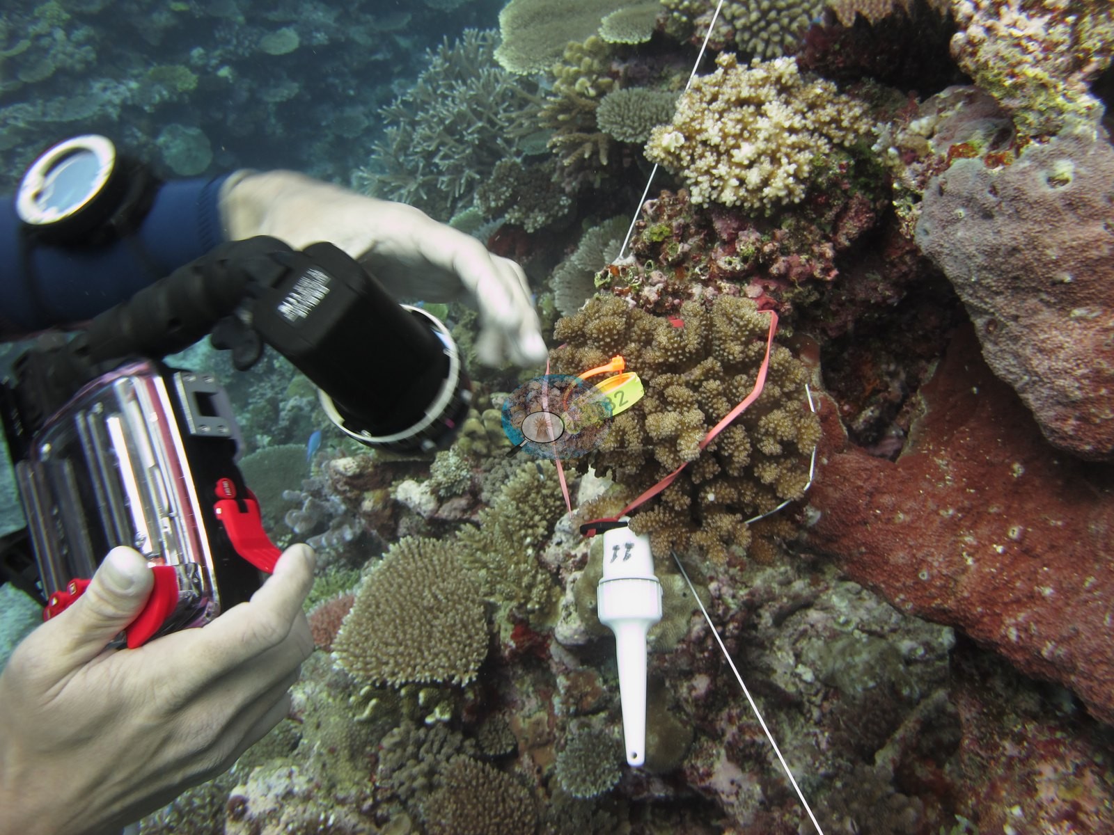
Anderson's notes record that these colonies were not collected — they were tagged and measured in place, not sampled.
CV model comparison
More info
The photo row below is a methods comparison only: it is never averaged with the transect and never used as the headline. It compares site means only: the photos come from the same survey dive as the transect but not from the same points.| Method | Hard coral cover (%) | Photos (n) ± SE |
|---|---|---|
| Transect mean (all transects) | 49.8 | |
| CoralSCOP edited | 41.4* | n = 18 · SE 3.6 |
| Claude (edited photos) | 41.0* | n = 18 · SE 4.4 |
The two computer-vision methods agree within 0.4 pp (41.0–41.4%) and read 8.4–8.8 pp below the in-water transect (49.8%); the headline is the in-water transect because a diver transect is independent of the photographs.
*Based on only 18 reef-habitat photos; this estimate is less certain.
See a more detailed, stand-alone version of this table.
Data & links
- Download benthic survey data (CSV) ↗
- Download all LOF-GRE data (933 sites) ↗
- Photo portfolio ↗
- Coral Reef Diagnostics page for this area ↗
All images from Coral Reef Diagnostics. Data are from the Khaled bin Sultan Living Oceans Foundation Global Reef Expedition and Coral Reef Diagnostics.
 © Coral Reef Diagnostics.