
Part of the regional summaries: Chagos Archipelago - Great Chagos Bank West / Brothers / Eagle and Chagos Archipelago
More info
Chagos (NOAA CRW virtual station)BIDI13, at Danger Island, is another algae-dominated reef (7.8% coral cover) with dense fleshy macroalgal beds over rubble, though scattered coral patches host a camouflaged grouper, a Pocillopora-type colony, and one of the best fish photographs of the expedition - an oriental sweetlips sheltering under a ledge. Three frames also recorded jellyfish, unusual for this reef series, and a lesioned massive coral was the site's one condition flag. Surveyed on the Living Oceans Foundation's Global Reef Expedition.
One other CRD dive site within 5 km shown in orange — click a marker to open its page.
🗺 Detailed map of the Global Reef Expedition dive sites, Danger Island, Chagos (Khaled bin Sultan Living Oceans Foundation; opens in a new window)
Site description
General benthic features
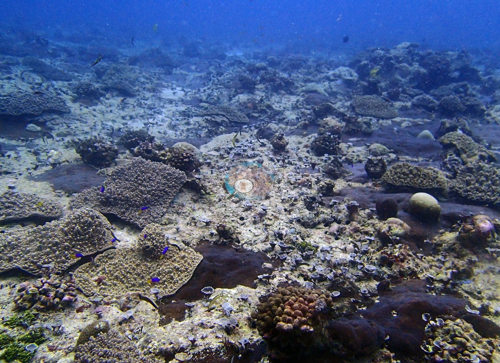
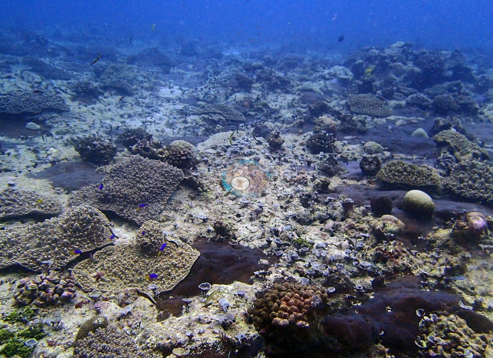
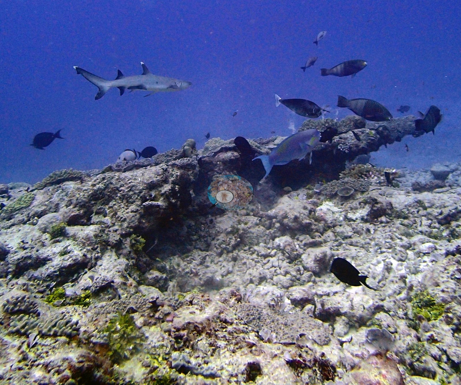
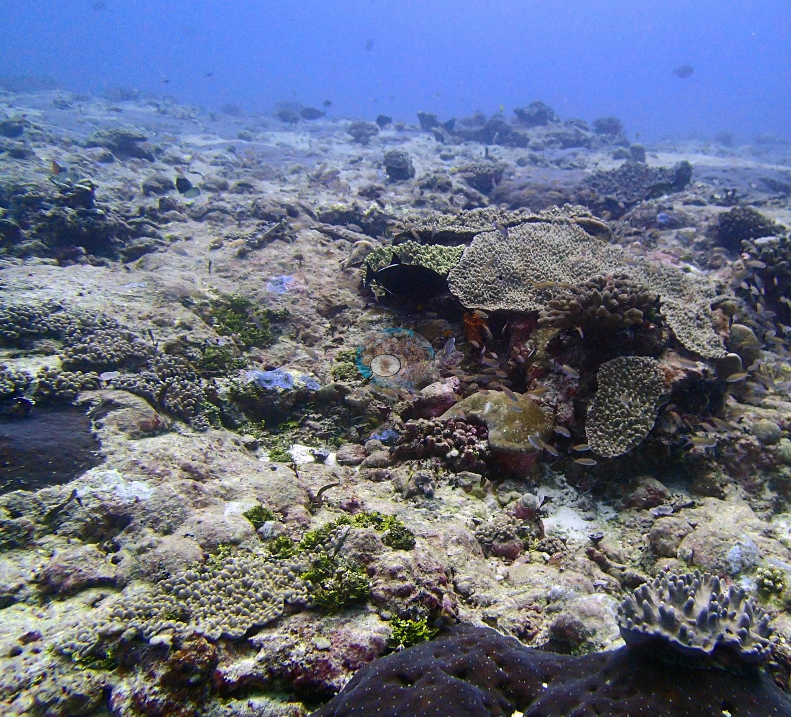
The bottom in these frames is low-relief pavement and rubble rather than coral framework. P3130347, P3130368 and P3130382 are the three cleanest wide views and all show the same thing: an expanse of pale, eroded consolidated carbonate and coarse rubble crusted with turf and pink and mauve coralline algae, carrying small, well-spaced colonies – pale tabular and foliose plates, low tan knobby and encrusting heads, a dark blue-gray massive at the bottom edge of P3130368 and another in P3130382, and scattered clumps of green macroalgae. P3130347 is dominated by a broad flat dark-brown mass low in the frame whose surface is entirely given over to turf, reading as old framework rather than living tissue. Nothing in these frames stands more than a colony's height above the substrate.
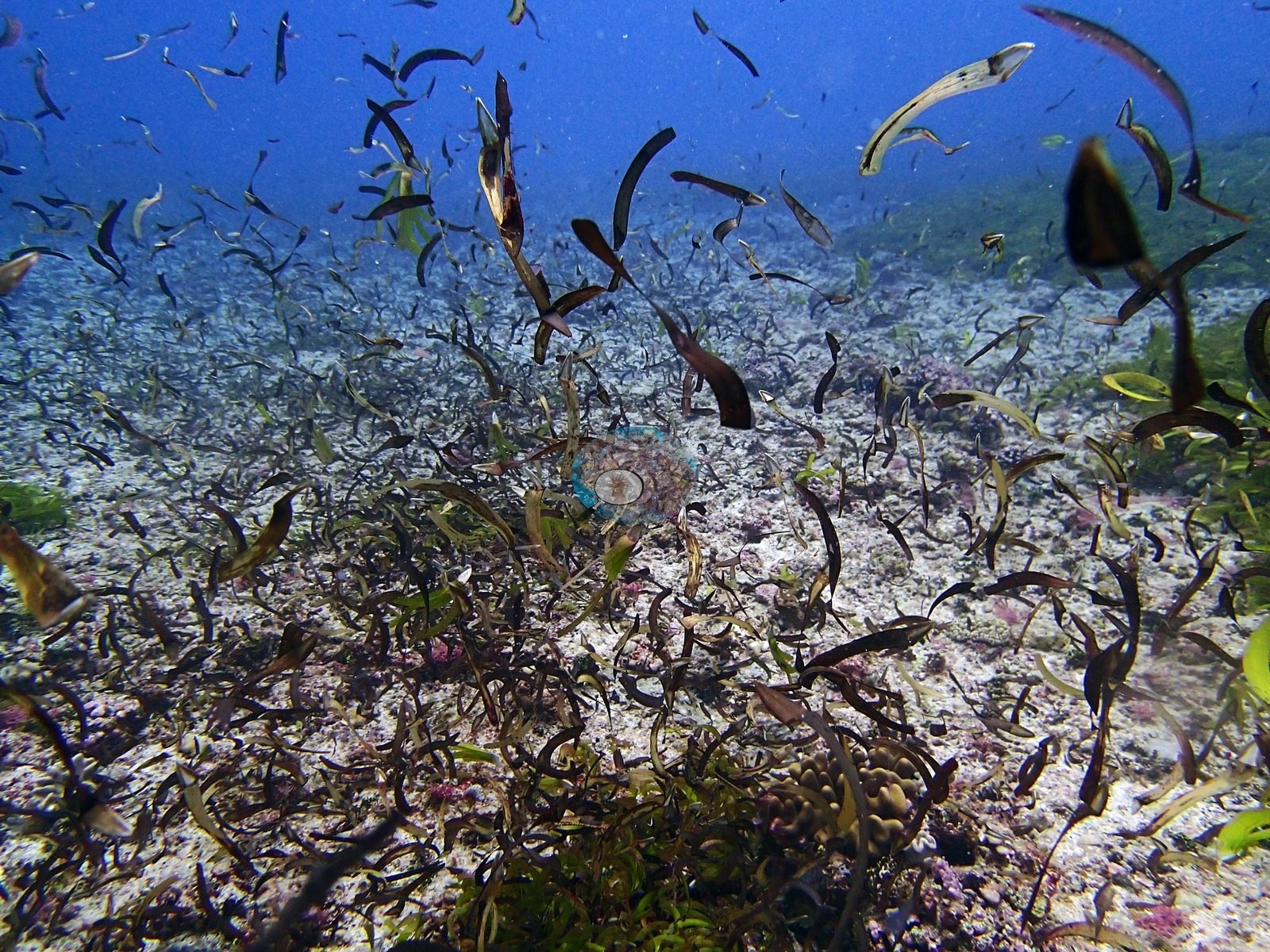
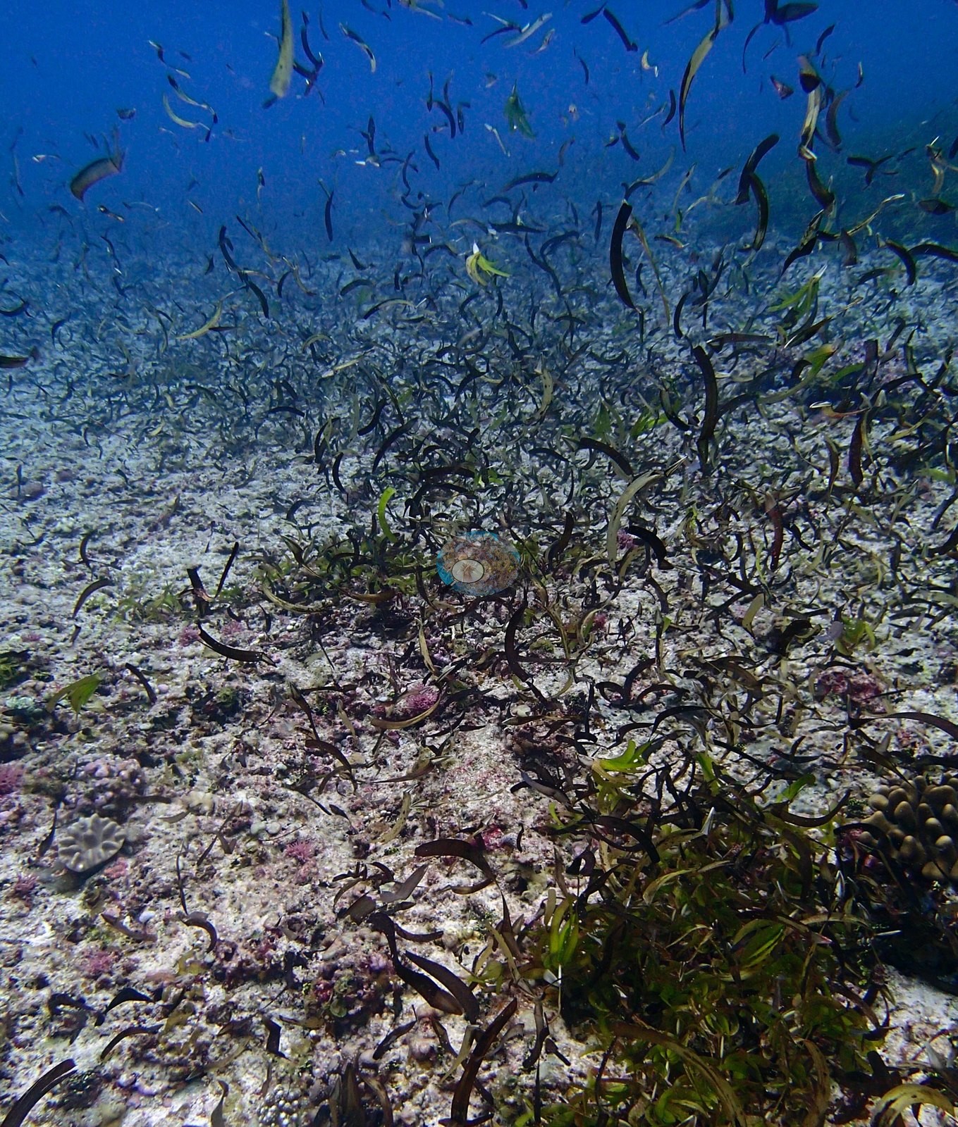
Two frames record a dense brown fleshy macroalgal bed. P3130388 and P3130389 show olive-to-dark-brown strap- and blade-like algae covering the seafloor over pale rubble, with only sparse coralline patches showing through and a single small tan coral head at the base of P3130389. This is a different alga from the bright yellow-green Caulerpa-type meadow photographed a day earlier at neighboring BIDI12 – it reads as a brown alga, compatible with Sargassum, Turbinaria the alga, or Lobophora, and cannot be taken further from these images. The ecological signal is worth Anderson's attention as a regional observation rather than a site quirk: two adjacent sites on this stretch of the Great Chagos Bank western rim, surveyed a day apart, both carry conspicuous fleshy macroalgae on a remote, unfished, unpolluted archipelago. That is a statement about what the frames show; the benthic composition of the site is the transect's to state, and the transect puts algae at 62% of the benthos.
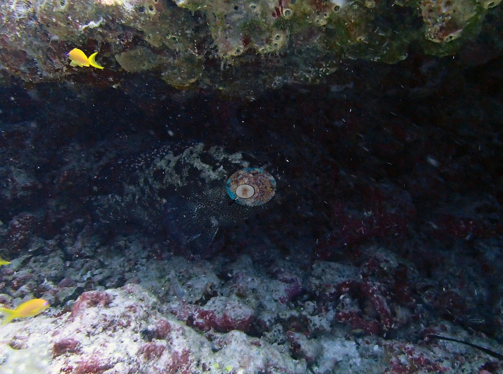
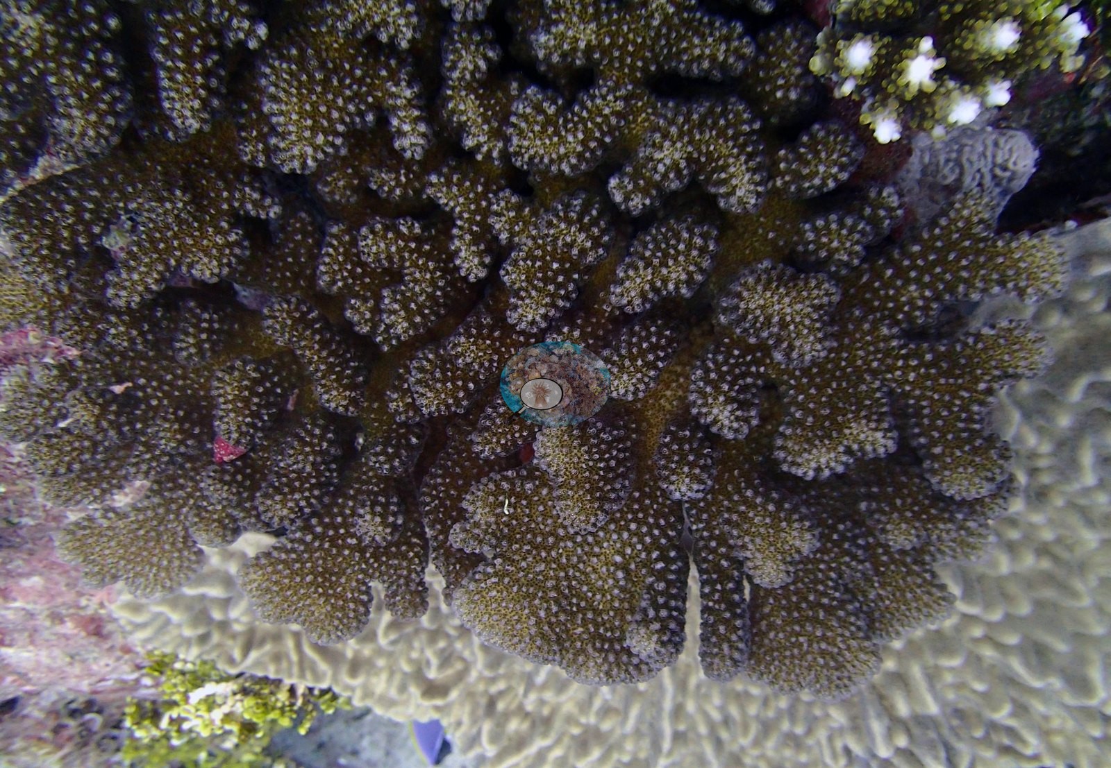
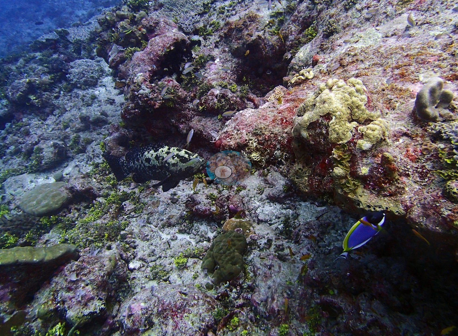
Where coral persists, the close frames are the informative ones. P3130357 packs the most into one field of view: a dark, camouflaged grouper compatible with Epinephelus wedged in a hollow, a powder-blue surgeonfish (Acanthurus leucosternon), yellow anthias, ruffled cream-yellow foliose colonies compatible with Montipora or Turbinaria, brown massive Porites and small olive-brown massives, and extensive red and pink crustose coralline algae. P3130367 is a clean close view of a brown, densely verrucose branching colony – Pocillopora or Stylophora, not separable here – set against a gray-white massive whose fine, even corallites are compatible with Porites.
Coral cover
The LOF benthic transect is this site's measurement. Coral cover at BIDI13 (Danger Island, Great Chagos Bank west, dived 13 March 2015) is 7.8% live hard coral, from the LOF BIOT-1 in situ transects (Mayfield & Dempsey 2026, Oceans, suppl. S1), against algal cover of 62% of the benthos. That diver-transect survey is the authoritative record of cover and community here, and the two figures together are the site in one line: this is an algal-dominated stretch of reef with coral persisting in patches. The computer-vision products and the frame-by-frame account below supplement the transect. No cover value may be read off a photograph, no image-derived product overrides the transect, and nothing in the imagery qualifies or revises it.
Method comparison – transects, points and masks. All 16 frames were paired between the two image products. Across methods the same reef reads transects 7.8% · CoralNet points 13.8% · Coralscapes masks 12.3% (16 frames). This site is the useful control for the multimethod comparison, and it is worth stating why. Here both photo methods read above the transect (+6.0 and +4.5 pp) but agree with each other to within 1.5 pp – which is precisely the archipelago-wide pattern documented in CHAGOS_MULTIMETHOD_README.md, where the photo-versus-transect gap is positive at low cover and strongly negative above 40%. The mechanism is straightforward: a photographer frames on coral, so at a coral-poor site the frames over-represent the colonies that are there, while a transect samples the pavement and rubble between them at full weight. BIDI12 is not – there the two photo methods split by nearly 18 pp (19.5% versus 1.7%), which no cover-dependent bias explains. So the BIDI12 flag should rest on the photo-method disagreement, not on CoralNet merely reading high: reading high at low cover is expected, and BIDI13 shows what that looks like when nothing is wrong. None of this touches the transect value, which stands at 7.8% regardless of how the image products behave. Here both photo methods read above the transect (+4.5 and +6.0 pp) but agree with each other to within 1.5 pp — which is precisely the archipelago-wide pattern documented in CHAGOS_MULTIMETHOD_README.md, where the photo-vs-transect gap is positive at low cover and strongly negative above 40%. BIDI13 is that pattern behaving normally. BIDI12 is not: there the two photo methods split by nearly 18 pp (19.5% vs 1.7%), which no cover-dependent bias explains. So the BIDI12 flag should rest on the photo-method disagreement, not on CoralNet merely reading high — reading high at low cover is expected, and BIDI13 shows what that looks like when nothing is wrong. Coral cover: 7.8%. Live hard coral measured on the in situ benthic transects of the LOF BIOT-1 expedition (Mayfield & Dempsey 2026, Oceans, supplemental S1), against algal cover of 62% of the benthos. The transect figure is this site's authoritative cover value; no image-derived product overrides it. Across methods the same reef reads: transects 7.8% · CoralNet points 13.8% · Coralscapes masks 12.3% (16 frames). These products have different sampling footprints and denominators and should not be averaged.
Stressors
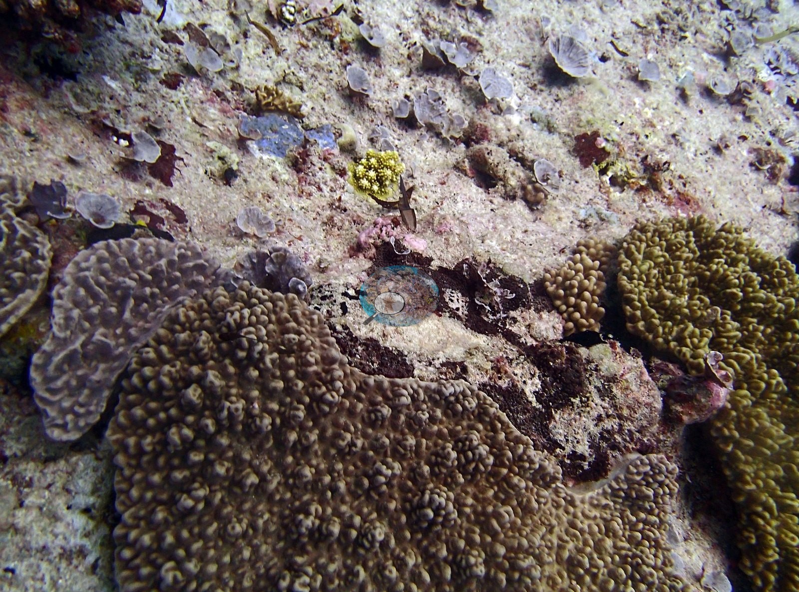
BIDI13 is that pattern behaving normally. P3130381 is the condition frame: two large brown knobby massive colonies sit either side of a distinct dark brown lesioned or dead patch, surrounded by white coralline-crusted substrate, with a single small yellow branching recruit compatible with Acropora settled nearby – loss and recruitment inside one field of view. The lesion is a real photographic record but cannot be diagnosed from an image.
Condition. No unambiguously bleached colony appears in any of the sixteen frames. The corals that are present read as normally pigmented brown, tan, olive, and cream. The one ambiguous feature is a small cluster of bright white branch tips at the upper right of P3130367, which at this magnification cannot be separated from ordinary bright growth tips; it is reported as seen and diagnosed no further. No bleaching percentage is read off this set and none is tested against it. Anderson's published per-site percentage bleached is the authoritative figure for this reef; the photographs record only that whitening was not conspicuous in the frames taken on this dive. The lesioned patch in P3130381 is mortality of some kind, not bleaching, and belongs alongside the other lesioned massives noted in the Danger Island group. No crown-of-thorns starfish (COTS) or other corallivores evident.
Fish and other fauna
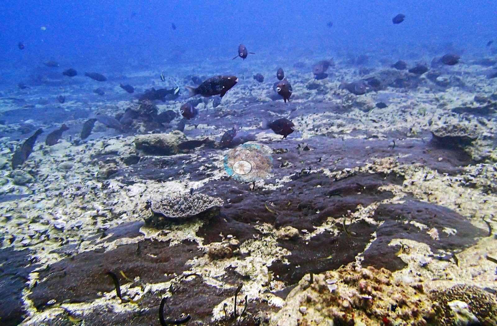
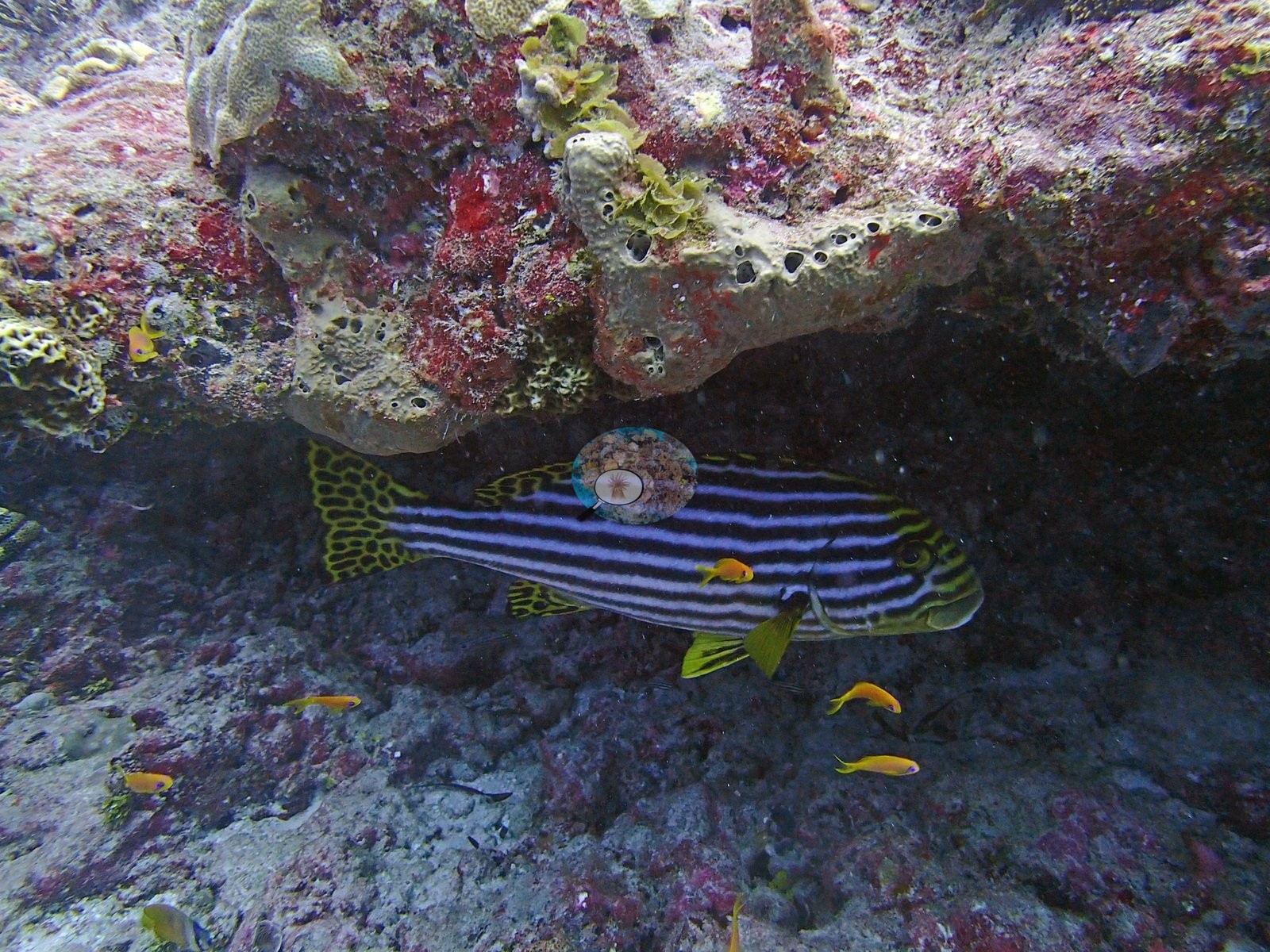
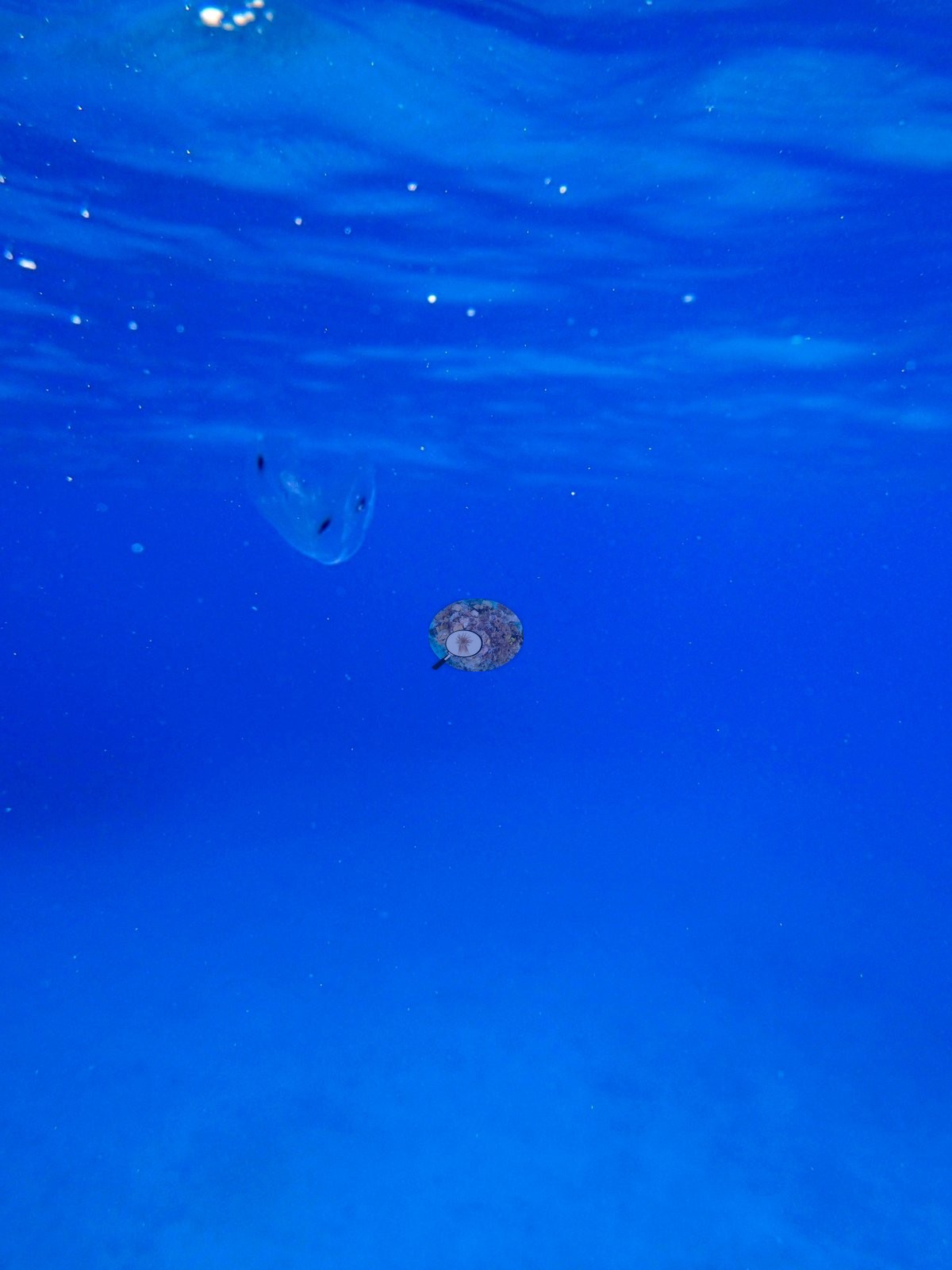
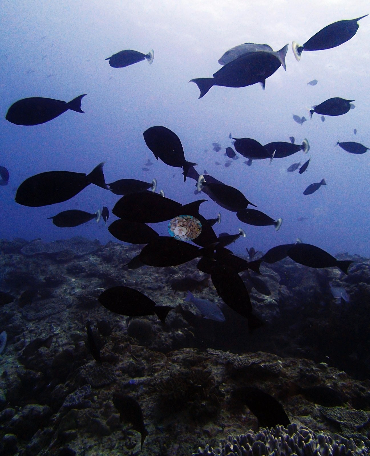
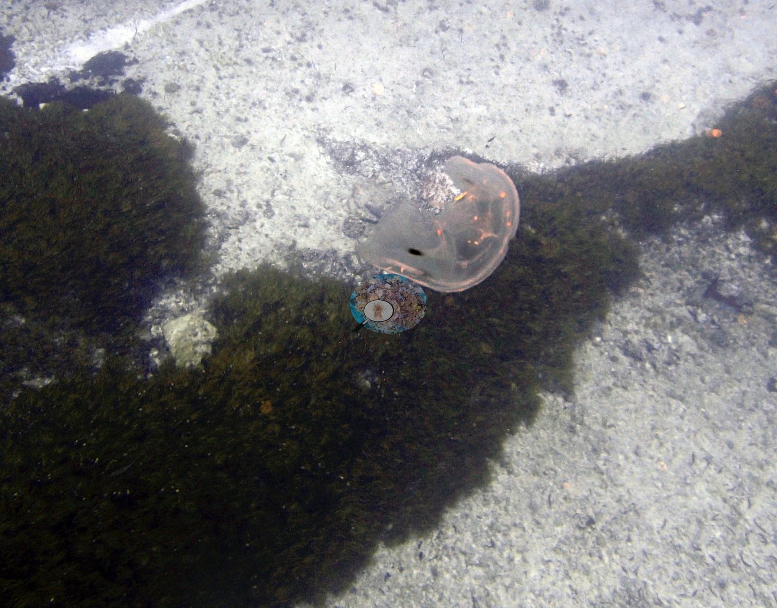
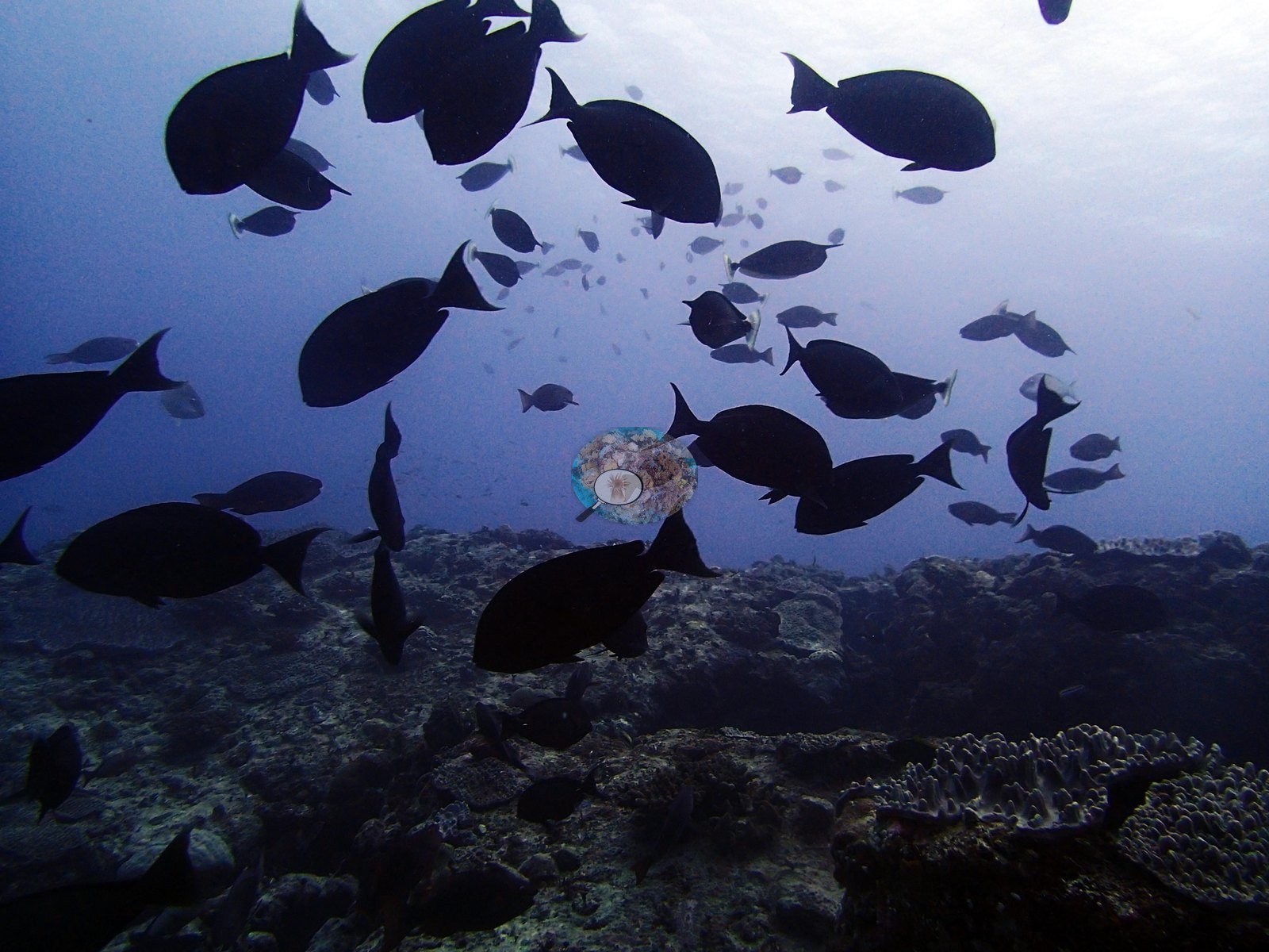
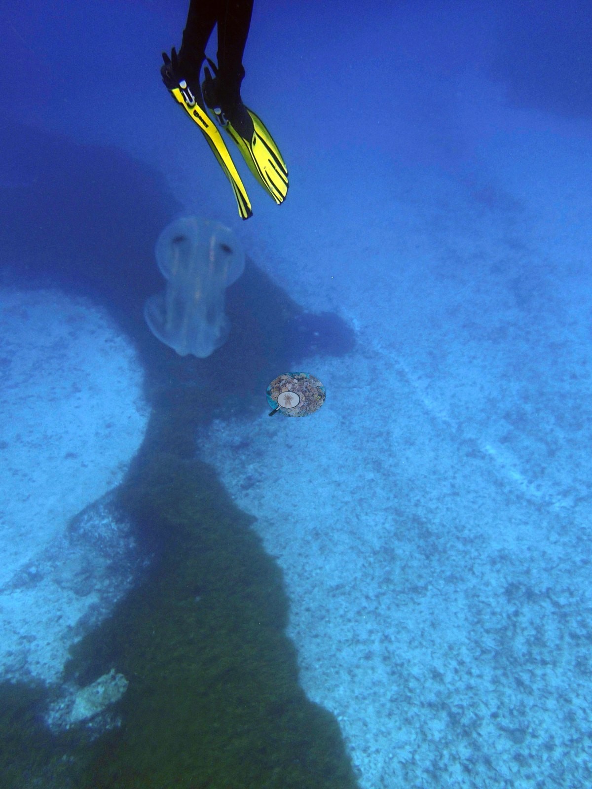
Fauna – a grazer-dominated dive, which fits an algal-rich site. Dense schools of surgeonfishes and unicornfishes compatible with Acanthurus, Ctenochaetus and Naso fill P3130347, P3130354 and P3130356, mostly as silhouettes against the surface, and parrotfishes compatible with Scarus or Chlorurus work the pavement in P3130347 and P3130355. A whitetip reef shark (Triaenodon obesus) cruises low over the rubble ridge in P3130355 – the second whitetip record after BIDI08. P3130358 is the best fish photograph of the expedition to this point: an oriental sweetlips, Plectorhinchus vittatus, sheltering under a coralline- and sponge-crusted ledge, white with crisp black longitudinal stripes and yellow, black-spotted fins – an unambiguous species-level identification, and one of the few frames in the batch that supports one. P3130359 is a deeply shadowed undercut with a single small yellow fish. Most unusually, three frames record jellyfish: a small translucent medusa resting against algal turf on pale sand in P3130390, a large bell with trailing oral arms drifting past a diver's yellow fins in P3130391, and a third in open blue water near the surface in P3130393 – the only jellyfish in the Chagos frames reviewed to date.
Comparisons with other data
Also on record for this site: Assemblage composition. The cover line carries the key numbers, and this site is the useful control for the BIDI12 flag.
Other notes
Image-by-image. Sixteen habitat frames survive from the dive, P3130347 to P3130393, all from Anderson's own folder BiDi13-finished, which carries no condition annotation. There are no sampled-colony frames. Visibility and natural color are better than most of the Chagos batch, but the effective benthic sample is smaller than 16: three frames (P3130390, P3130391, P3130393) are jellyfish and water-column subjects, two (P3130354, P3130356) are fish silhouettes against the surface with the bottom barely lit, and P3130359 is a deeply shadowed undercut with essentially no benthic information. The genuinely informative benthic frames are P3130347, P3130355, P3130357, P3130367, P3130368, P3130381, P3130382, P3130388, and P3130389. No quadrat, transect line or scale object appears anywhere.
CV model comparison
More info
Those are point counts on the photographs; this is a diver swimming a transect. The models and the reference never saw the same thing. ⚠ SITE-LEVEL ONLY. The 2,500 frames were not associated with any transect taken within 200 m of them, so transect-vs-image comparisons are site means, never frames. The transect value is already in the pin `cover` field (109 of 112 pins, sourced to the paper's suppl. S1) and is read from there, never re-imported. The transect is THE coral-cover value for this site and stands alone. The rows below it are a methods comparison only: no computer-vision value is averaged with it, feeds the headline, or carries over to the site's cover figure (Anderson, 2026-09-22).| Method | Hard-coral cover (%) |
|---|---|
| Transect (published) | 7.8 |
| CoralNet | 20.5 |
| In-house DINOv3 | 18.2 |
| CoralSCOP edited | 24.9 |
| Claude | 17.8 |
Data & links
- Read the full site report ↗
- Download benthic survey data (CSV) ↗
- Download all LOF-GRE data (933 sites) ↗
- CoralNet image repository for this region ↗
- Photo portfolio ↗
- Coral Reef Diagnostics page for this area ↗
All images from Coral Reef Diagnostics. Data are from Coral Reef Diagnostics.
 © Coral Reef Diagnostics.