
Part of the regional summaries: Chagos Archipelago - Victory Bank / Great Chagos Bank North and Chagos Archipelago
More info
Chagos Archipelago, UK (NOAA CRW virtual station)An Acropora-dominated bank reef on the northern Great Chagos Bank caught in an active crown-of-thorns outbreak, with at least eleven starfish in five frames, including about three on freshly killed white skeleton. Coral cover is 49%, halving from about 53% in the shallows to 24% at depth. Surveyed on the Living Oceans Foundation's Global Reef Expedition.
CTD-cast midpoint; the cast read 29.8°C throughout
🗺 Detailed map of the Global Reef Expedition dive sites, Nelson Island, Chagos (Khaled bin Sultan Living Oceans Foundation; opens in a new window)
6 of the 22 photographs on this page are shown beside the text that discusses them under "Site description" below.
Site description
Setting
BINI80 is a protected submerged bank reef on the north of the Great Chagos Bank, filed to Nelson Island, dived twice on 21 April 2015.
General benthic features
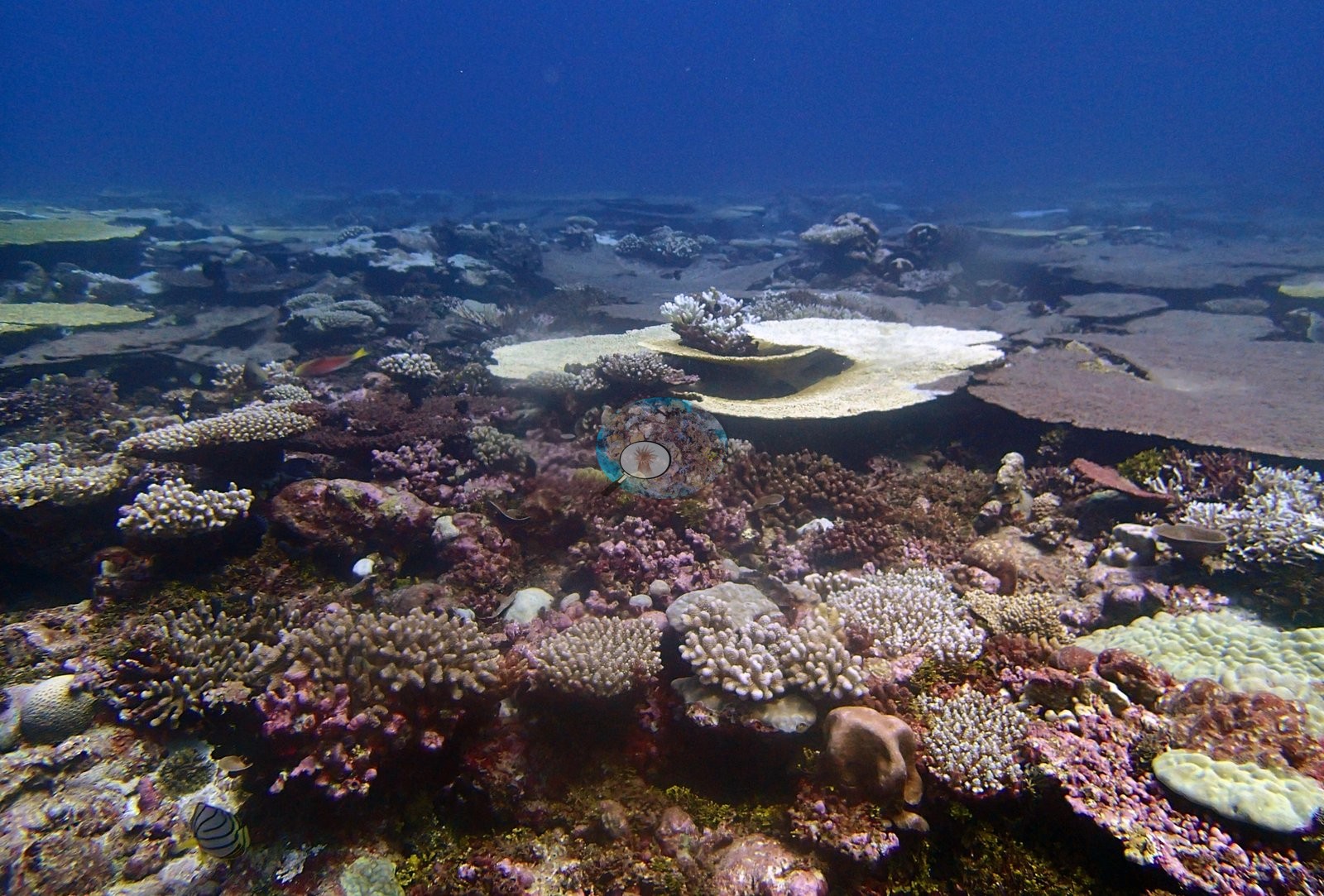
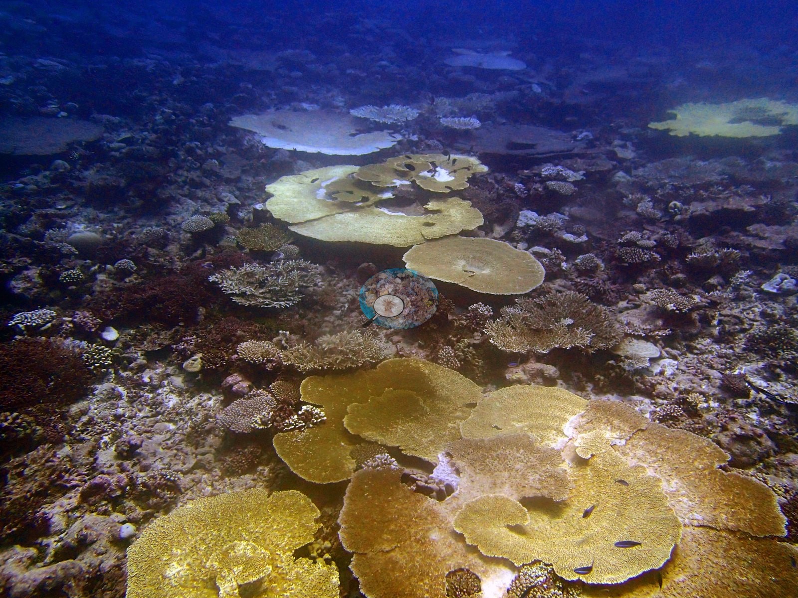
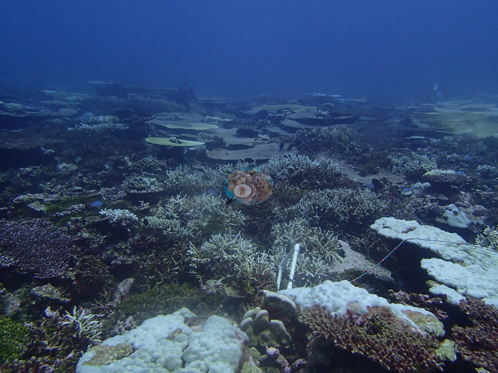
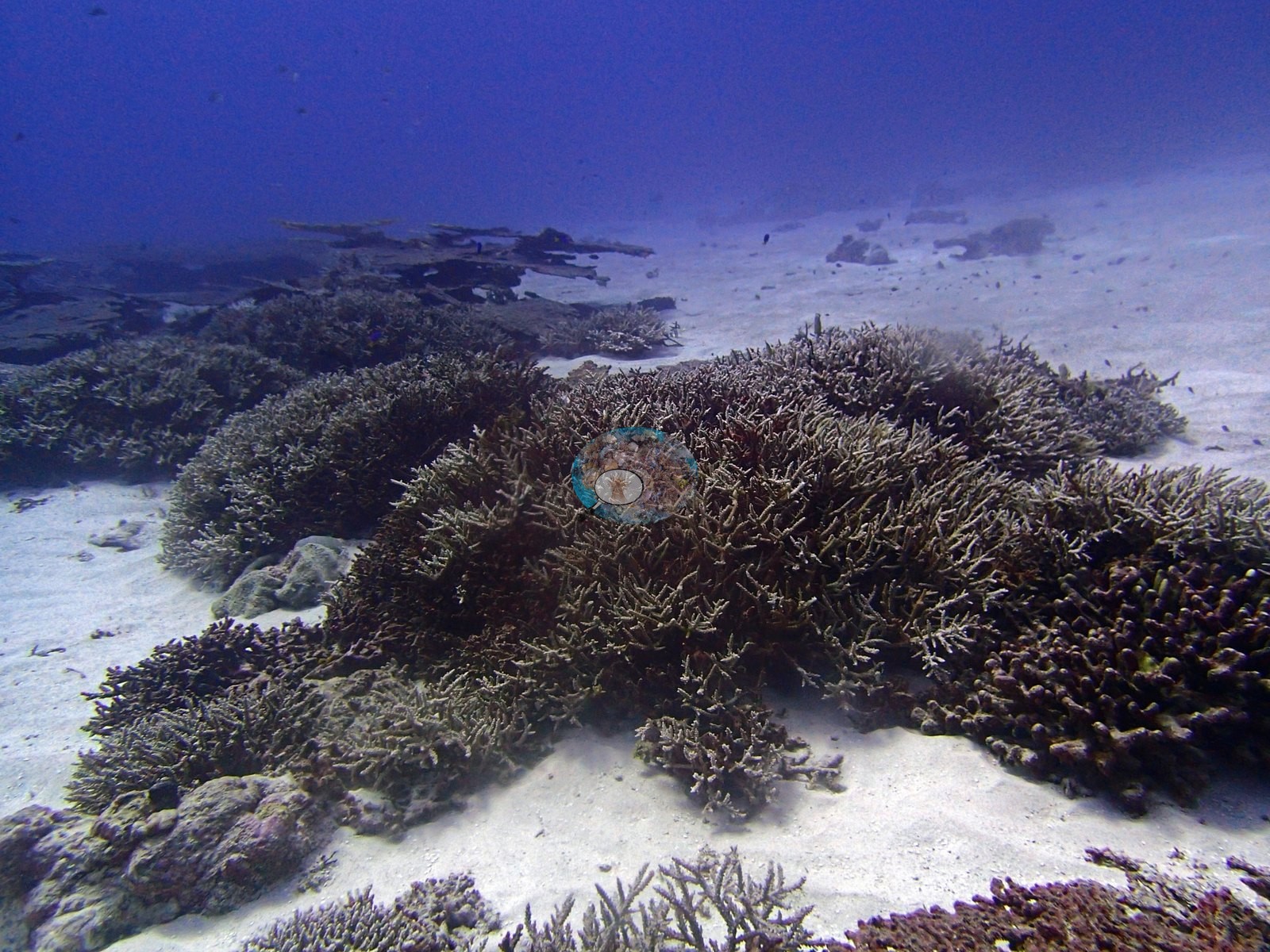
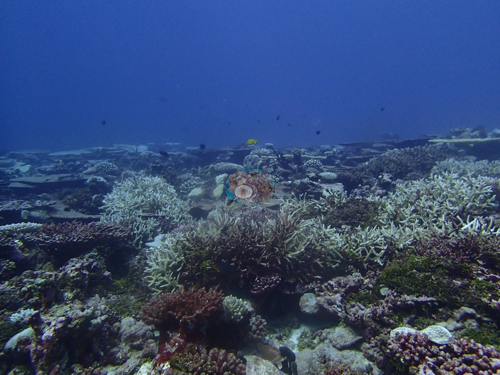
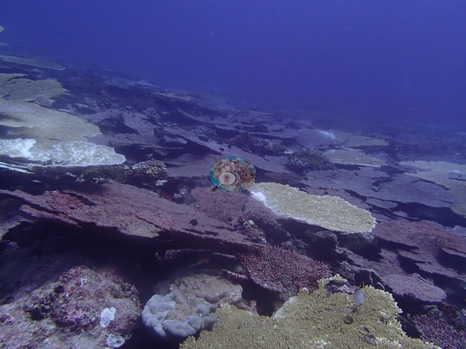
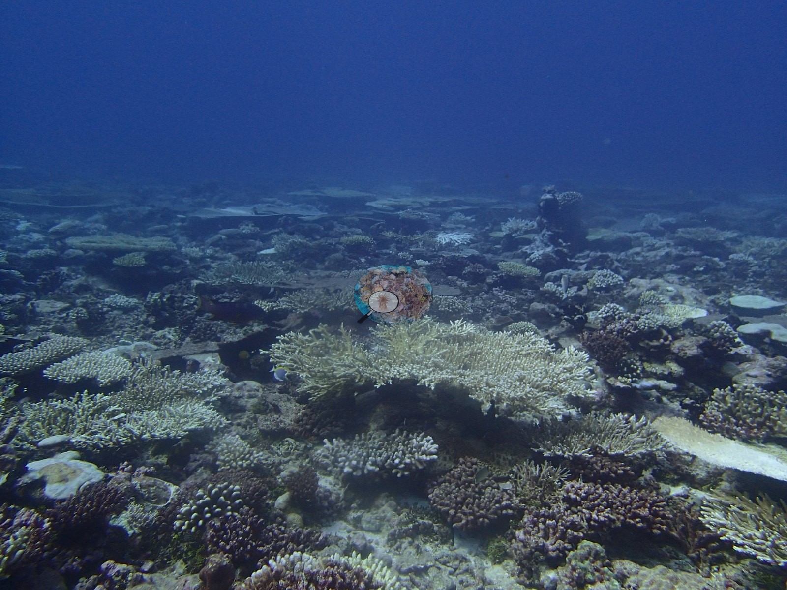
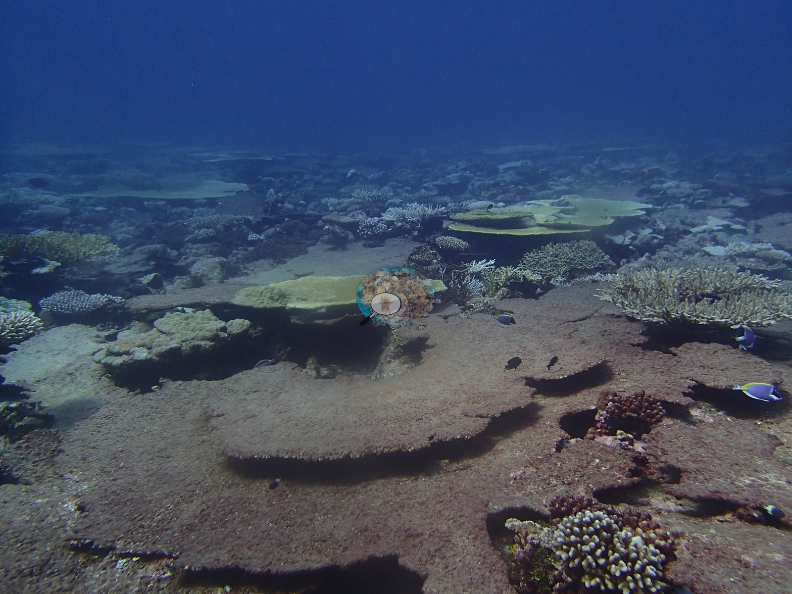
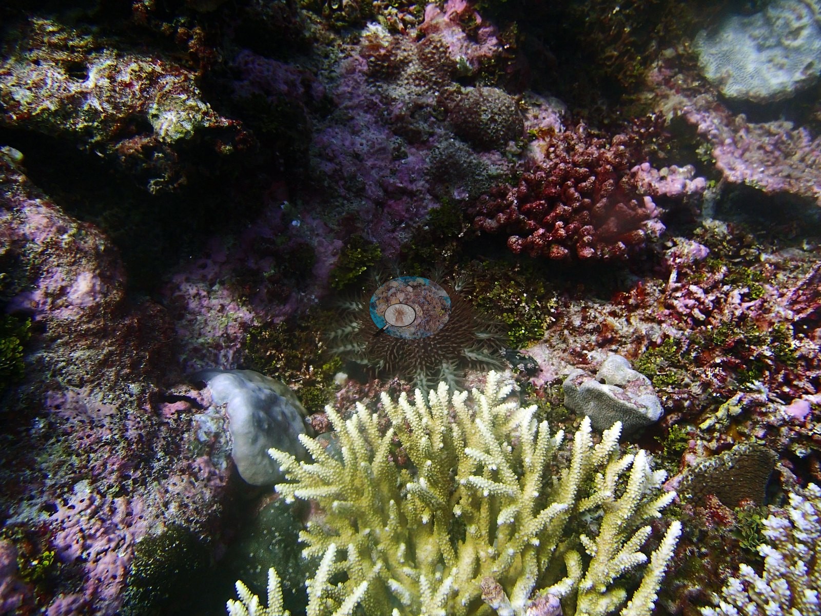
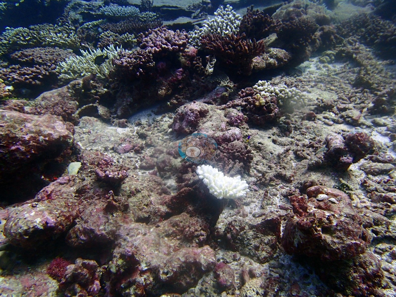
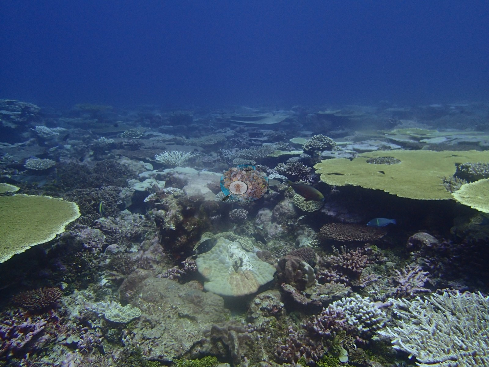
The assemblage is overwhelmingly monogeneric: across 20 genera, Acropora alone is 38% of the benthos, 77% of every coral on the site (e.g., Image 2, Image 3).
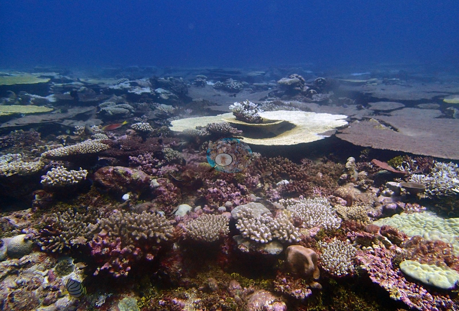
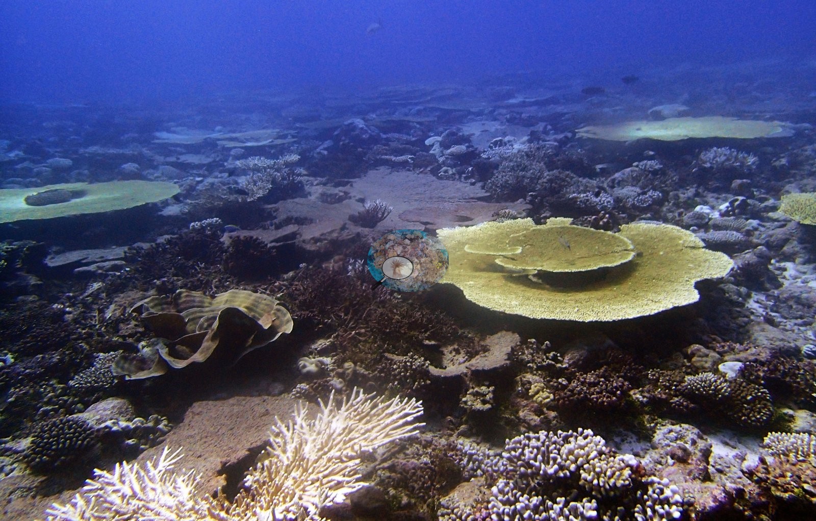
There is no genus handover with depth — Acropora leads every station, 79%, 77%, and 63% of transect coral — while absolute cover halves, from 53% shallow to 24% deep.
Coral cover
- Reference: Mayfield & Dempsey (2026)
- CoralNet submission package
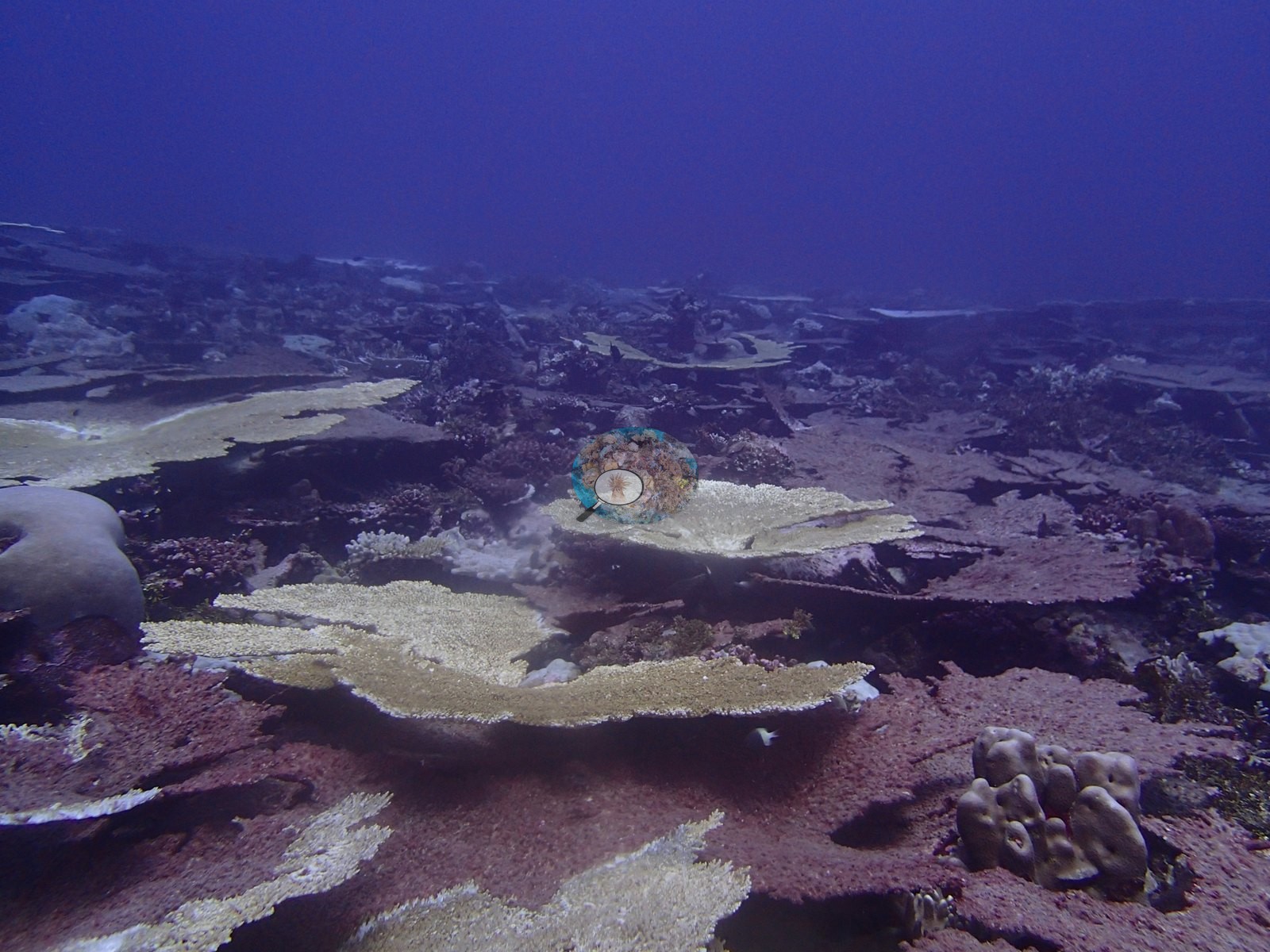
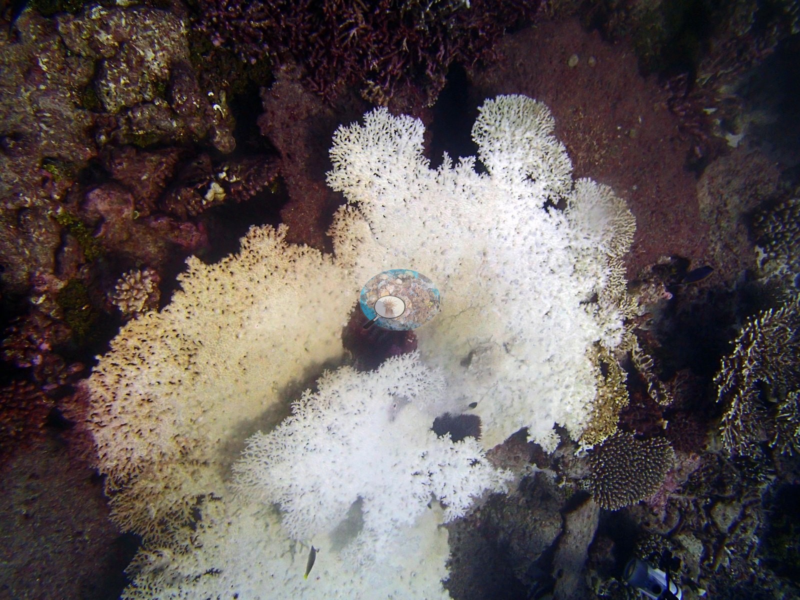
- Formal transects recorded 49% coral against 43% algae, with fleshy turf at 23% of the benthos, only 7.7% coralline algae and 5.8% bare substrate (e.g., Image 1).
- Transect mean 49 ± 5.6% SE (n = 18 transects; Living Oceans Foundation per-transect benthic data); the headline above is this all-transect mean, and the published station value, which weights each depth equally, is 43% (Mayfield & Dempsey 2026, Oceans, supplemental S1).
- Image-based estimates (CoralNet on 14 photographs, Claude, DINOv3, CoralSCOP) are listed in the method table for comparison only; the transect figure is this site's cover value.
- Live hard coral measured on the in situ benthic transects of the LOF BIOT-1 expedition (the mean of all of this station's transects), against total algal cover (all algal groups) of 43% of the benthos.
- The transect figure is this site's authoritative cover value; no image-derived product overrides it.
Stressors
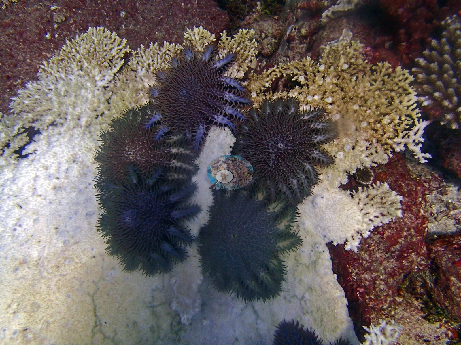
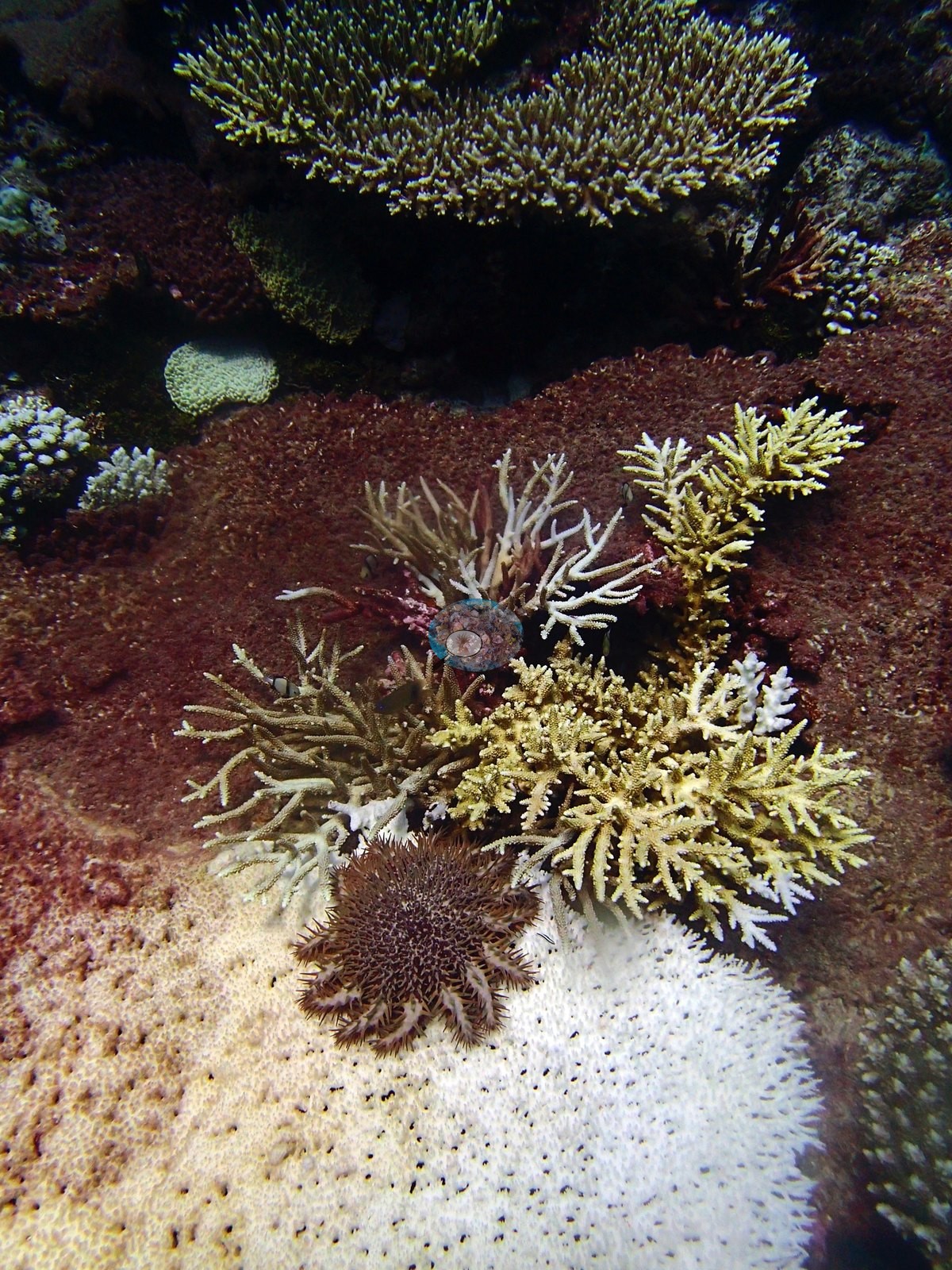
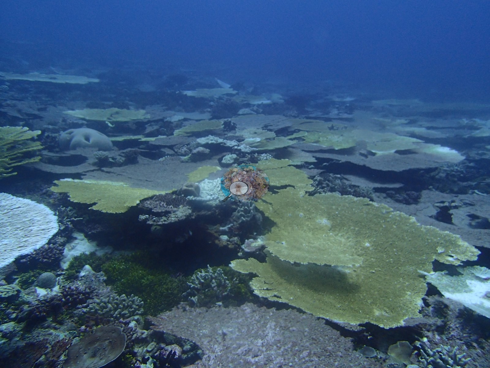
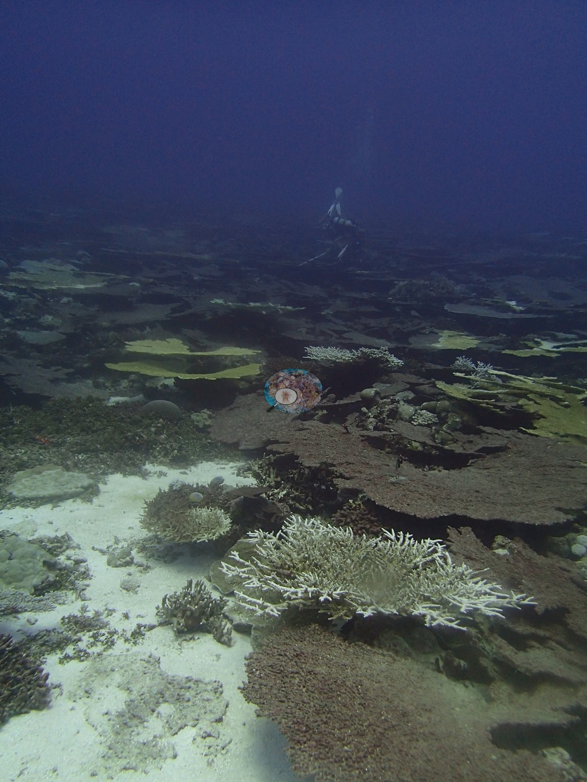
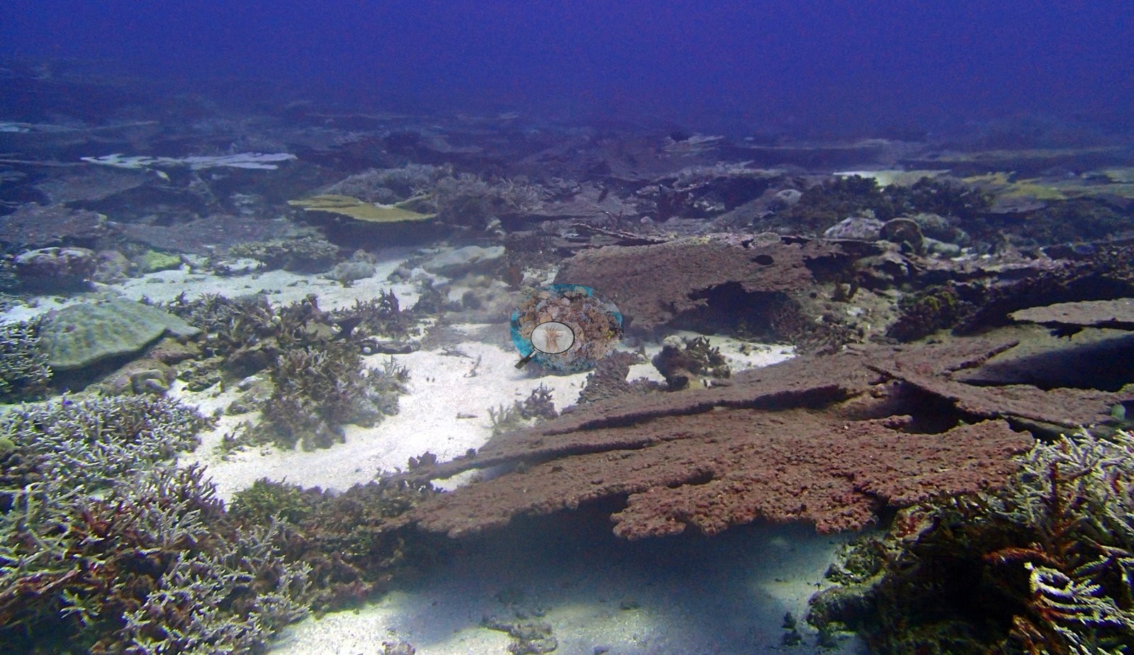
Crown-of-thorns predation is the finding, and it is unambiguous: at least eleven sightings across five frames in one day, including about three animals on freshly exposed white table skeleton and one starfish sitting exactly on the boundary between living plate tissue and fresh kill, on a reef where their preferred prey, Acropora, makes up three-quarters of the coral (e.g., Image 4).
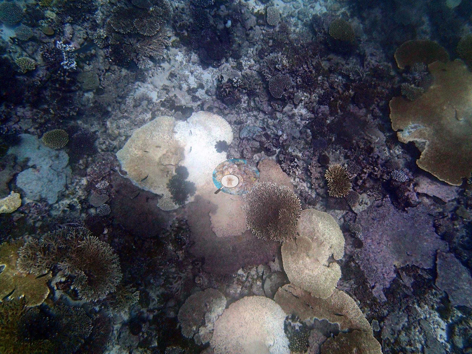
No bleaching figure was published for this site, and none is estimated here, since white Acropora on a reef with active feeding fronts cannot be told from bleached tissue in a single still. The expedition's field report describes an active crown-of-thorns outbreak at this station (NI80): 194 starfish were counted within a 10 × 50 m area, concentrated around the large table corals, and about 30% of the tables showed recent crown-of-thorns feeding, 10% were dead and 10% had white syndrome (Bruckner 2015, LOF Global Reef Expedition BIOT Field Report, p. 15). Anderson's sampling sheet records four colonies sampled here: Seriatopora hystrix ChSe96 (28.9 m, 50% bleached (outer branches)), ChSe97 (26.8 m, >90% bleached), ChSe98 (30.2 m, >90% bleached, likely unhealthy), and ChSe99 (25.0 m, >90% bleached). It also records four other pocilloporids sampled for a collaborator (species by field call): Pocillopora sp. ChJo205 (15.6 m, 20% bleached), ChJo207 (13.0 m, 25% bleached), and ChJo208 (13.0 m, large, healthy, unscathed in a crown-of-thorns-damaged area); Pocillopora verrucosa ChJo206 (13.8 m, 70% bleached). Pocillopora acuta colonies from this archipelago were analyzed in Thomas et al. (2026, Global Ecology and Biogeography 35:e70305, https://doi.org/10.1111/geb.70305). The Living Oceans Foundation transects here recorded no cyanobacteria and 0.2% recently dead coral.
Fish and other fauna
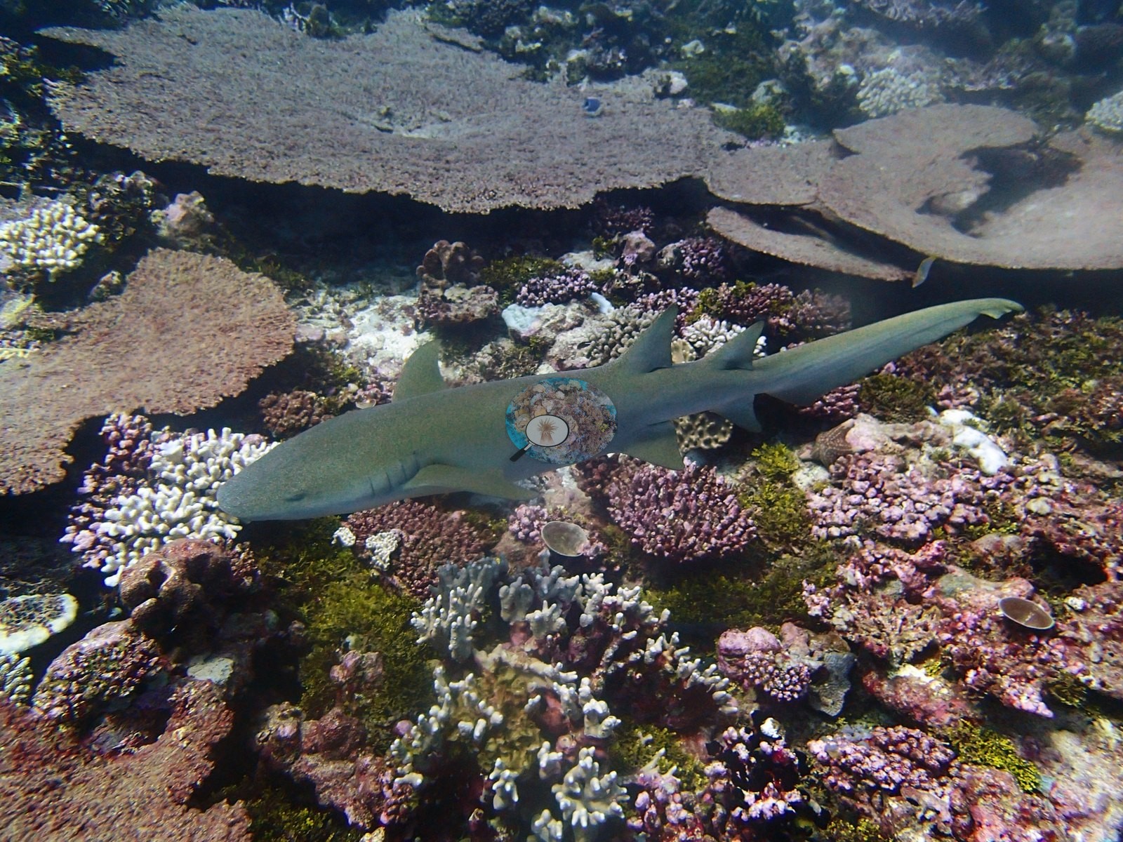
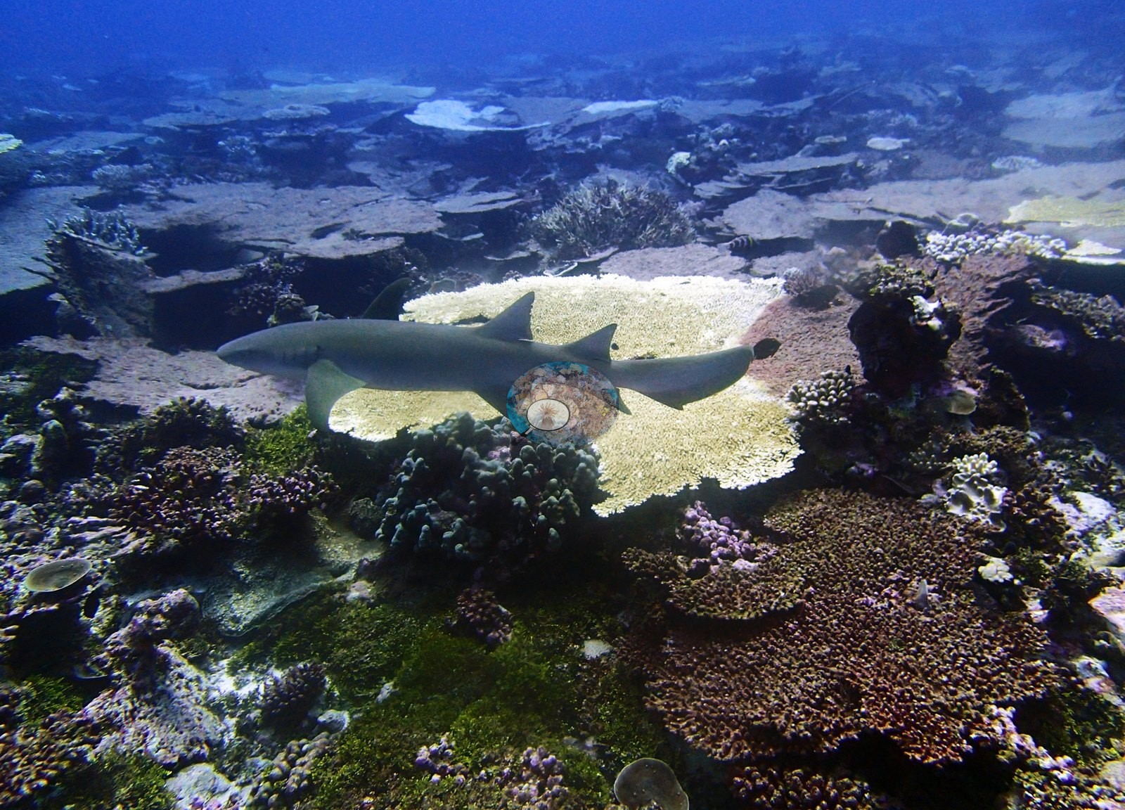
One large shark rests among the tables on the first dive (e.g., Image 15, Image 16): a stout body with two similar-sized dorsal fins set far back and no white fin tips, the shape of a nurse shark; no shark appears in the second dive's frames.
Comparisons with other data
Apart from the Living Oceans Foundation survey shown here, we found no other surveys at this site as of 2026. If you would like to share yours, please email us.
CV model comparison
More info
Those are point counts on the photographs; this is a diver swimming a transect. The models and the reference never saw the same thing. ⚠ SITE-LEVEL ONLY. None of the Chagos photographs (2,500 frames across all stations) can be matched to a transect taken within 200 m, so transect-vs-image comparisons use site means, never individual frames. The transect value is the mean of all of this station's own transects in the expedition's per-transect benthic data. The transect is THE coral-cover value for this site and stands alone. The rows below it are a methods comparison only: no computer-vision value is averaged with it, feeds the headline, or carries over to the site's cover figure.| Method | Hard coral cover (%) | Photos (n) ± SE |
|---|---|---|
| Transect mean (all transects) | 49.1 | |
| CoralNet | 26.8* | n = 14 · SE 3.3 |
| In-house DINOv3 | 36.9 | n = 24 · SE 3.1 |
| CoralSCOP edited | 34.9 | n = 24 · SE 2.5 |
| Claude | 29.4 | n = 24 · SE 3.3 |
The four computer-vision methods disagree by 10.1 pp (26.8–36.9%) and read 12.2–22.3 pp below the in-water transect (49.1%); the headline is the in-water transect because a diver transect is independent of the photographs.
*Based on only 14 reef-habitat photos; this estimate is less certain.
See a more detailed, stand-alone version of this table.
Data & links
- Download benthic survey data (CSV) ↗
- Download all LOF-GRE data (933 sites) ↗
- CoralNet image repository for this region ↗
- Photo portfolio ↗
- Coral Reef Diagnostics page for this area ↗
All images from Coral Reef Diagnostics. Data are from the Khaled bin Sultan Living Oceans Foundation Global Reef Expedition and Coral Reef Diagnostics.
 © Coral Reef Diagnostics.
References
- Mayfield AB, Dempsey AC (2026). Using tourist diver images to estimate coral cover and bleaching prevalence in a remote Indian Ocean coral reef system. Oceans 7(1):1. PDF
- Bruckner AW (2015). Global Reef Expedition: British Indian Ocean Territory. Field Report. Khaled bin Sultan Living Oceans Foundation, Annapolis, MD. 36 pp. PDF