
Dongsha Atoll - Site 6 (branching)
Part of the regional summaries: Taiwan - Dongsha and Taiwan
More info
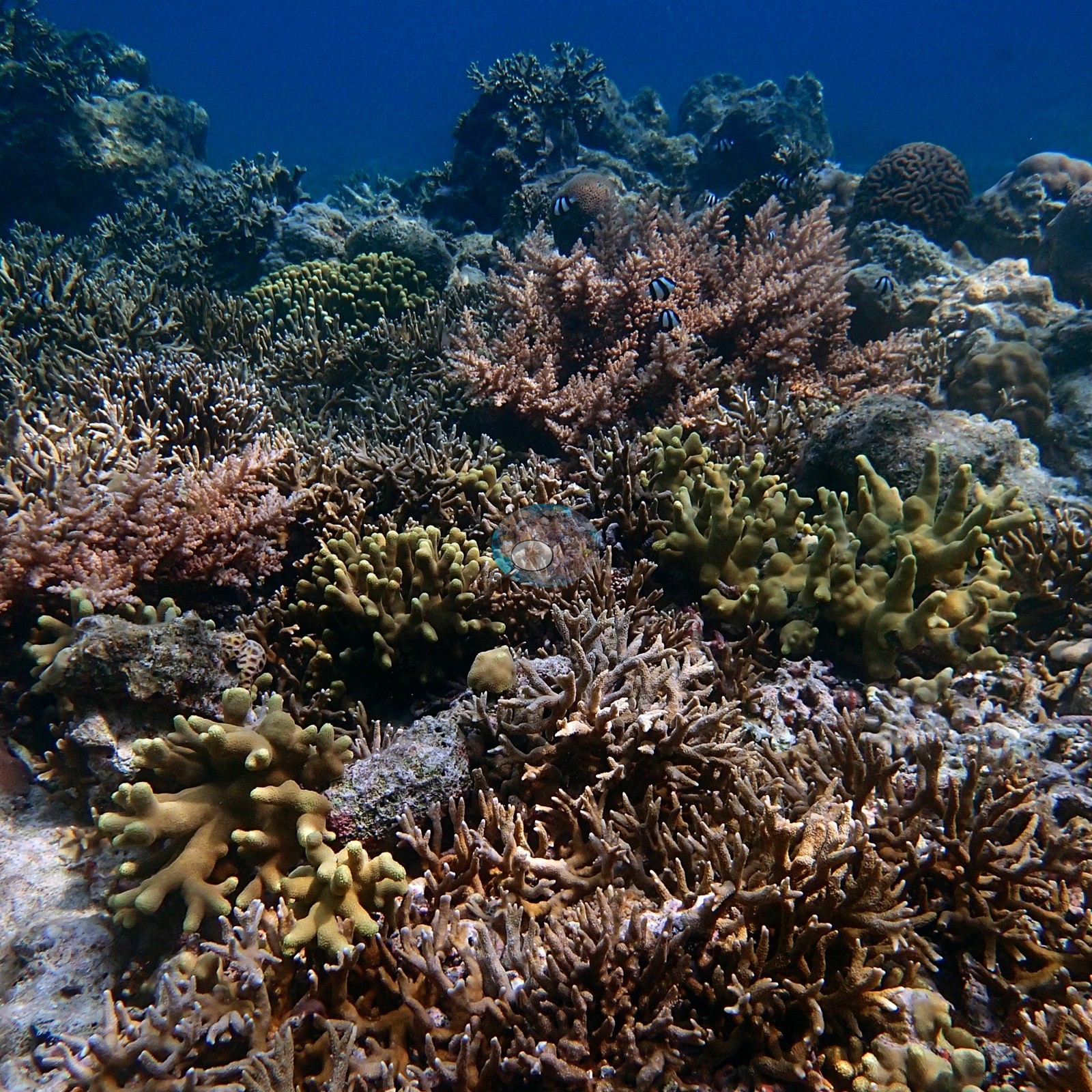
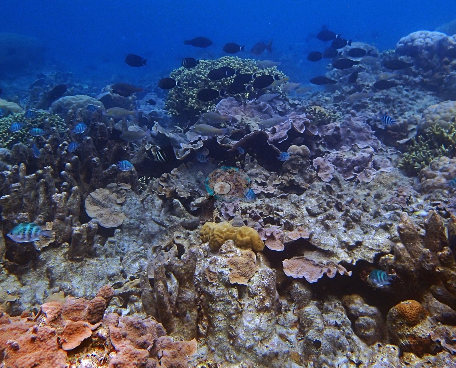
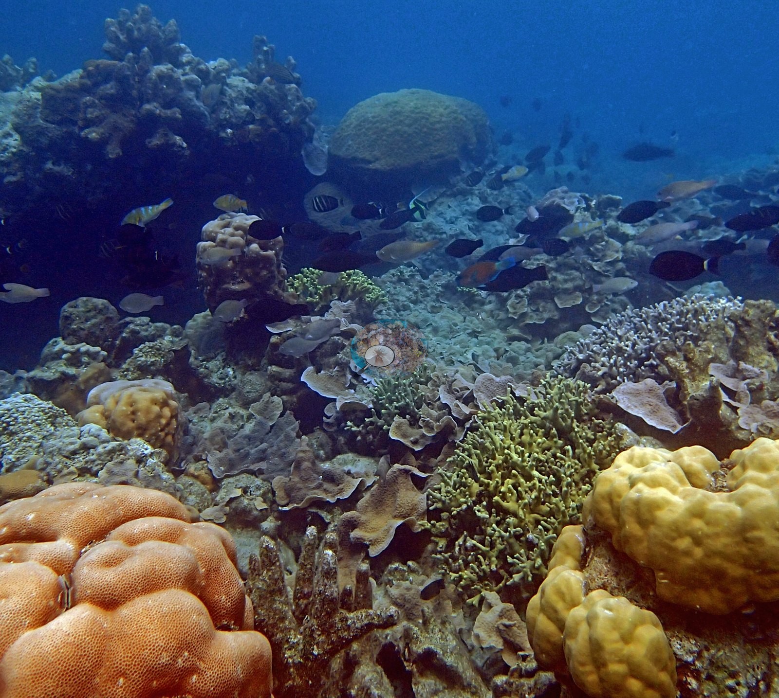
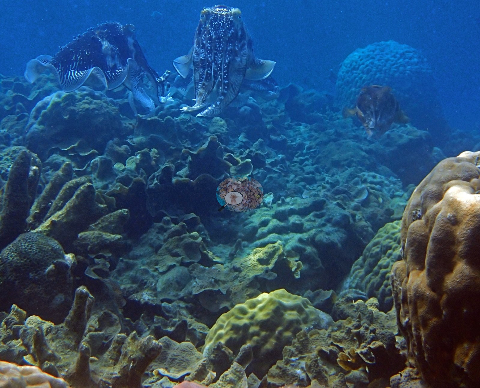
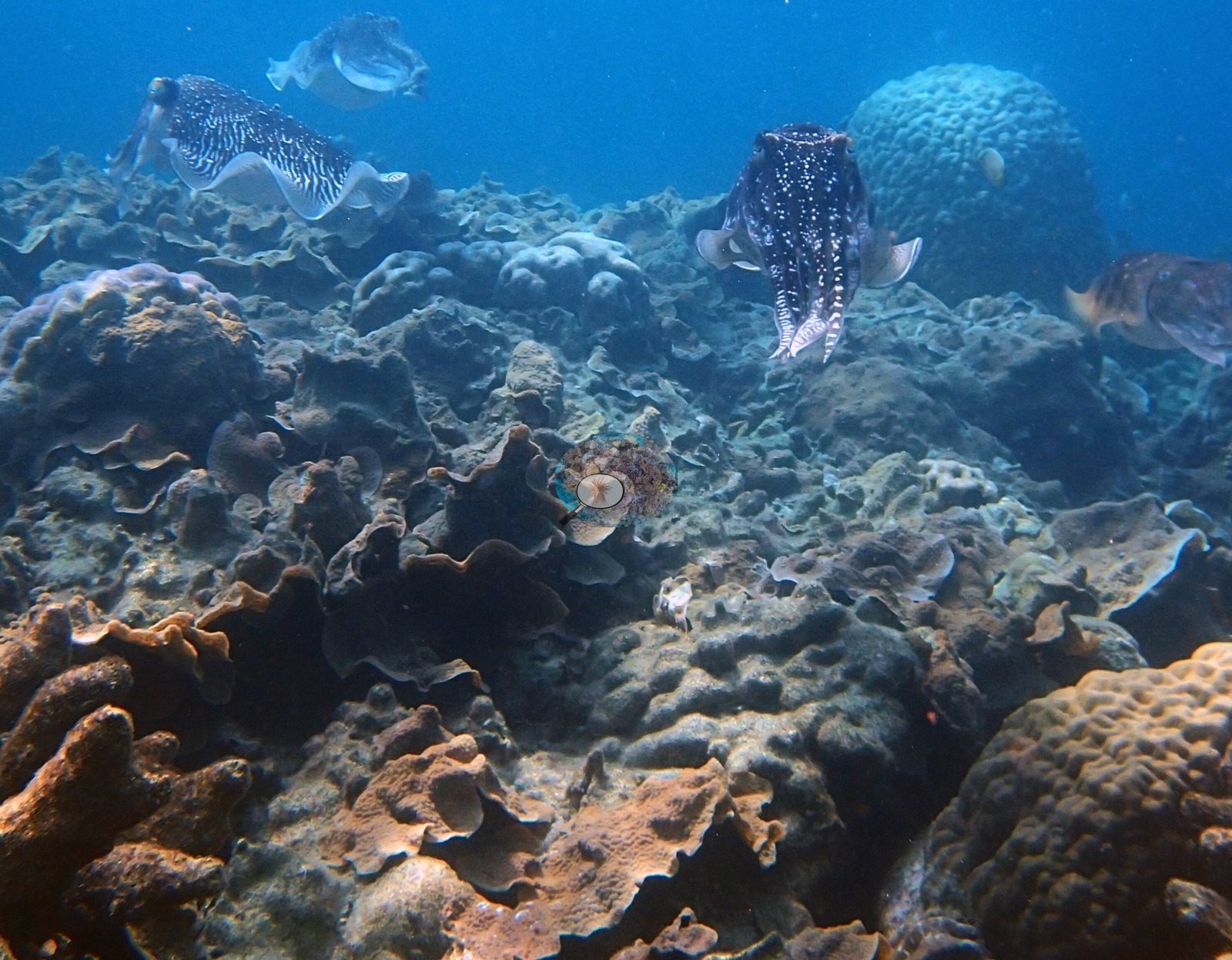
Dongsha, Taiwan (NOAA CRW virtual station)Site 6 at Dongsha Atoll is a high-relief, wall-to-wall hard coral reef of branching and table Acropora, giant Porites domes, sea fans, and broad plate-coral fields, with a loose group of large cuttlefish and dense damselfish schools over the reef. Anderson's sample sheet records storm damage and old partial mortality among the eight colonies sampled here.
東沙環礁 6 號測站是一處高地形起伏、石珊瑚滿覆的礁區,由分枝狀與桌形 Acropora、巨大的 Porites 團塊、海扇及大片片狀珊瑚組成,礁上有一群鬆散聚集的大型烏賊與密集的雀鯛魚群。Anderson 的採樣紀錄表記載,在此採樣的八株珊瑚中可見風暴損傷與舊的部分死亡。
本摘要由 AI 翻譯;以英文版本為準。
One other CRD dive site with its own page within 5 km shown in orange — click a marker to open its page.
This page shows 71 of the 123 photographs from this site, including every one the text cites.
Site description
General benthic features
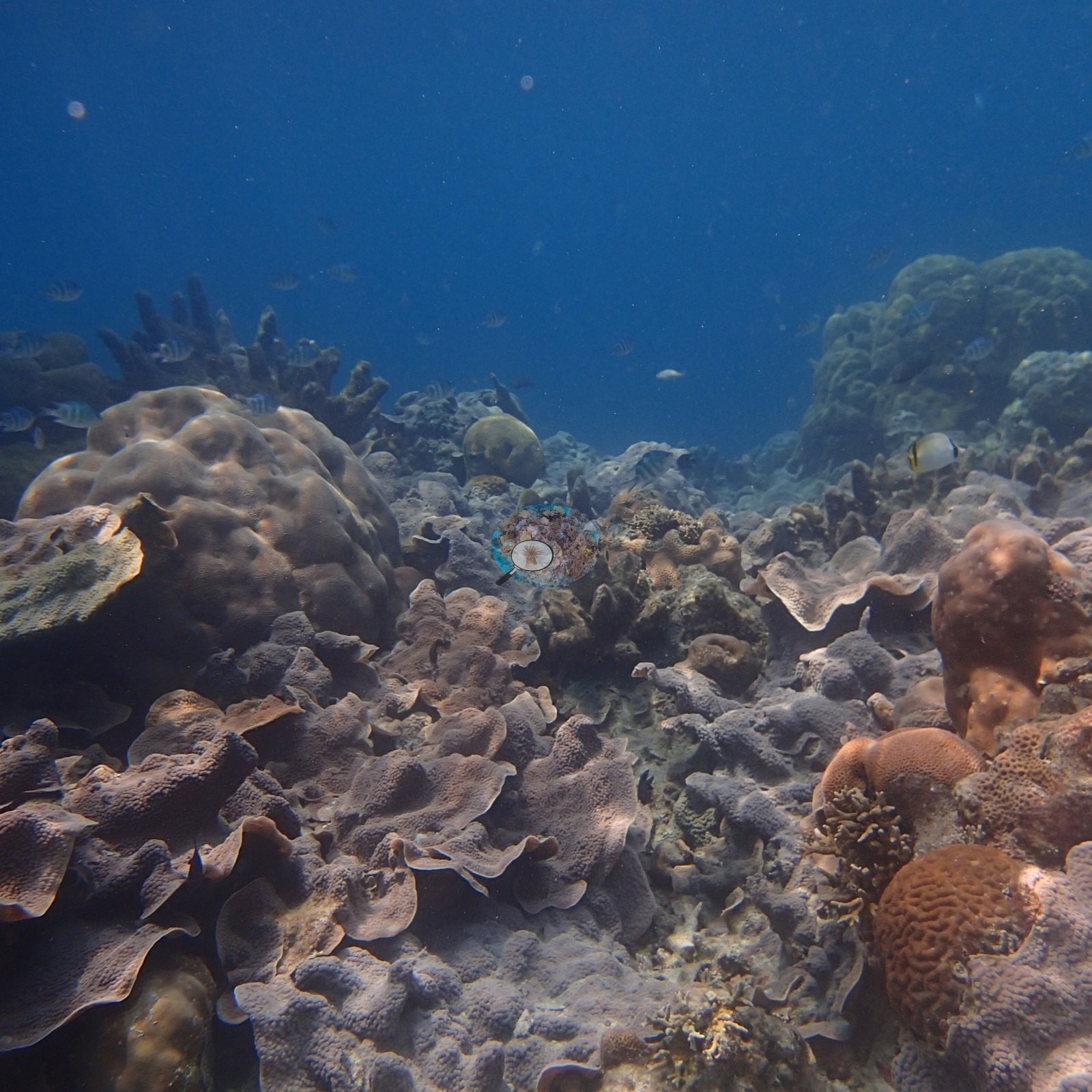
The 5 April dive is a high-relief, structurally complete hard coral reef (e.g., Image 22, Image 44).
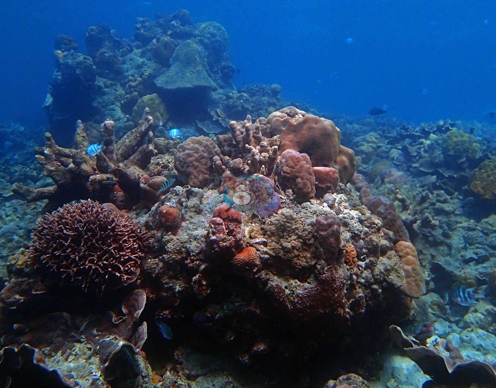
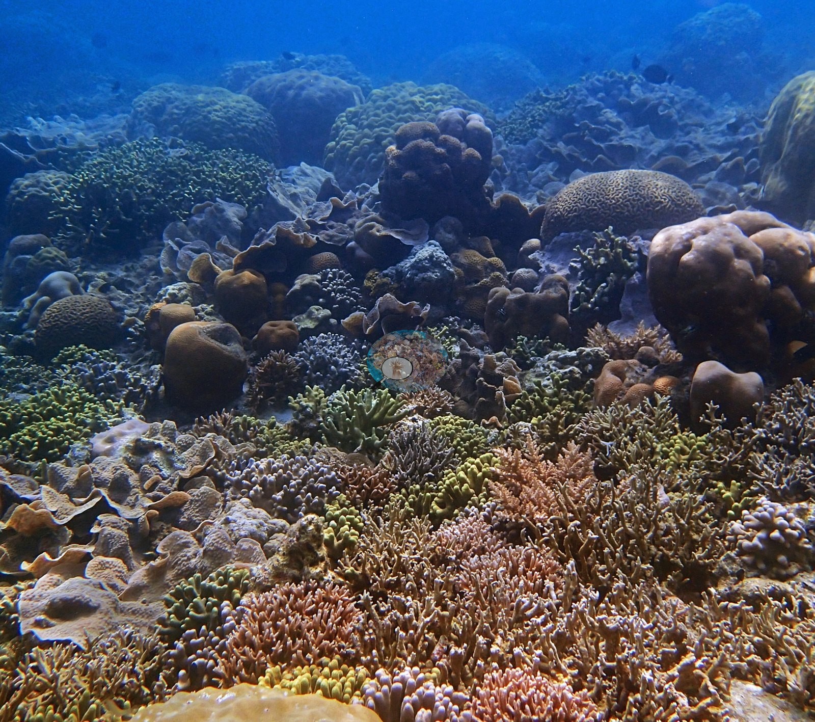
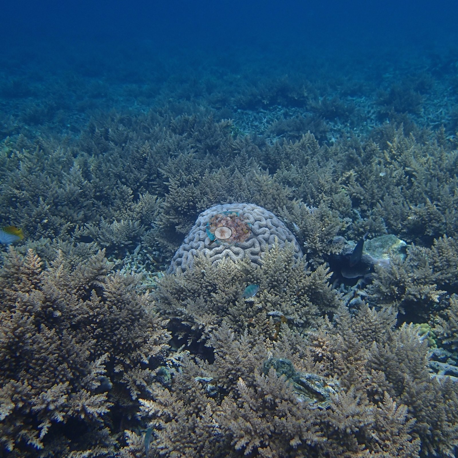
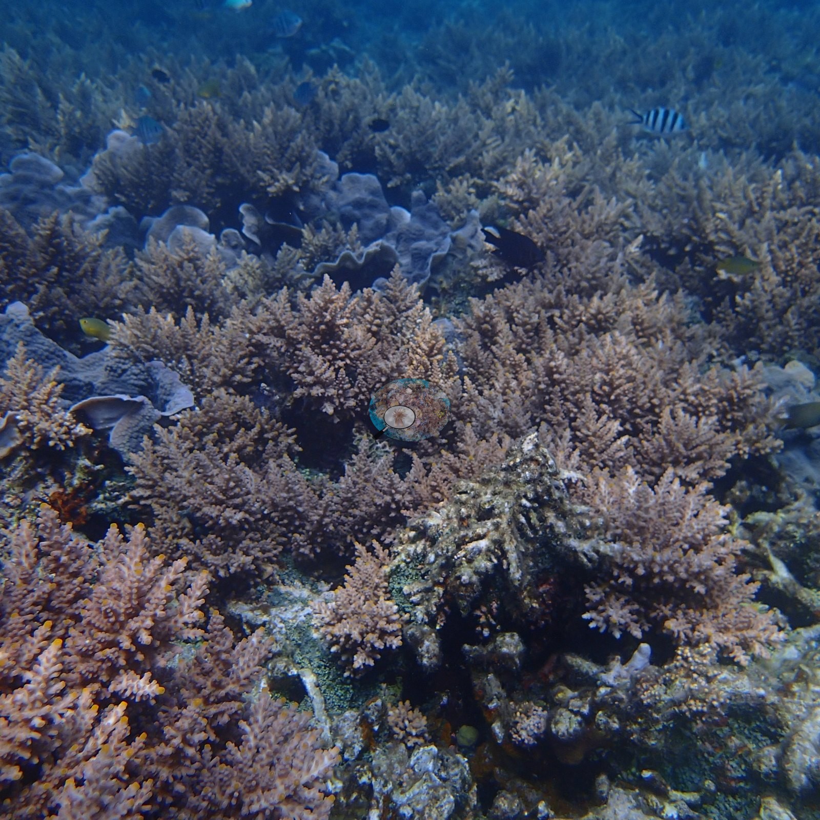
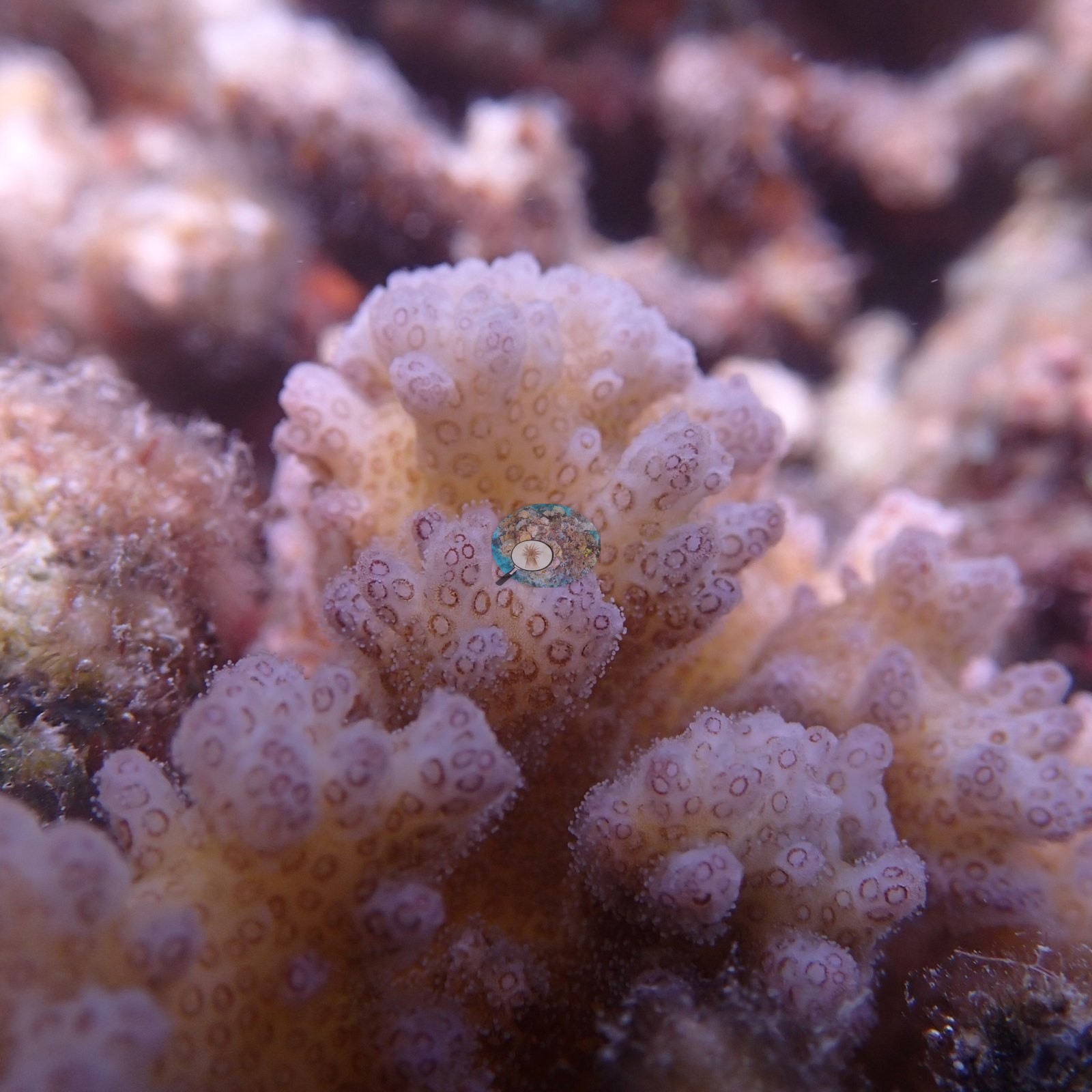
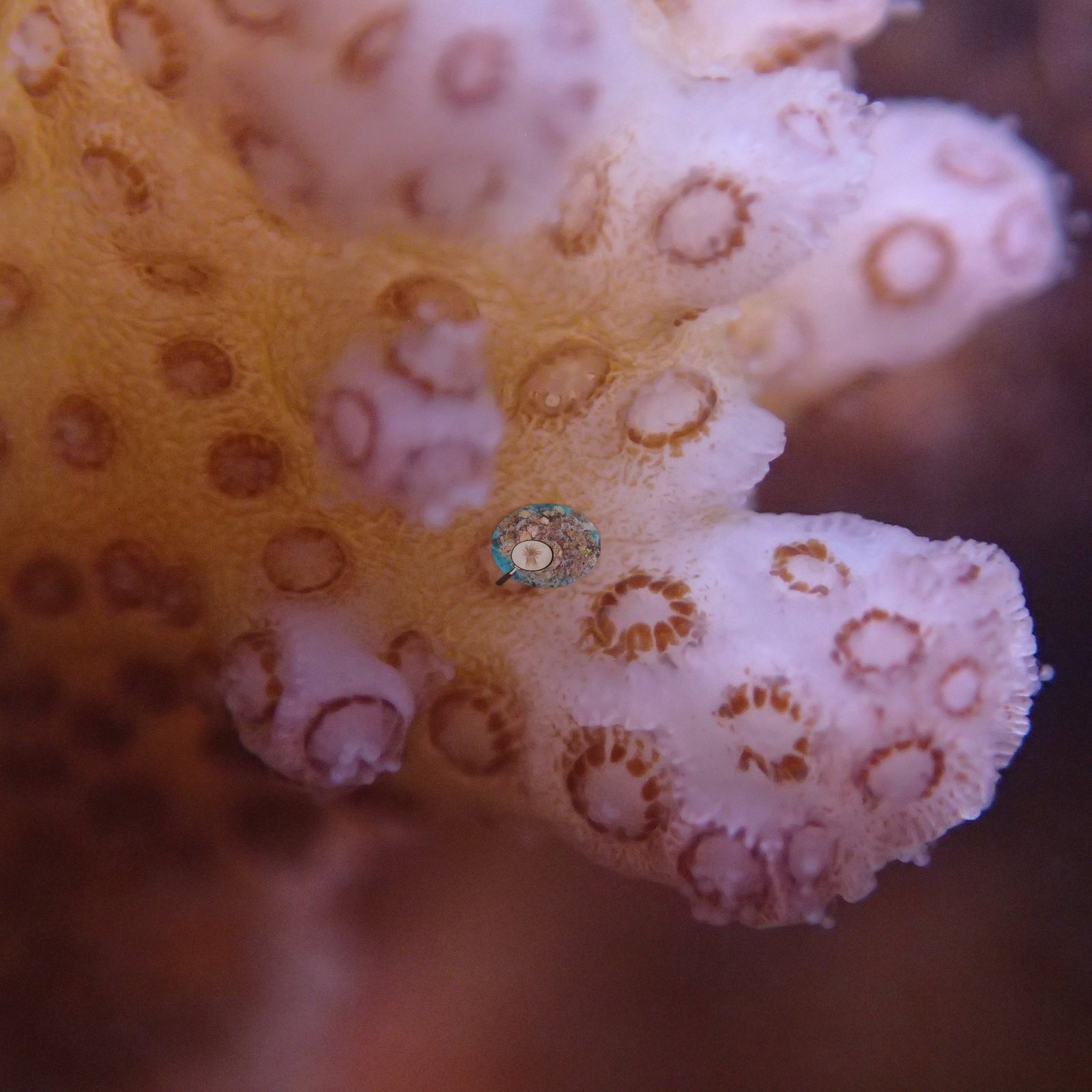
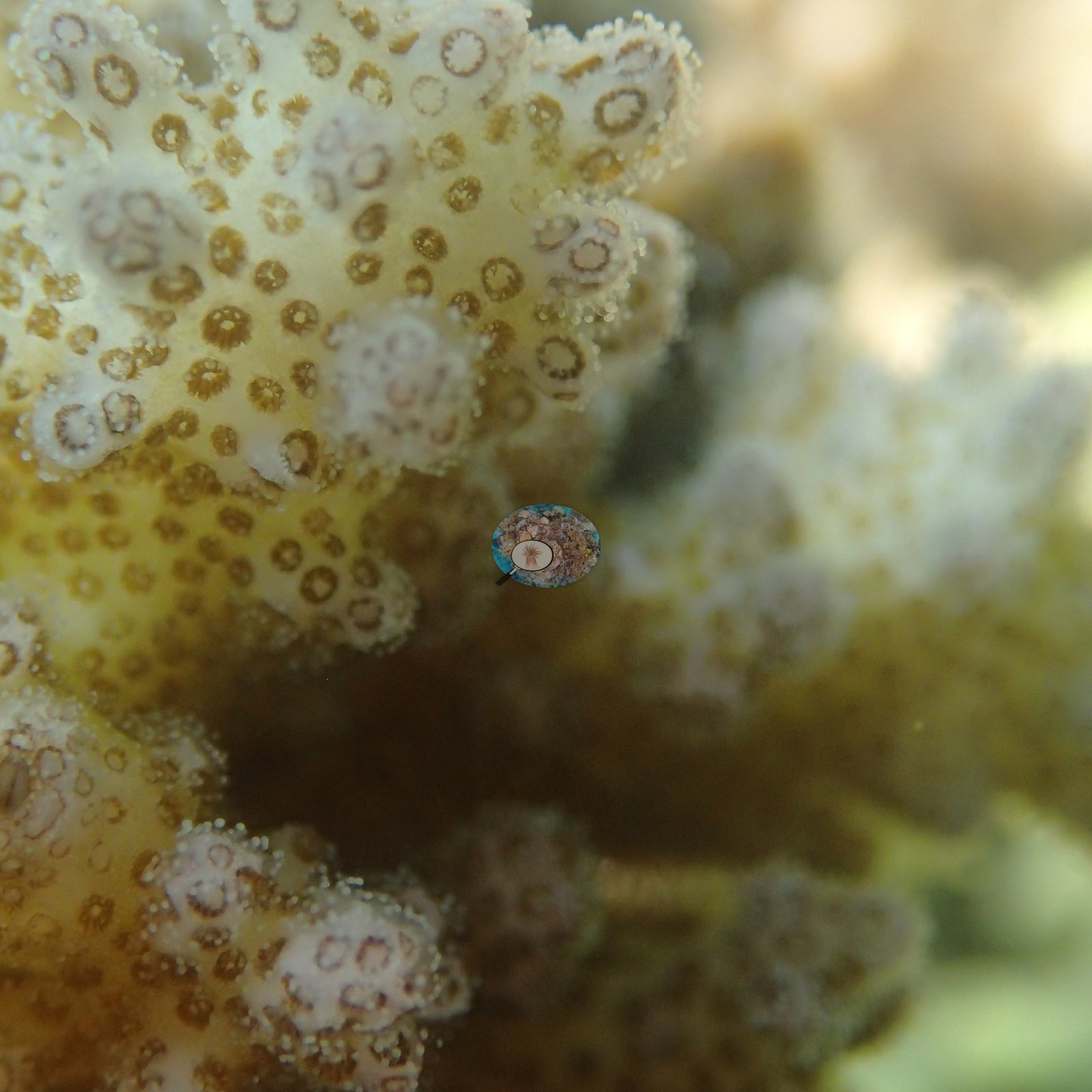
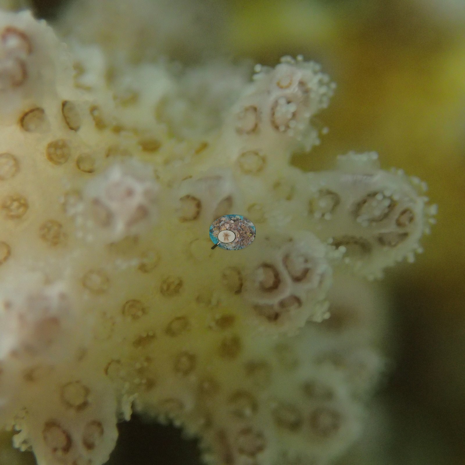
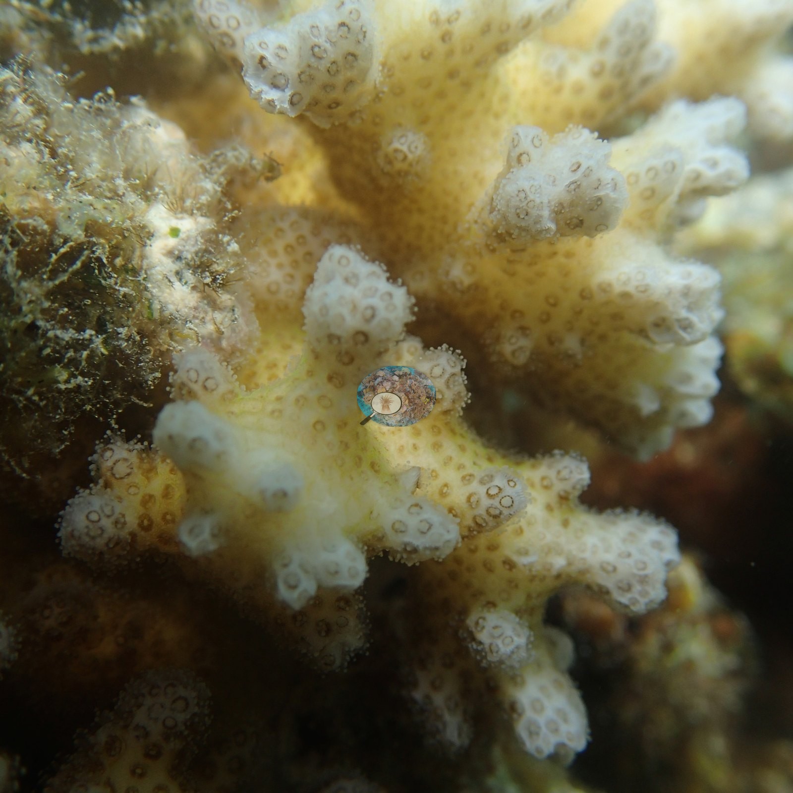
Salmon-pink, tan, and olive corymbose and digitate Acropora interlock with tabulate Acropora plates (e.g., Image 1, Image 24, Image 36, Image 43), blue-gray and mauve encrusting and plating Montipora-type colonies, yellow and gold knobby Porites domes over a meter across (e.g., Image 49, Image 53), brain corals of the Platygyra and Symphyllia growth form (e.g., Image 45), and scarlet and orange encrusting sponge (e.g., Image 35), with almost no open substrate between colonies across long runs of frames (e.g., Image 33, Image 46).
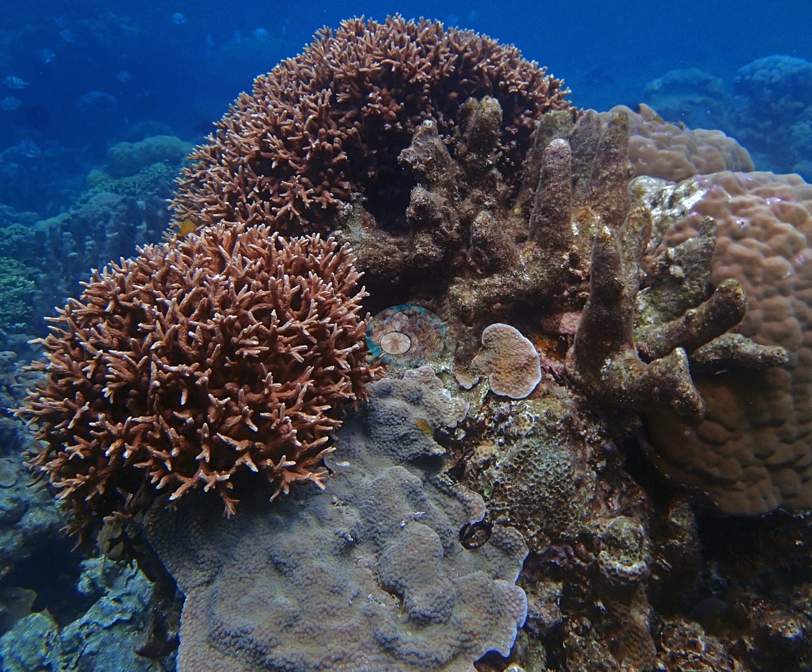
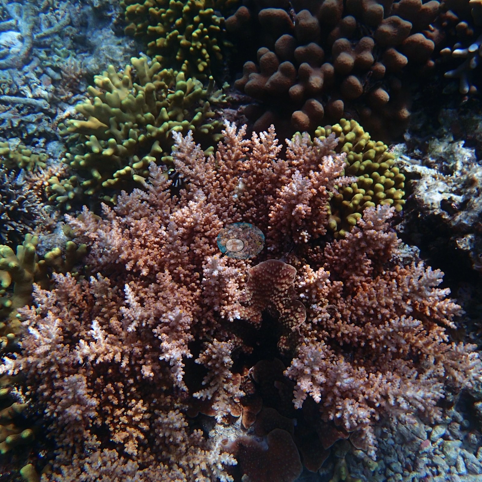
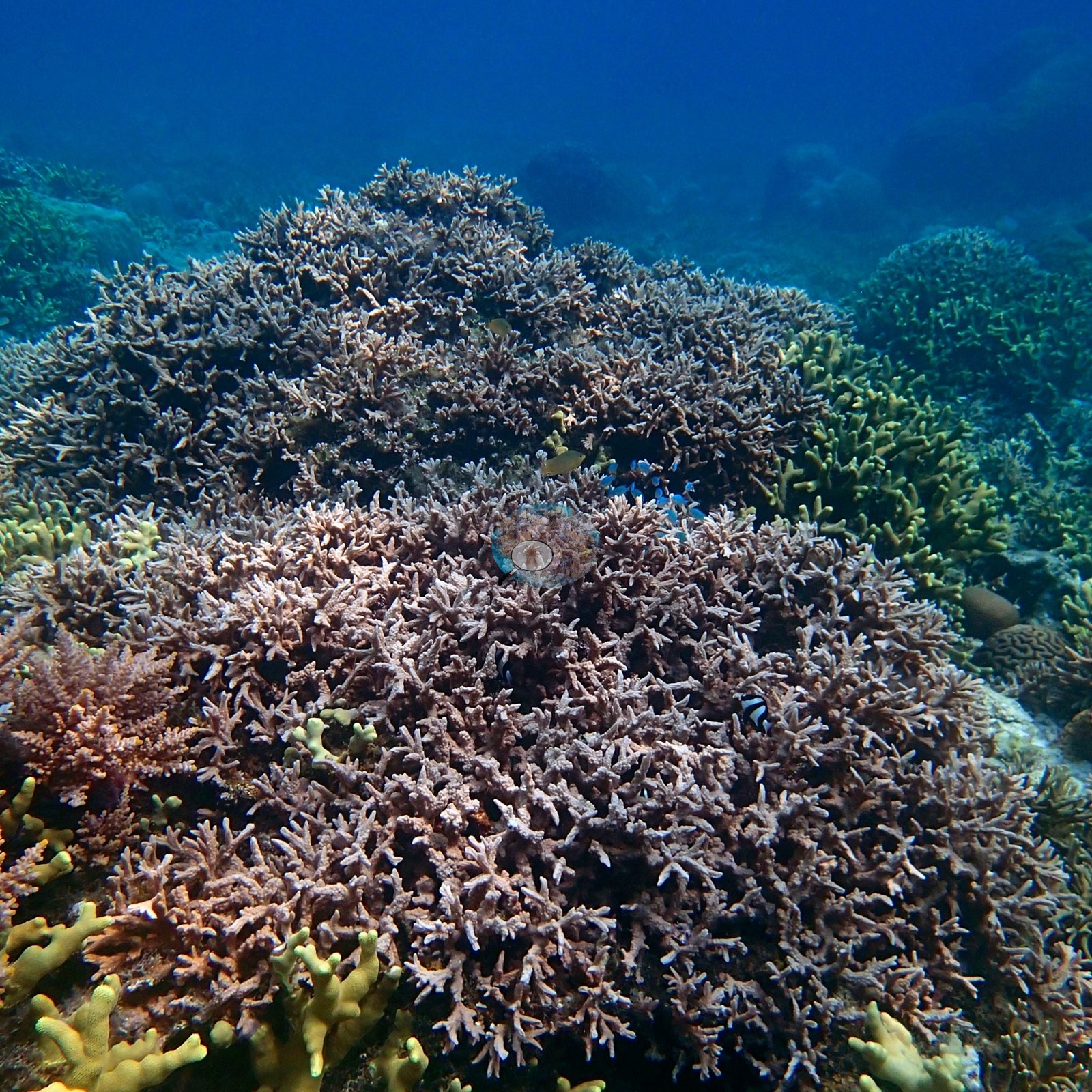
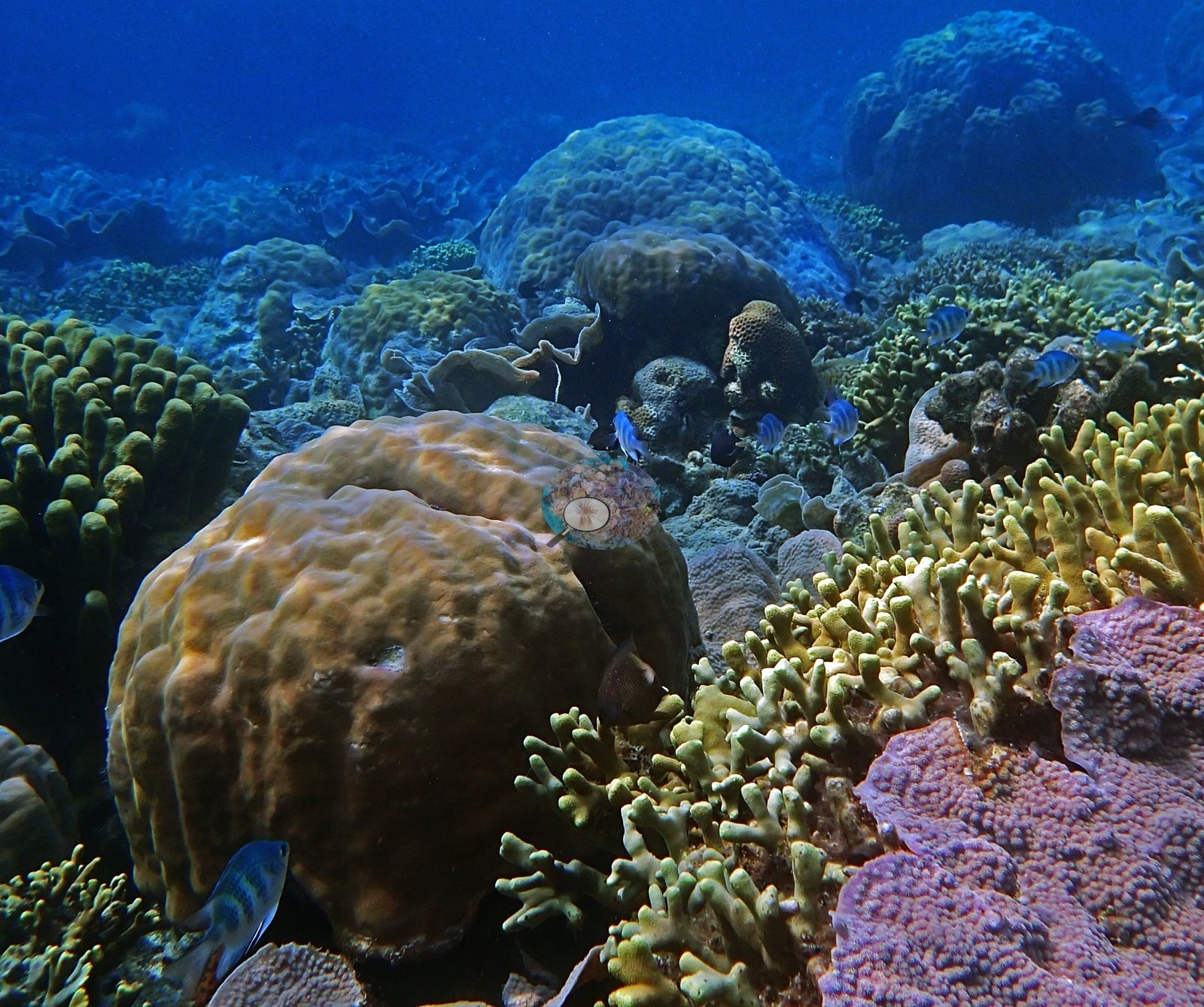
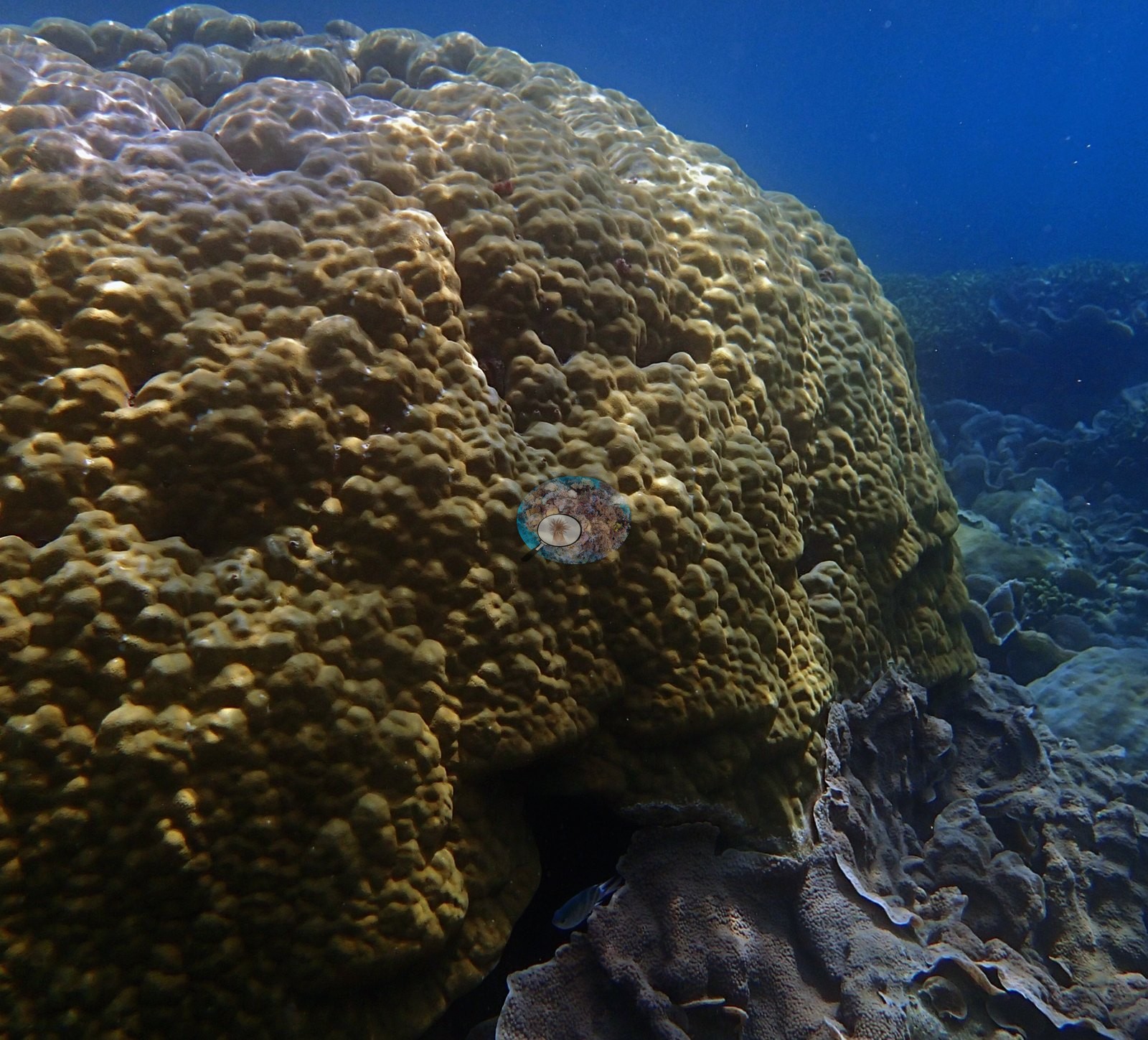
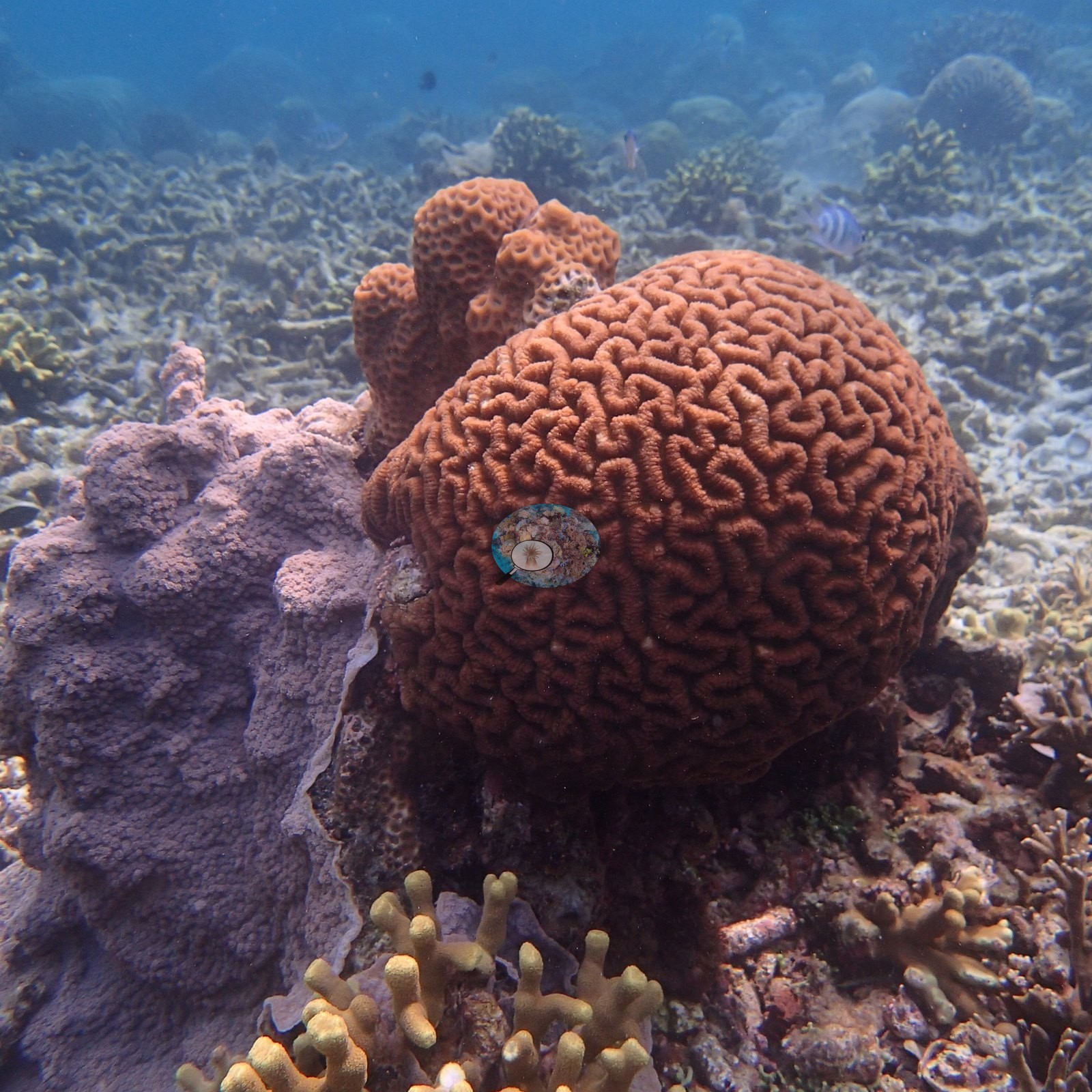
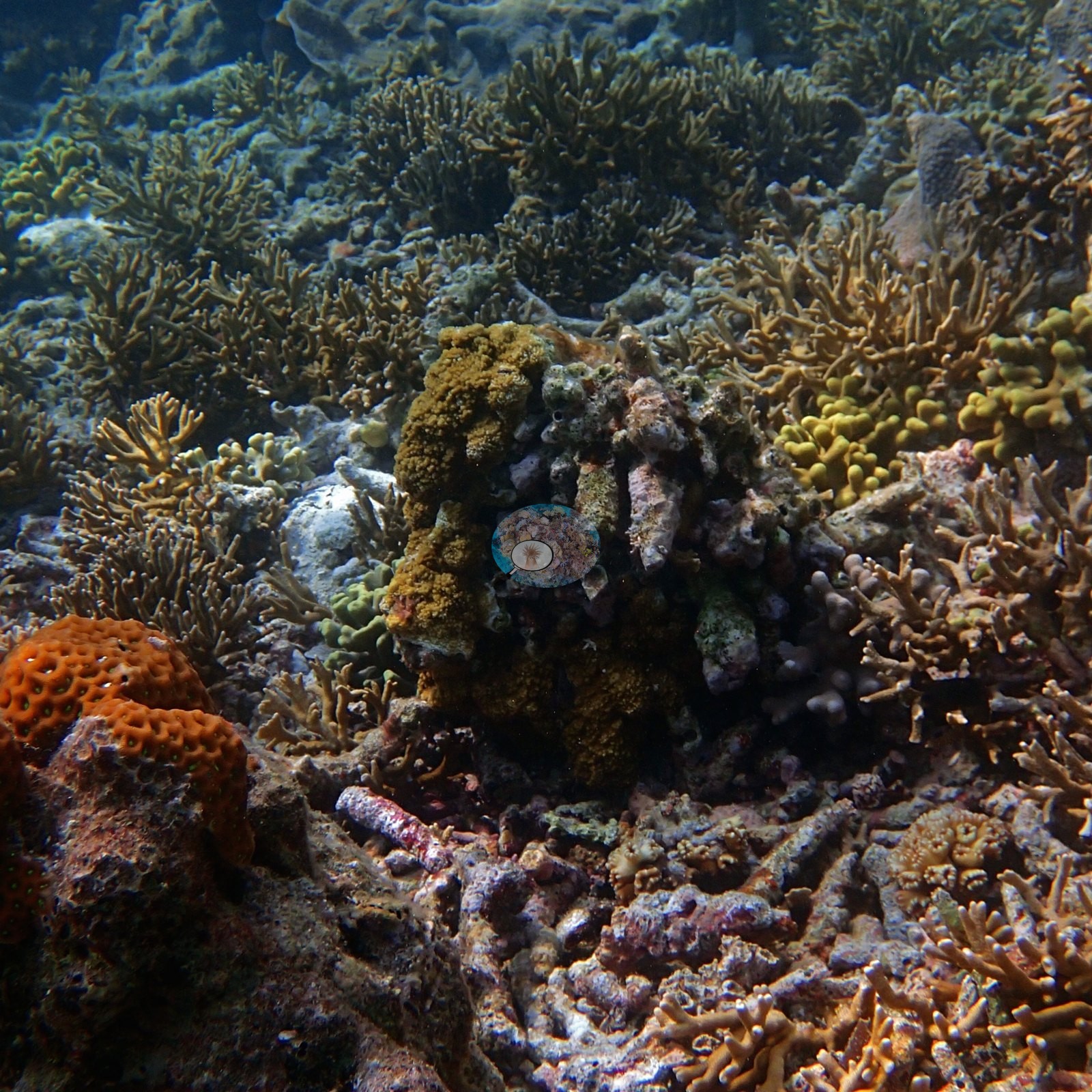
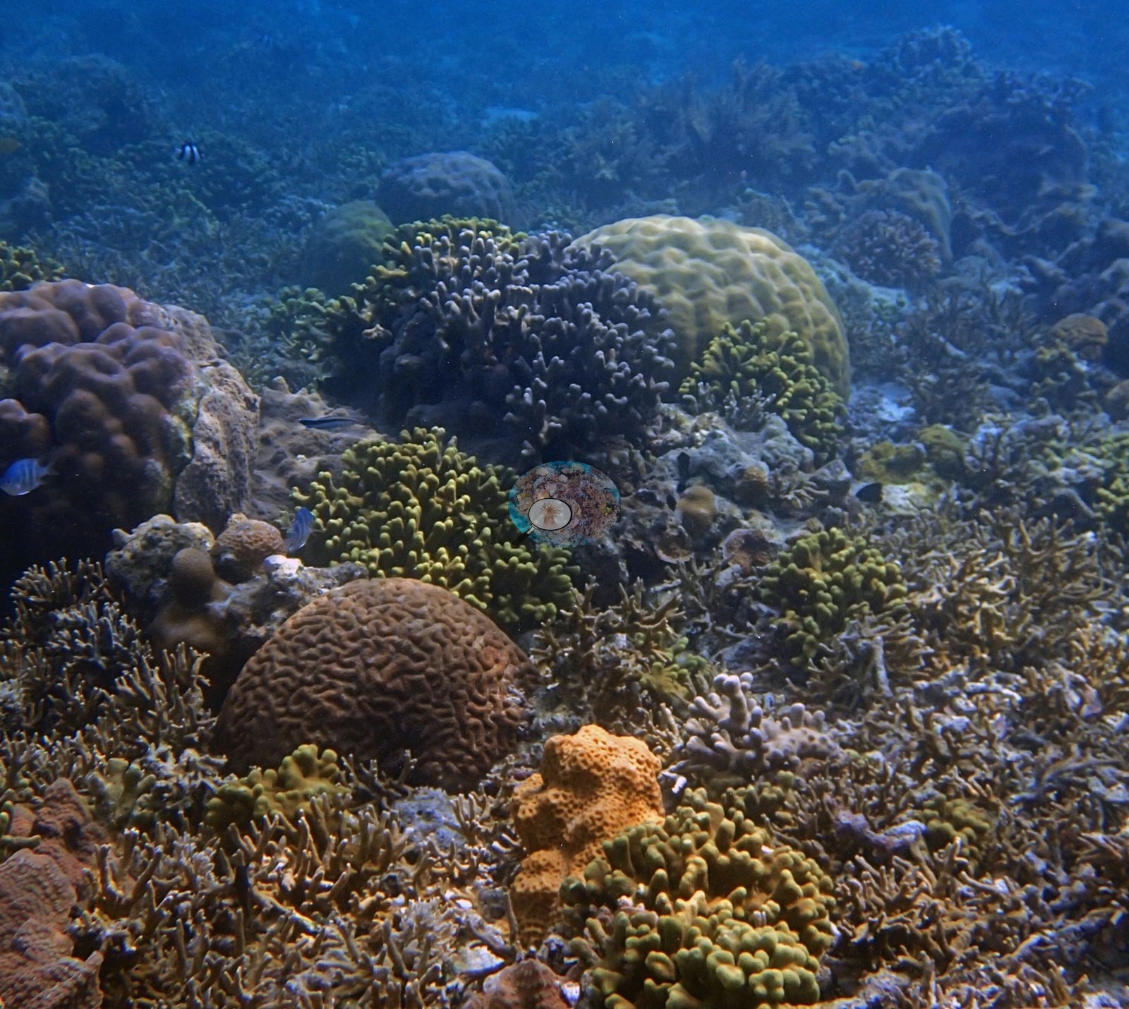
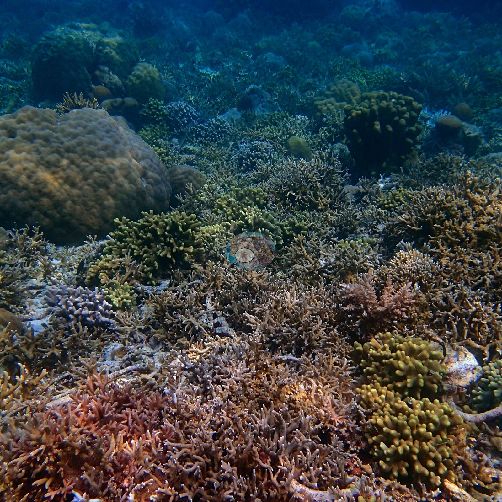
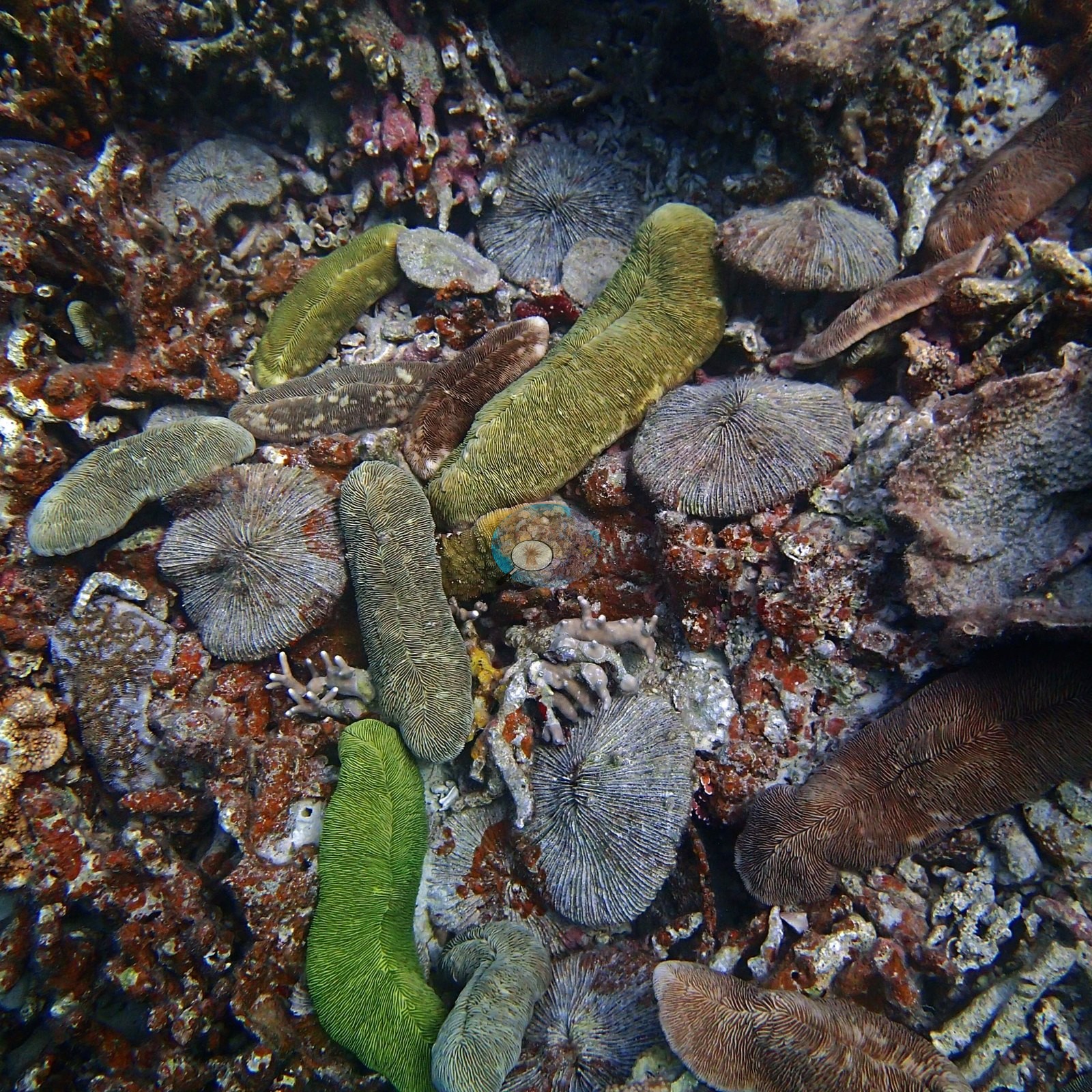
Three features of this dive stand out. A free-living mushroom-coral bed (Fungiidae, including a vivid green disc and elongate slipper forms) sits on rubble in one frame (e.g., Image 9).
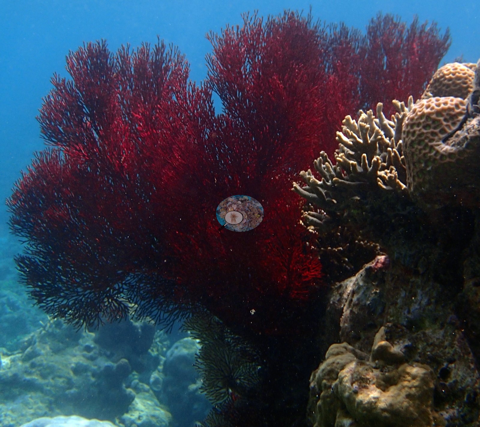
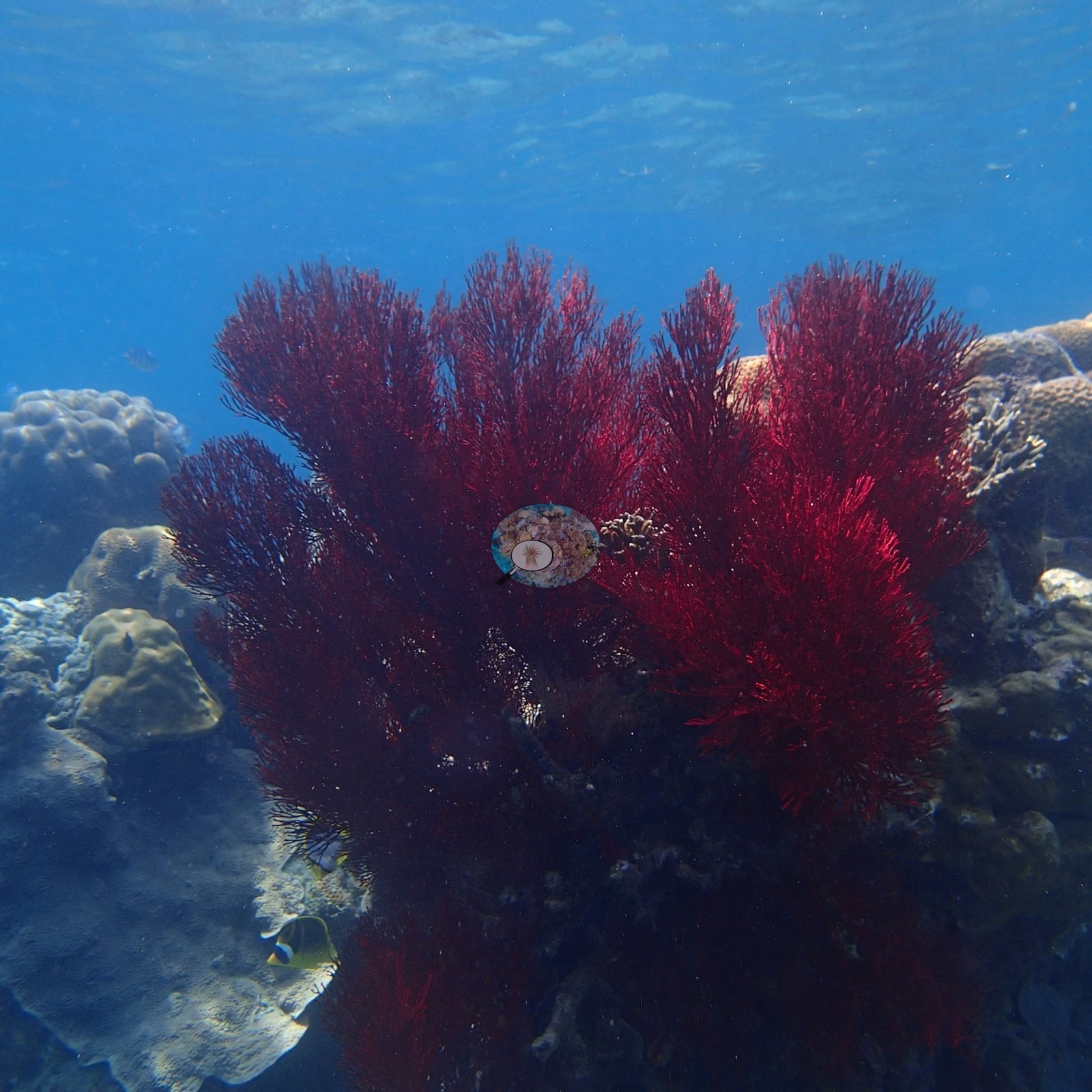
Two large deep-red gorgonian sea fans stand clear of the reef in two others, one with a crinoid at its base and a butterflyfish beneath it (e.g., Image 32, Image 112).
And beyond the mid-reef the station opens into extensive foliose plate-coral fields — thin whorled brown-gray plates, of the Turbinaria, Pachyseris, or Echinopora growth form, tiled across the substrate to the limit of visibility through fifteen or more frames (e.g., Image 61, Image 62, Image 63, Image 64, Image 65, Image 109, Image 110, Image 111, Image 113, Image 114, Image 119, Image 120, Image 121, Image 123).
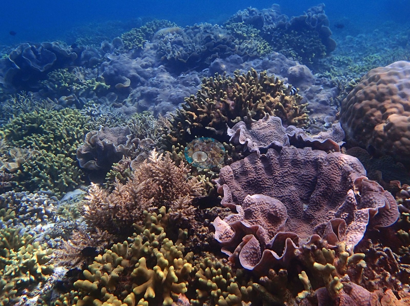
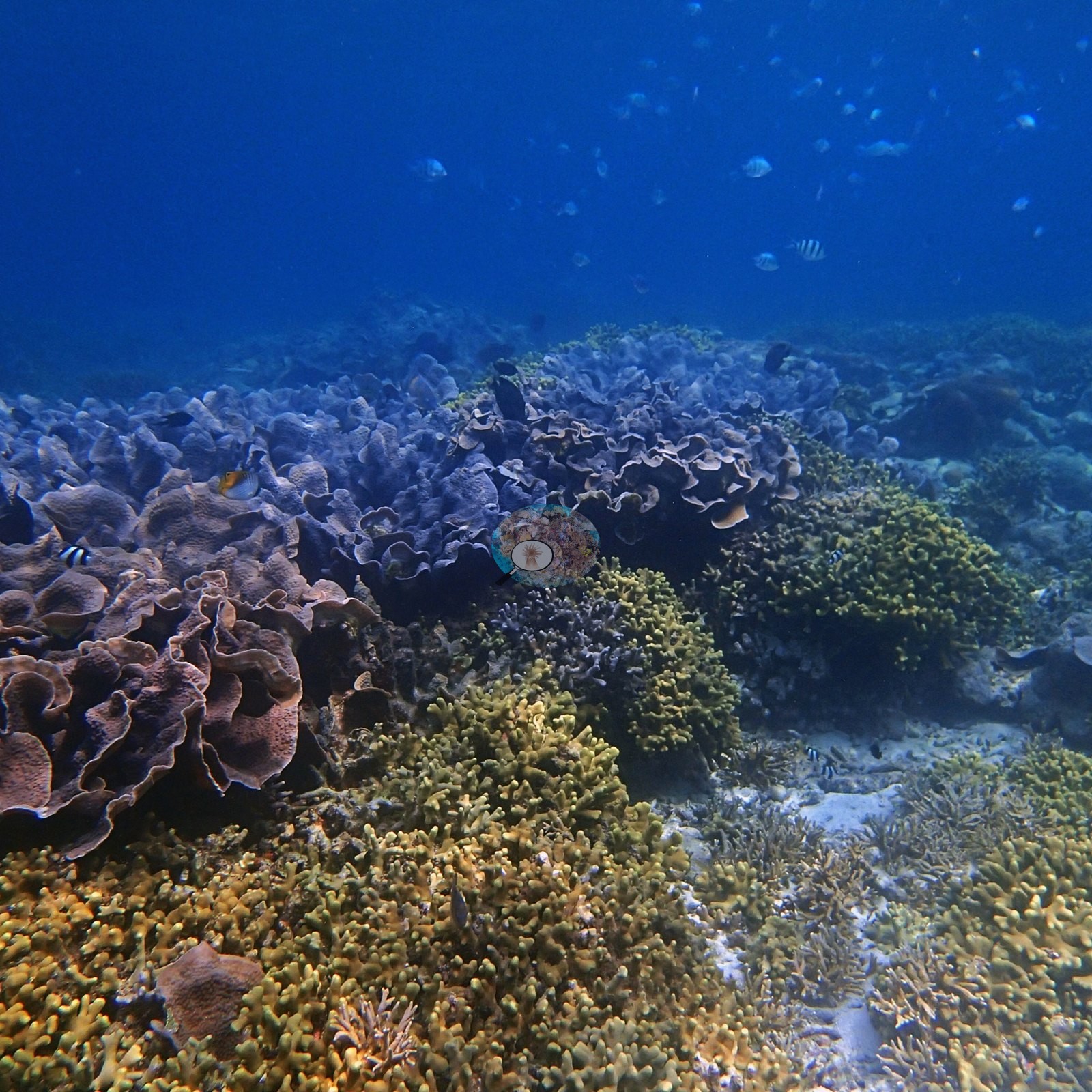
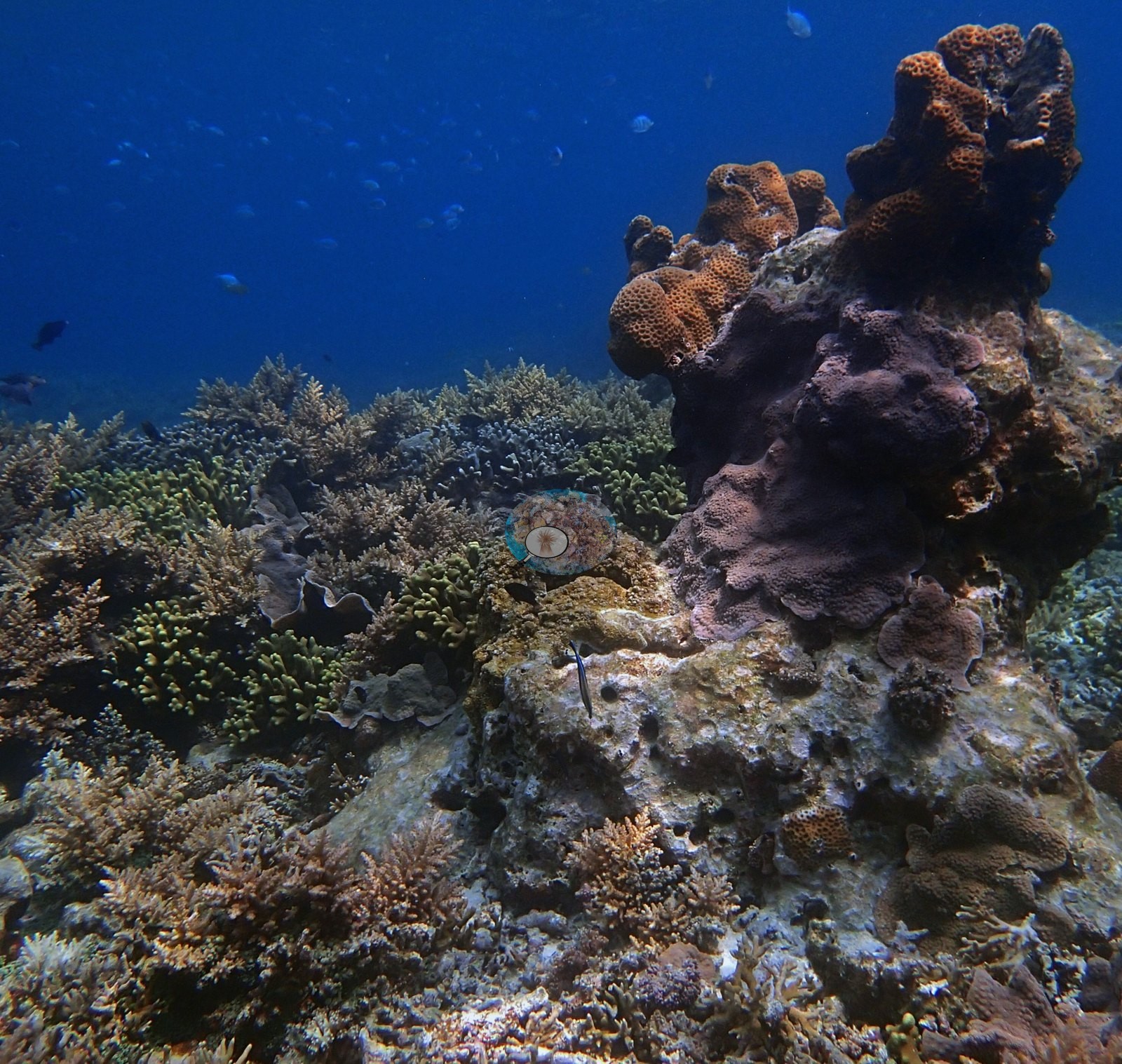
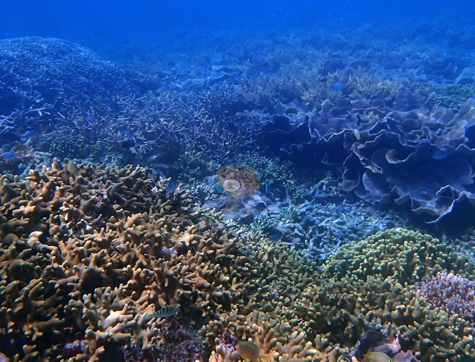
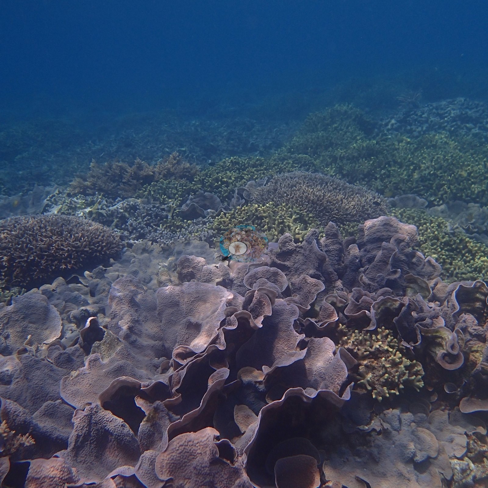
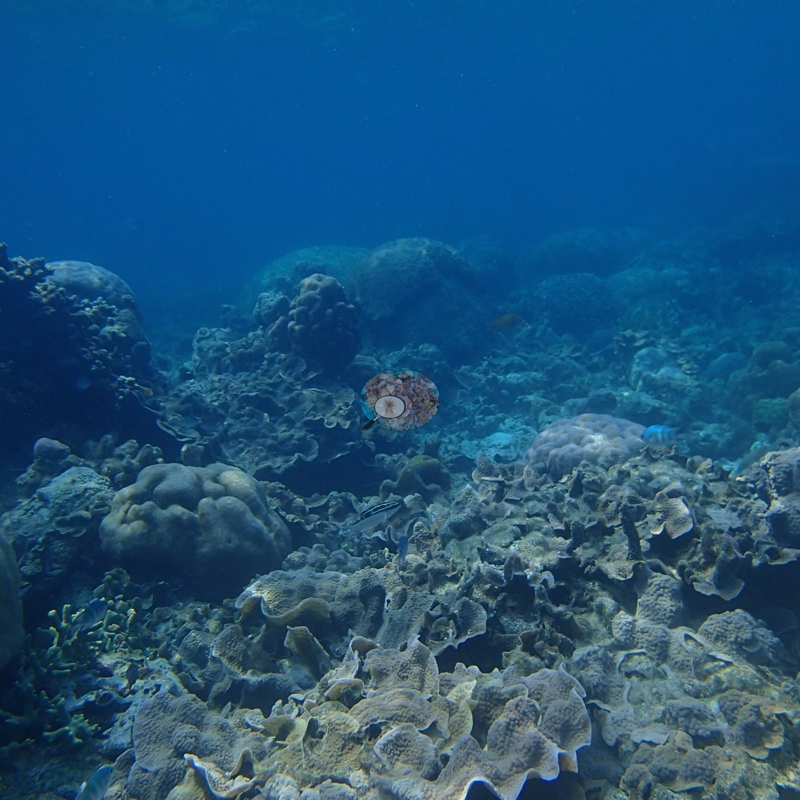
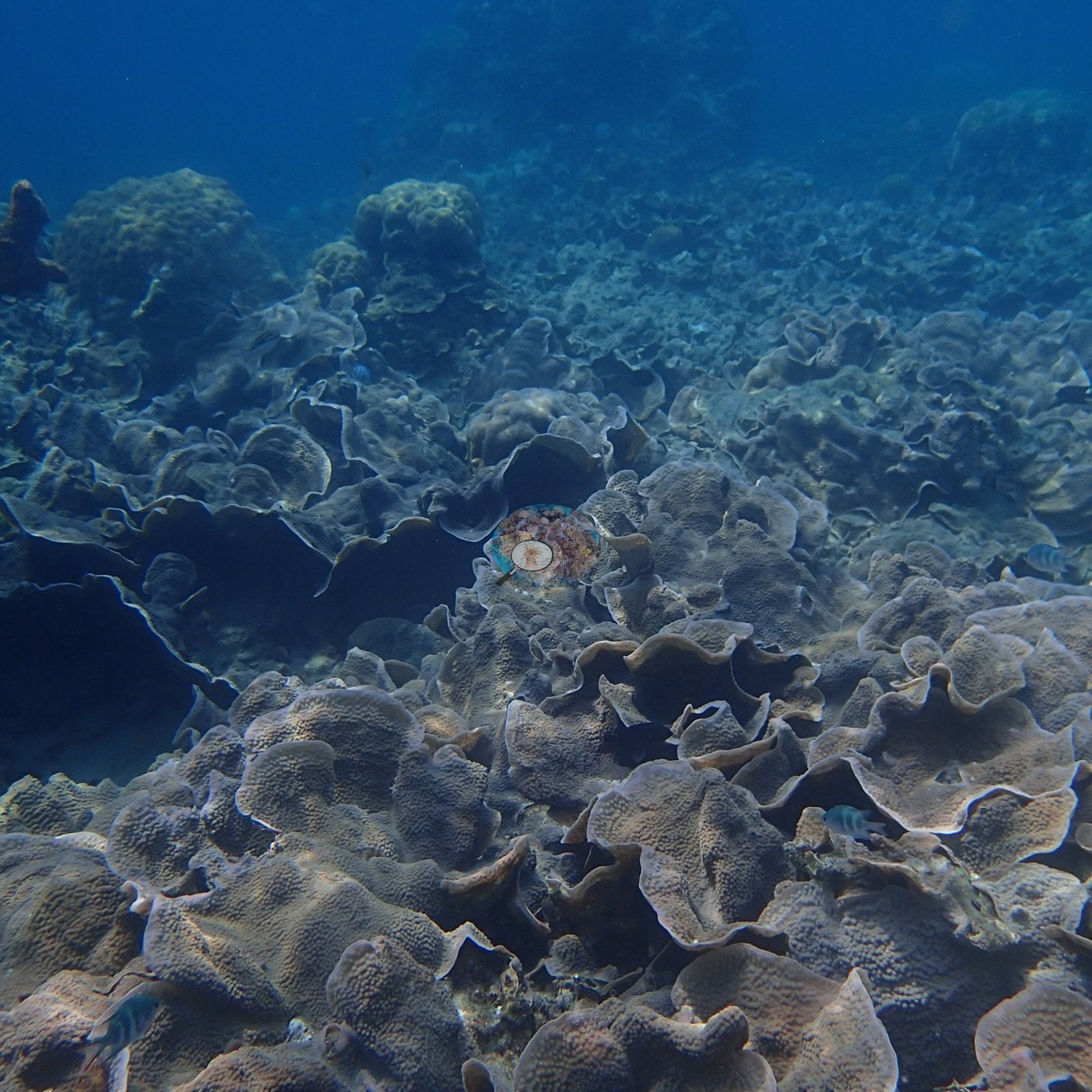
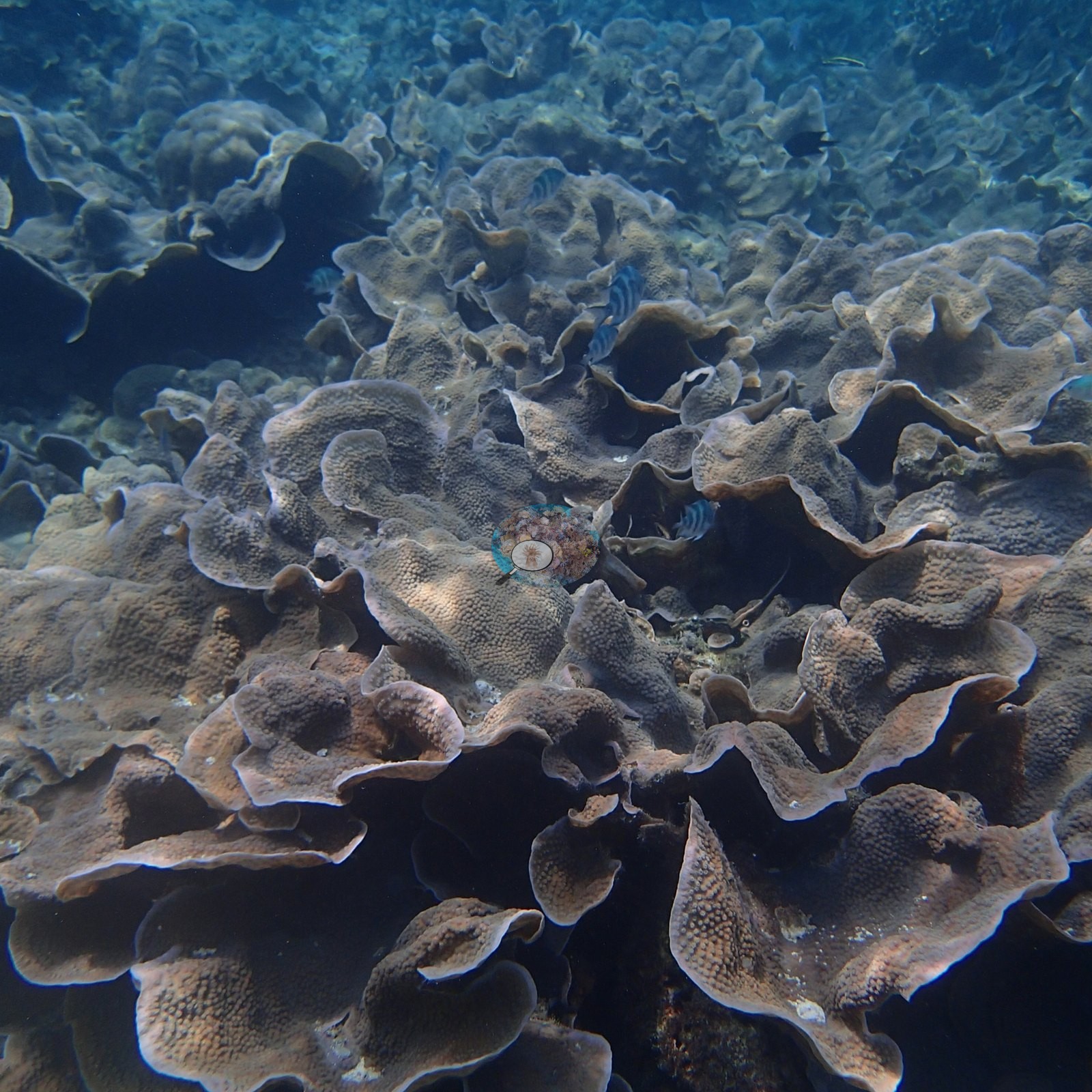
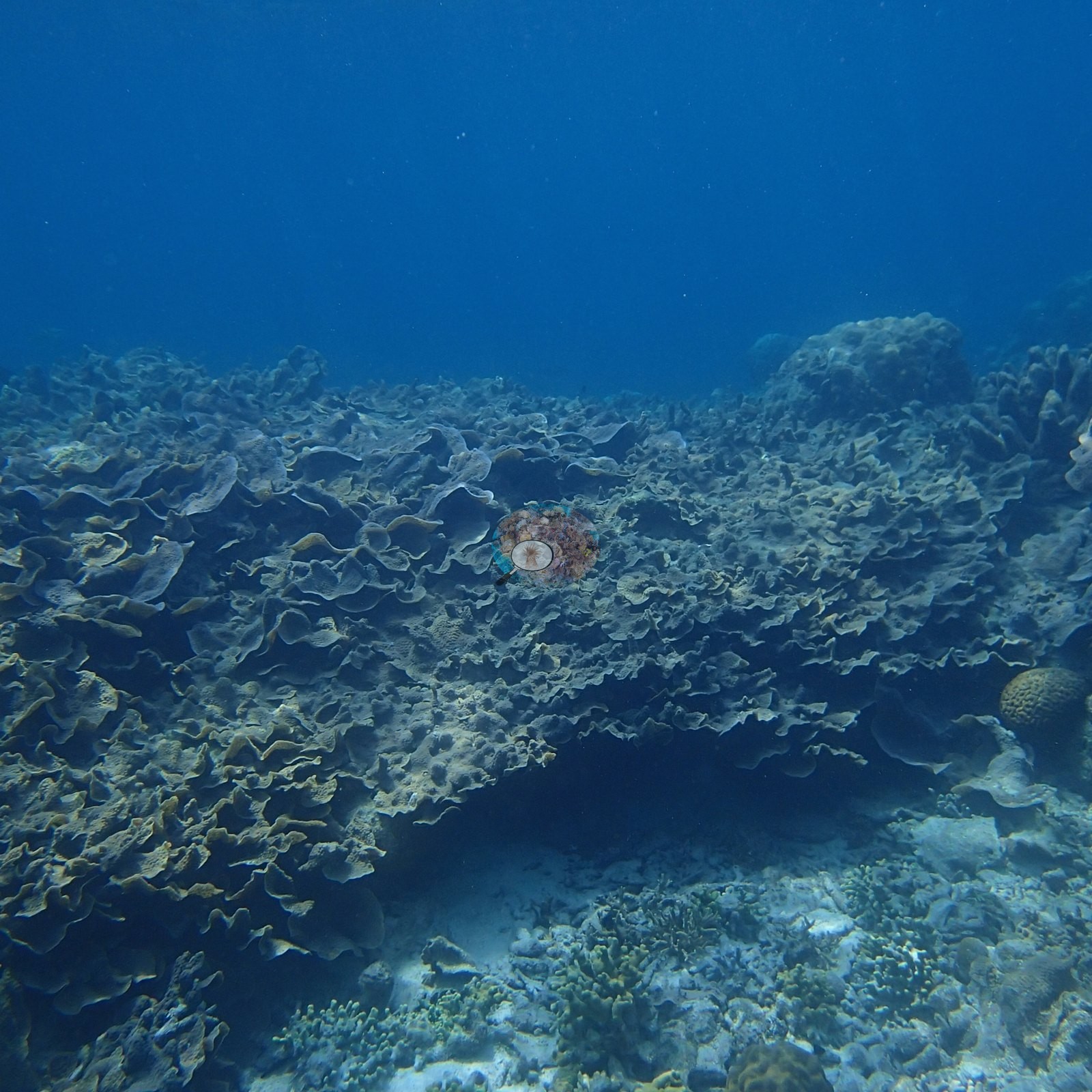
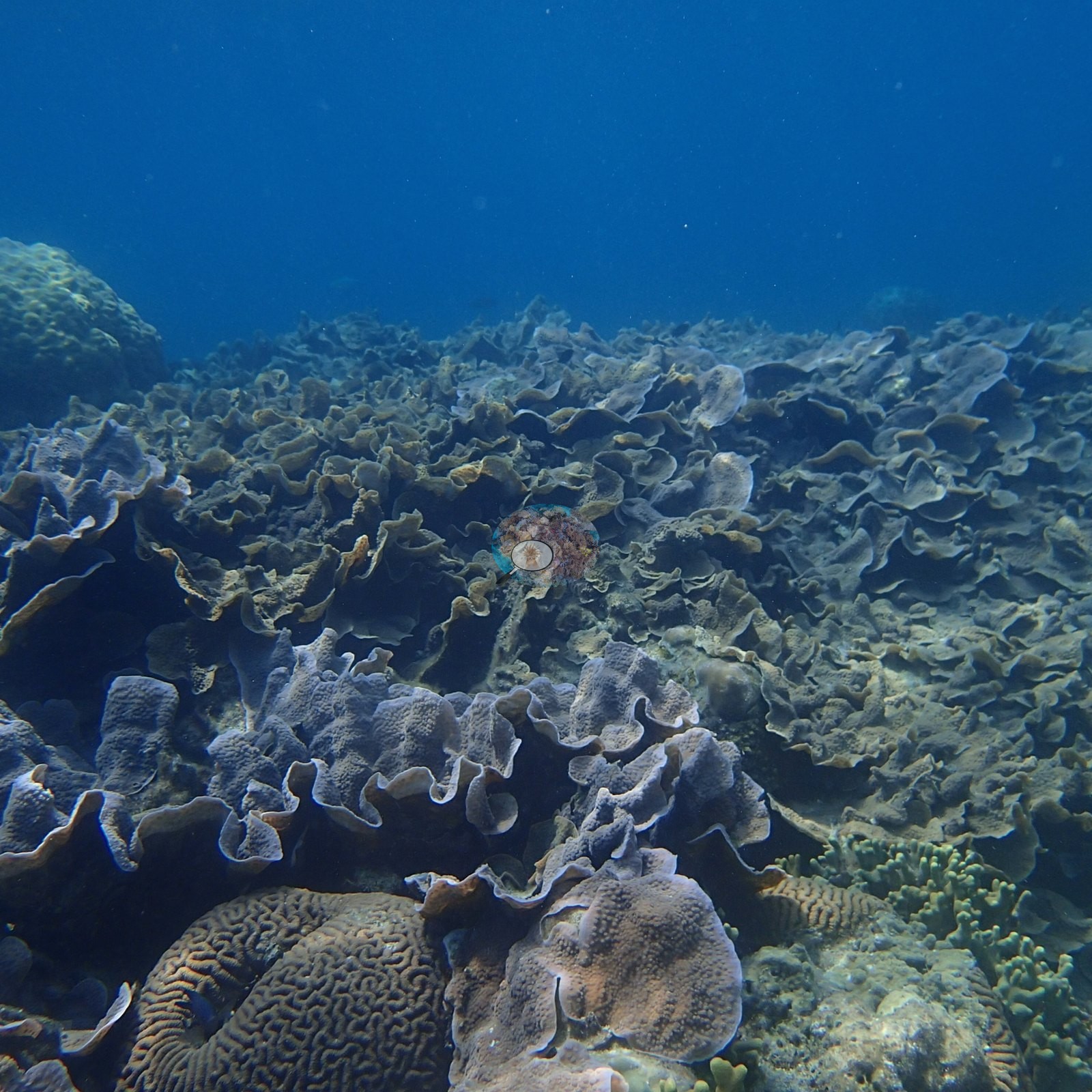
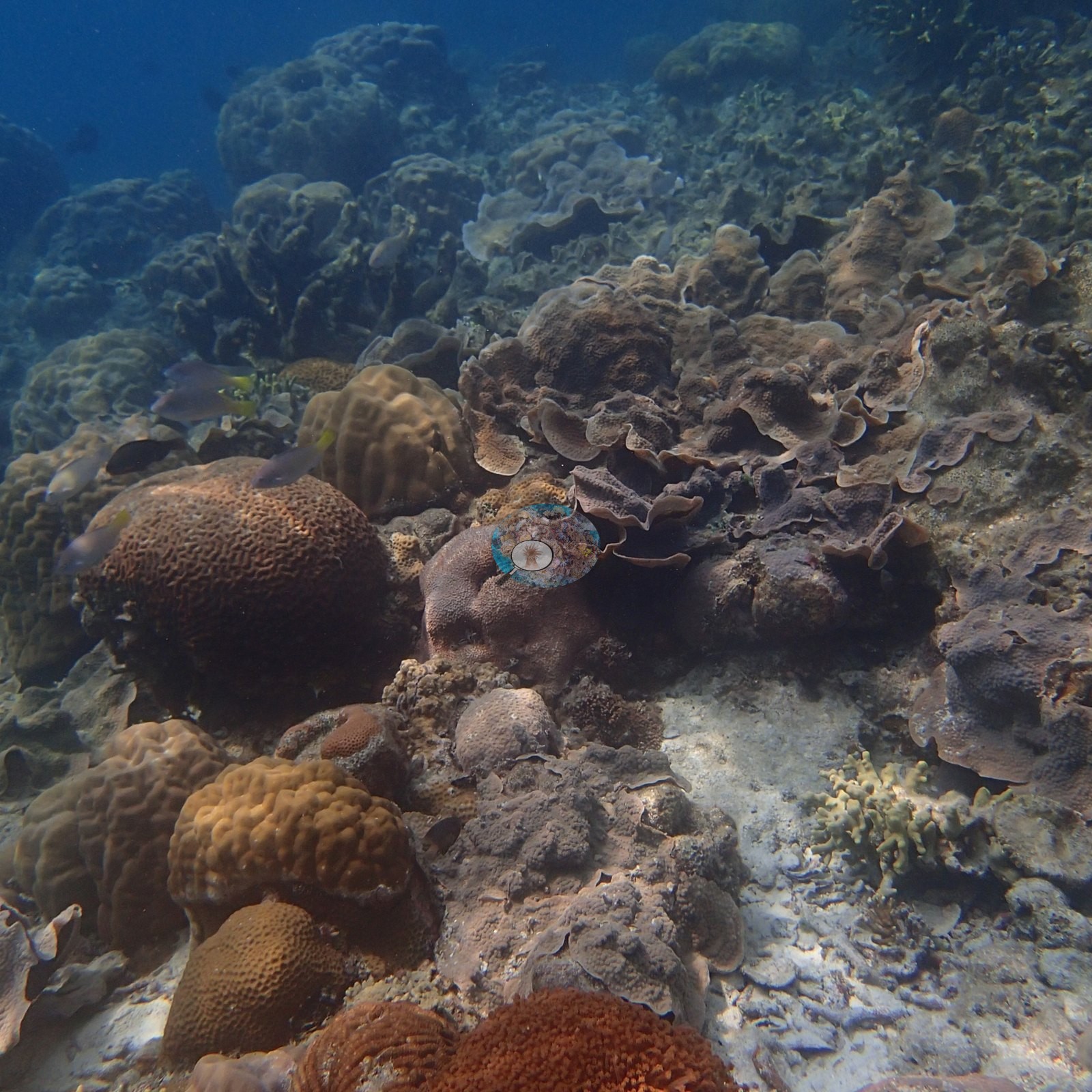
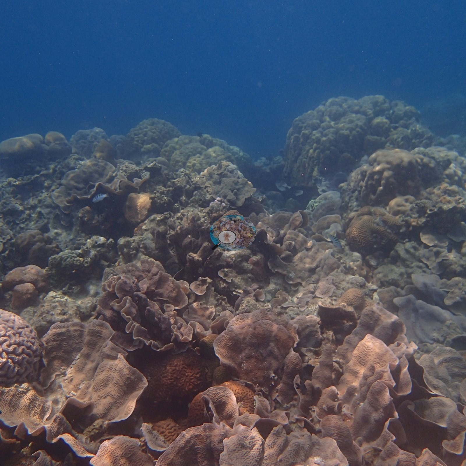
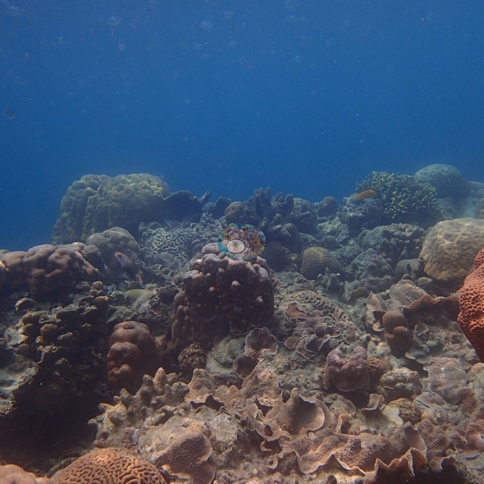
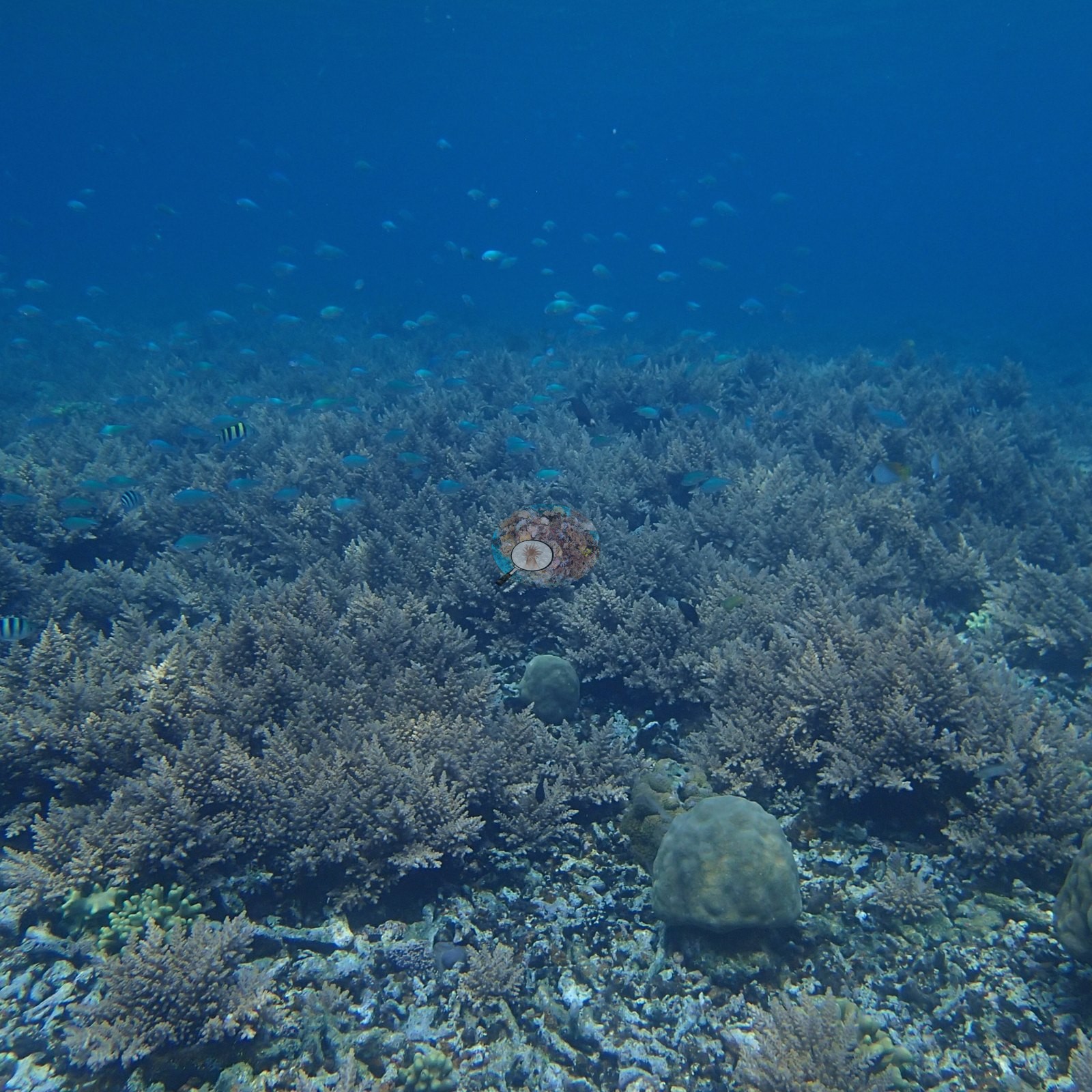
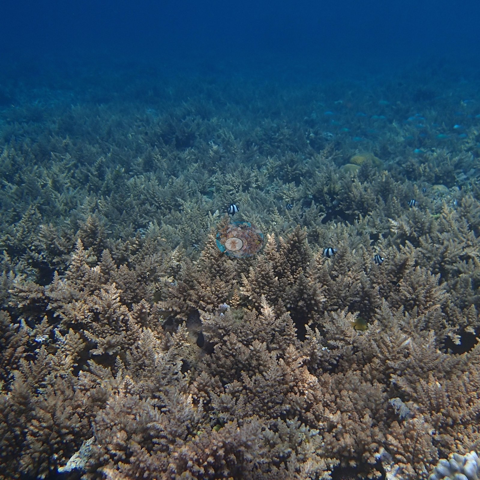
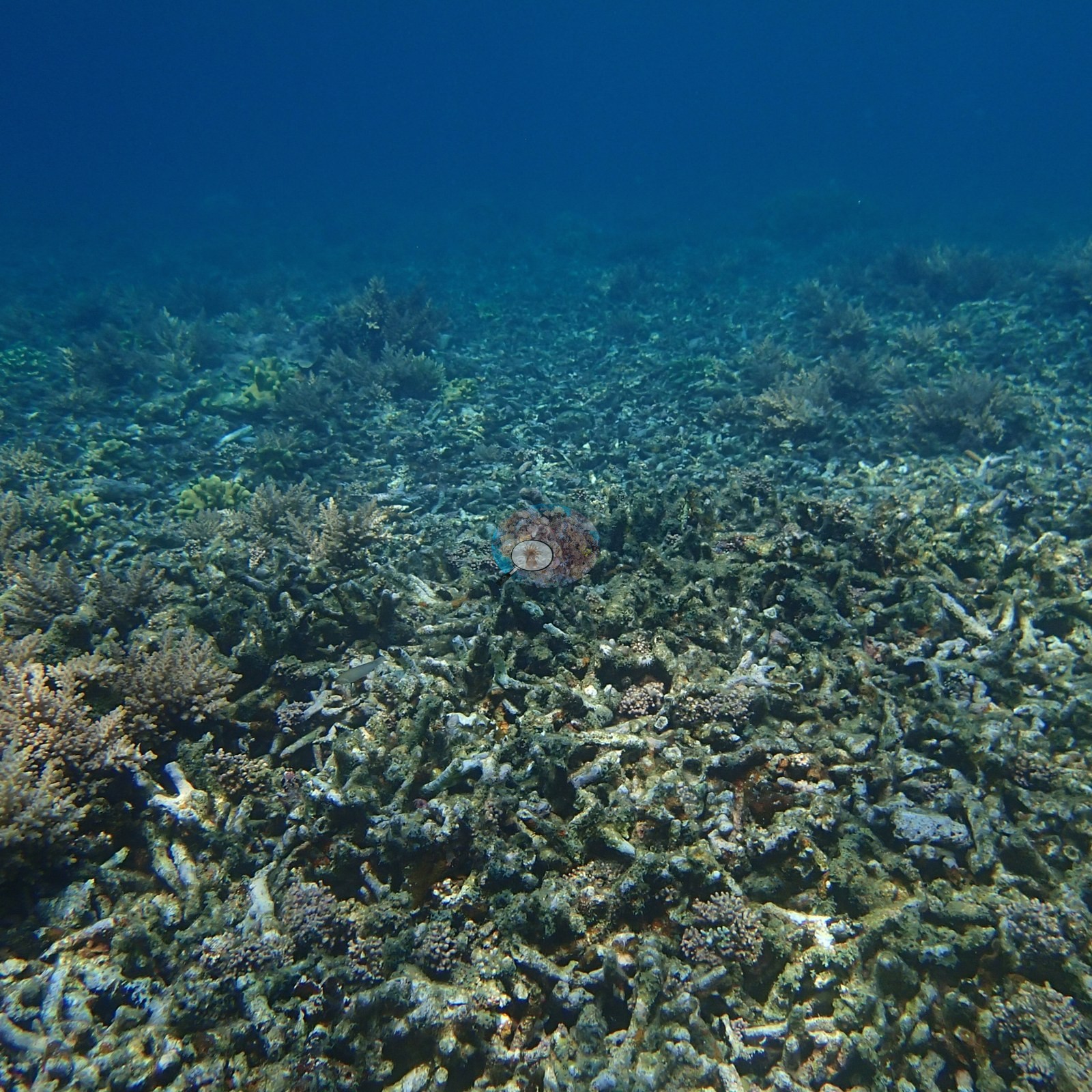
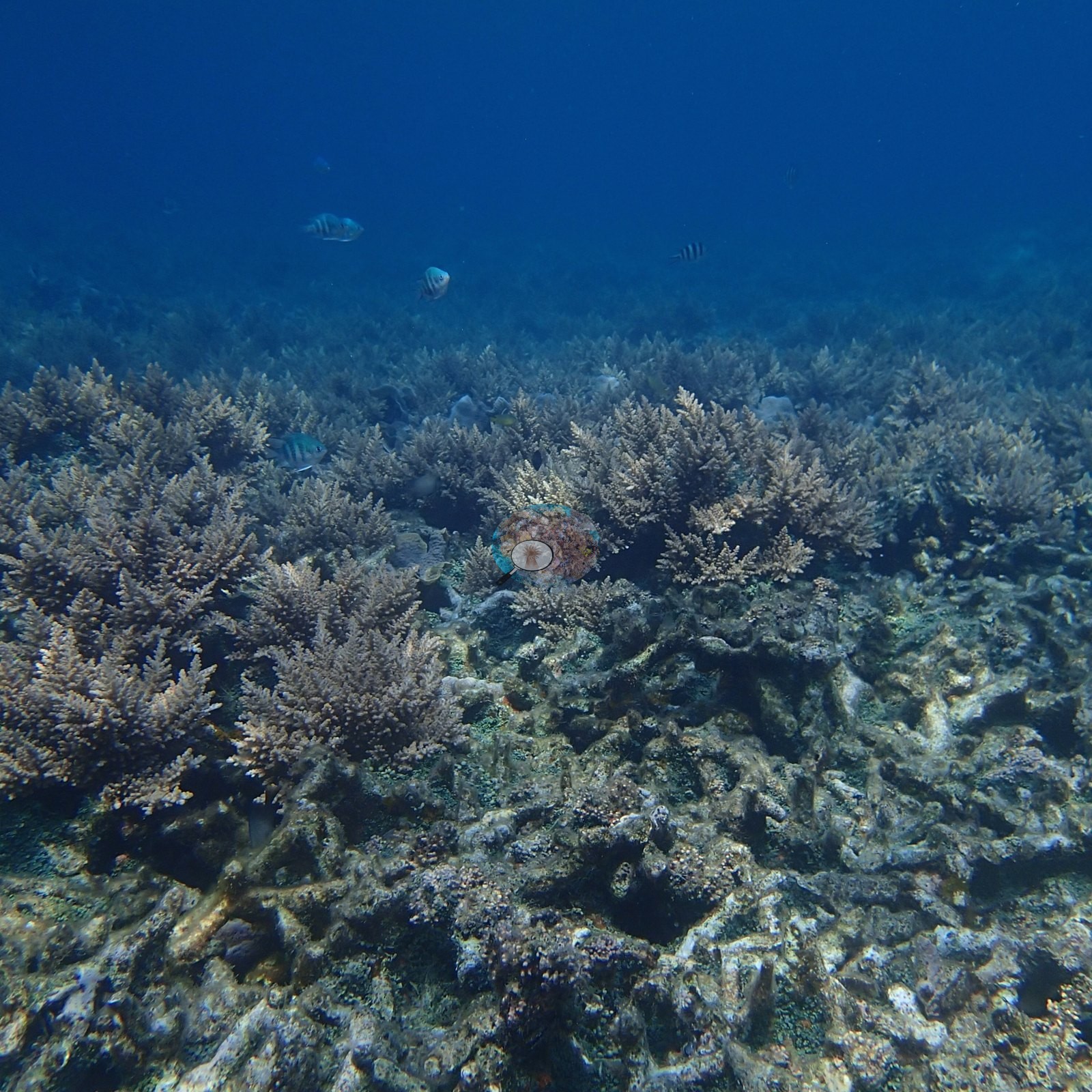
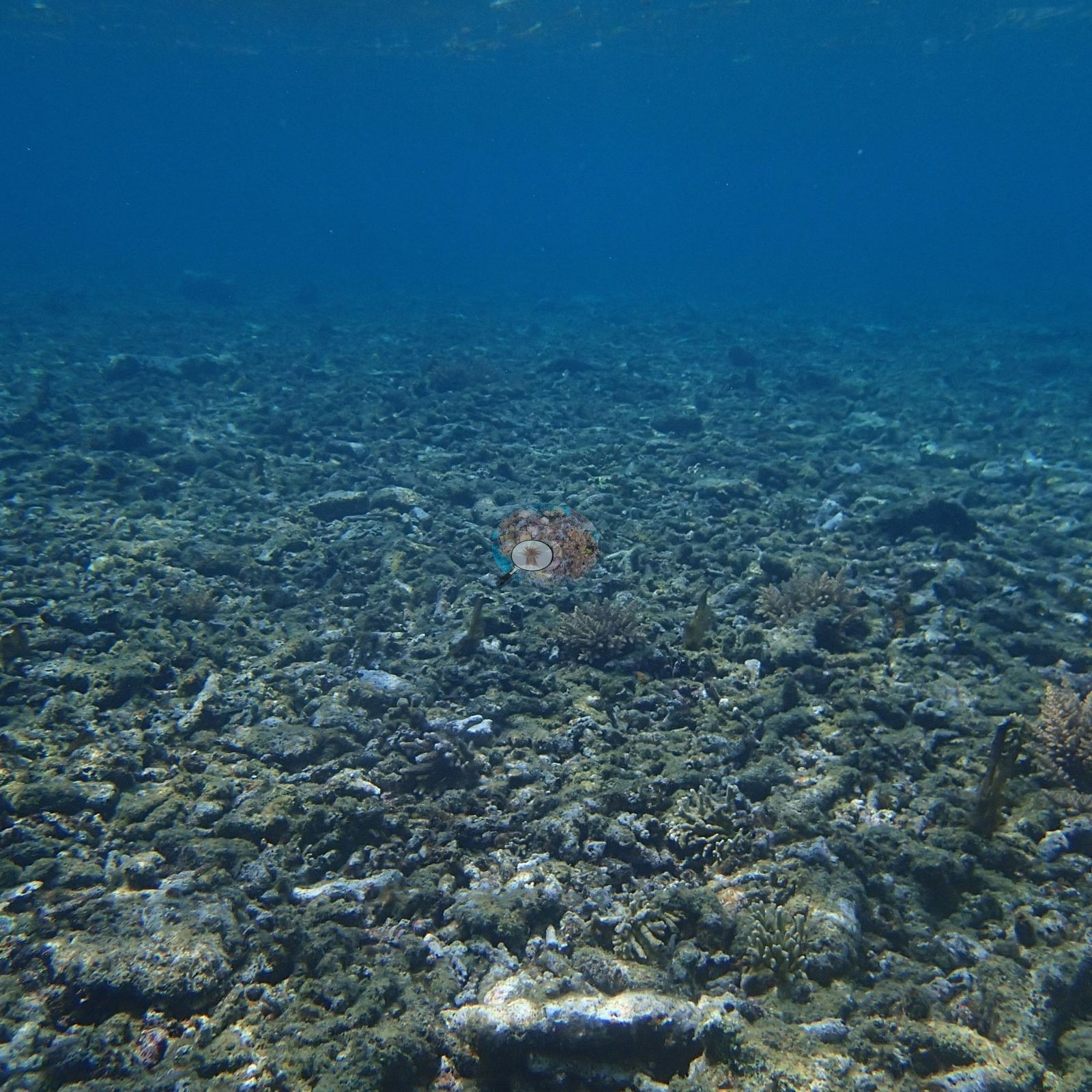
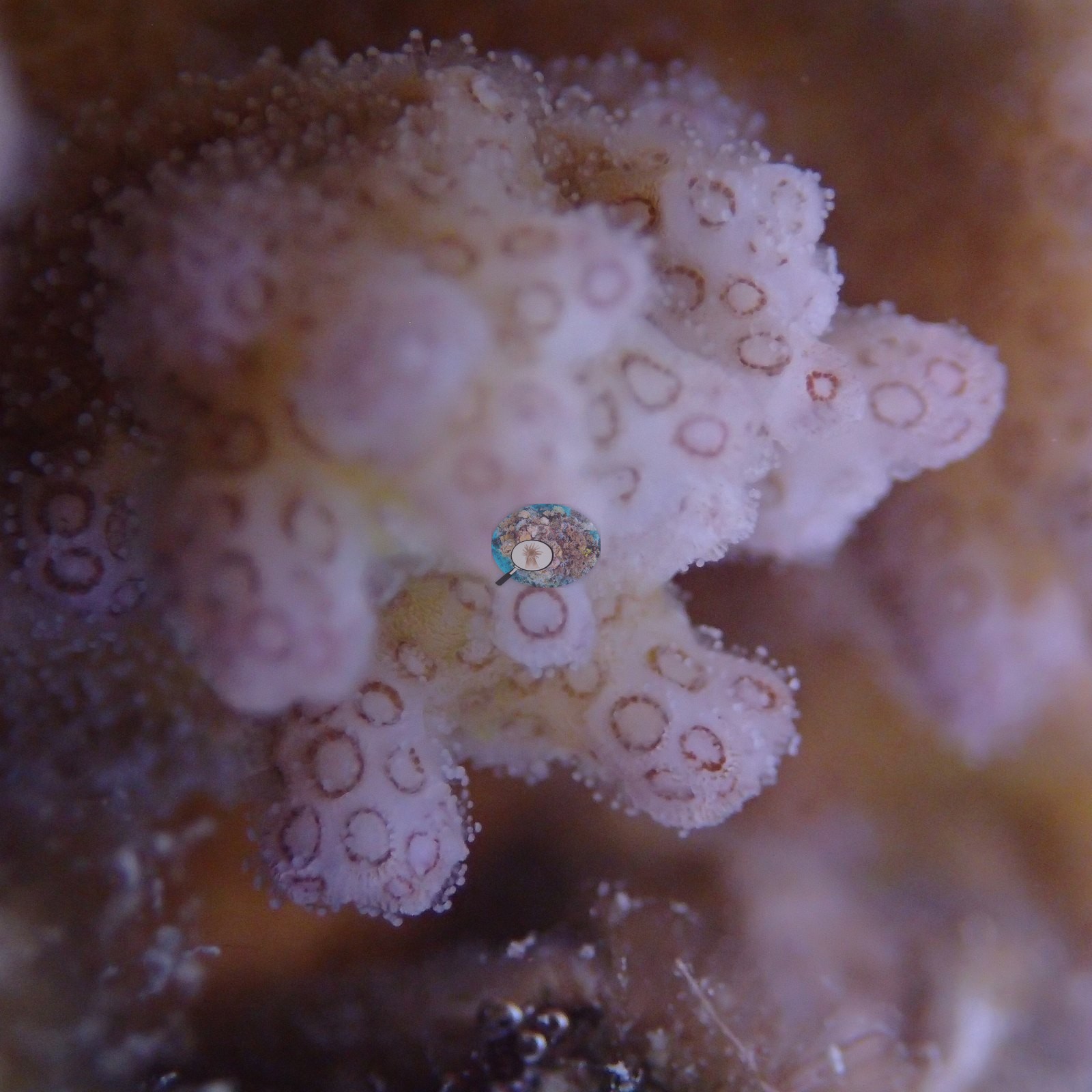
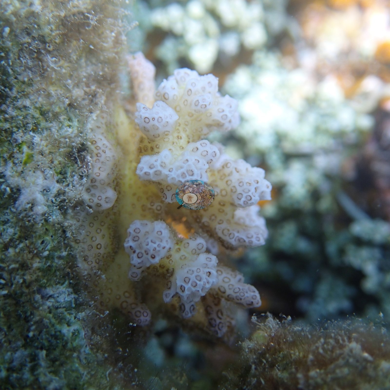
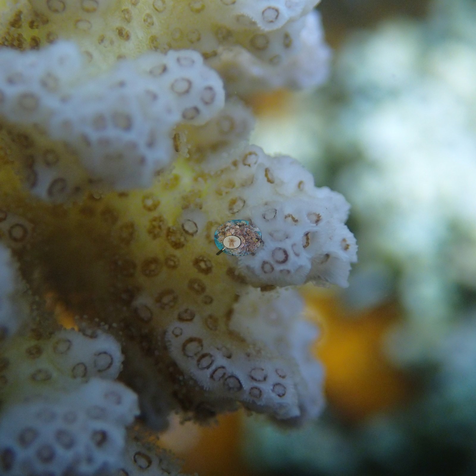
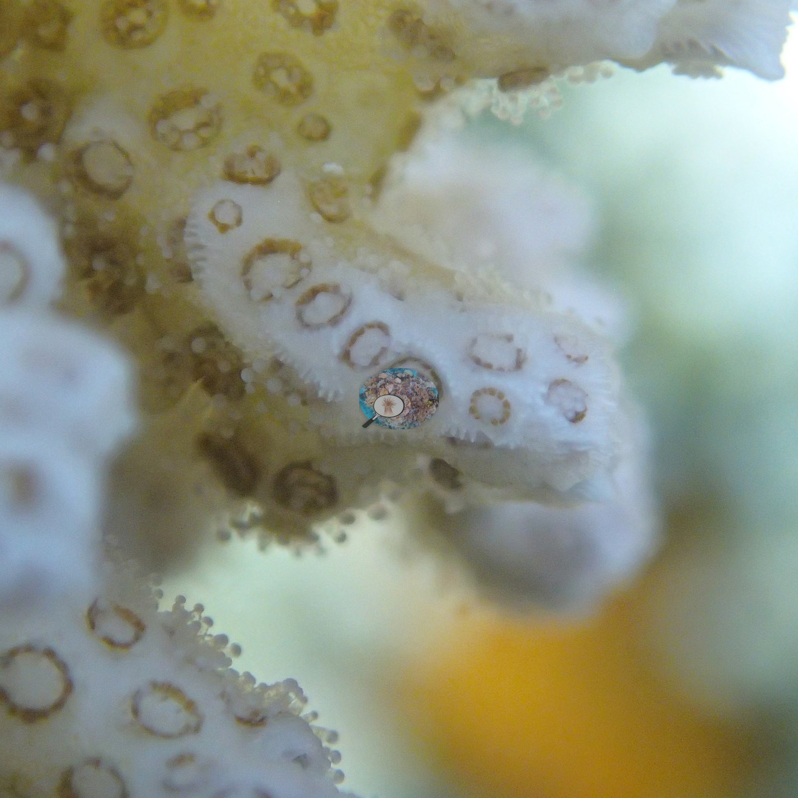
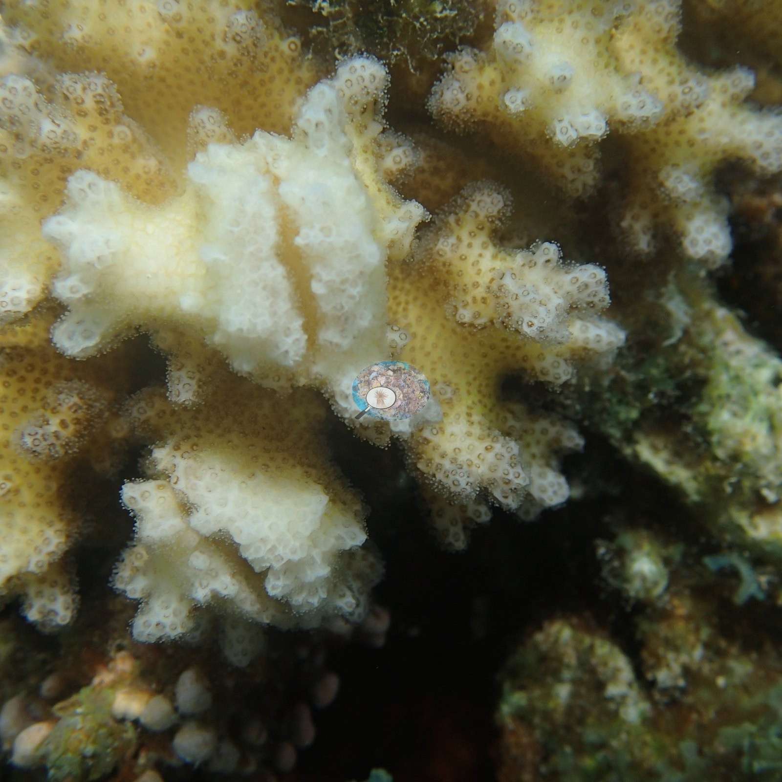
Standing branching Acropora thickets recur throughout (e.g., Image 86, Image 87, Image 88), so the station's "branching" label is earned, and its counterpoint is there too: three or four frames of consolidated, turf-coated dead branching rubble (e.g., Image 2, Image 72, Image 93), in one of which a live thicket has re-established on top of the dead framework (e.g., Image 73).
Coral cover
- Reference: Claude CV point count (this site's photos)
- Coral cover: 57% — a blind Claude computer-vision point count of 115 of Anderson's photographs from the 5 April dive, 20 edited and 95 unedited camera originals (20 points per photo; water, fish, and obstructions excluded); median 59%. Other computer-vision methods are listed in the method table.
Stressors
Anderson's sample sheet for the eight branching colonies sampled here on 5 April 2016, at 1.2–1.9 m, records storm damage and old mortality: one colony grew in 'a shallow area with significant storm damage, some algal smothering (~10%)', one had old and new mortality, two had about 50% old partial mortality, and others grew in rubble-dominated areas with old mortality (the Dongsha sample sheet). No crown-of-thorns starfish (COTS) or other corallivores were evident.
Fish and other fauna
Fish are conspicuous: dense sergeant major, chromis, and humbug damselfish aggregations, squirrelfish, and butterflyfish (e.g., Image 25, Image 27).
Two frames hold a loose group of three or four large cuttlefish over the reef (e.g., Image 29, Image 30).
Comparisons with other data
We found no other surveys confirmed at this site as of 2026. If you would like to share yours, please email us.
Dive record
Anderson photographed Site 6 on one dive, on the morning of 5 April 2016 (123 frames, 09:33-11:01); the 70 frames from two dives on 6 April 2016 that were filed with this site came from a different site whose position was not recorded, and they are on their own page, Dongsha, 6 April 2016. Anderson's sample sheet logs the 5 April dive at this site (Branching 6) as in at 09:30, 90 minutes, maximum depth 11.3 m; no water temperature was recorded for this site.
Other notes
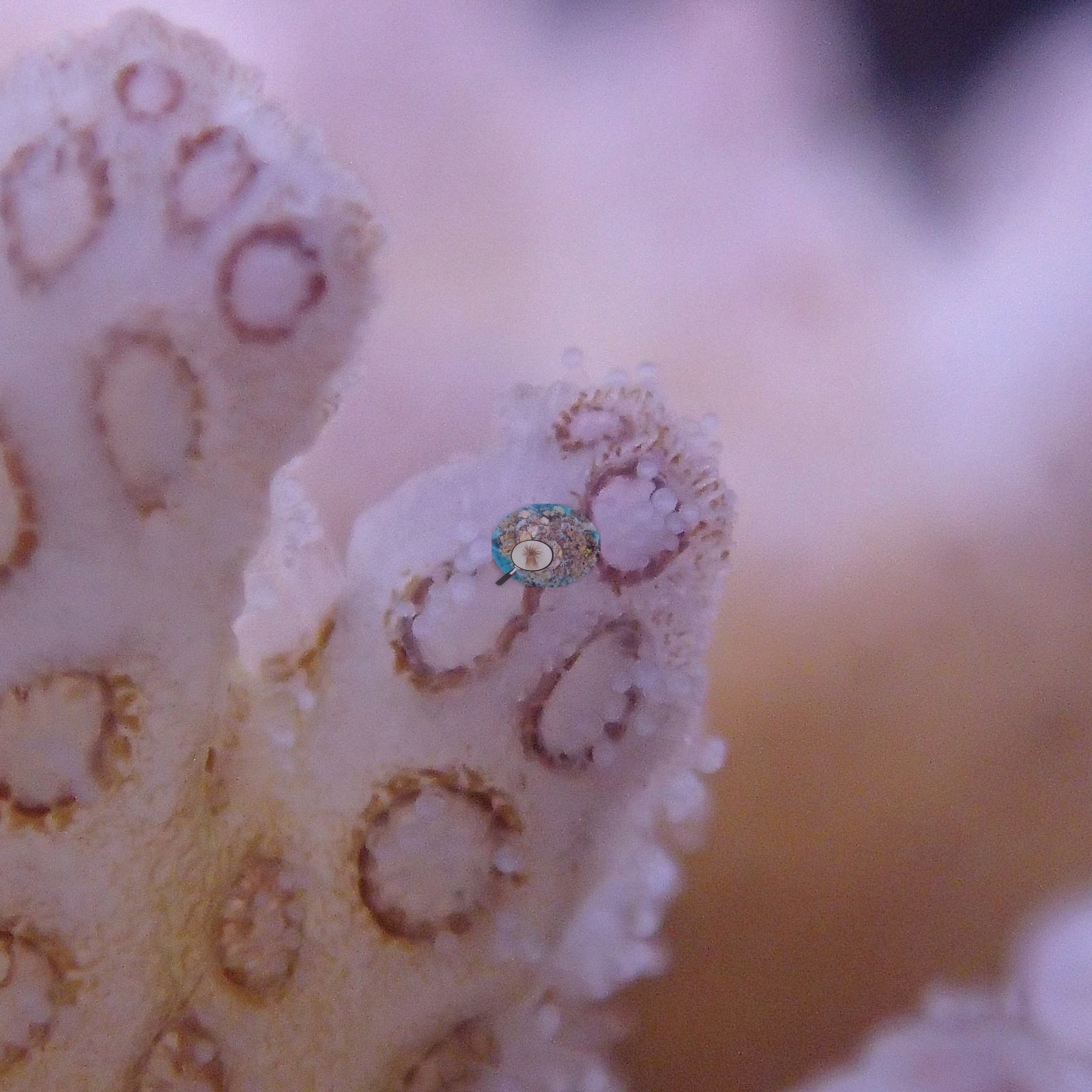
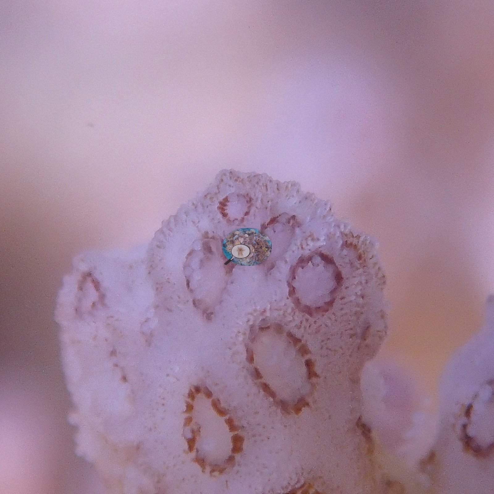
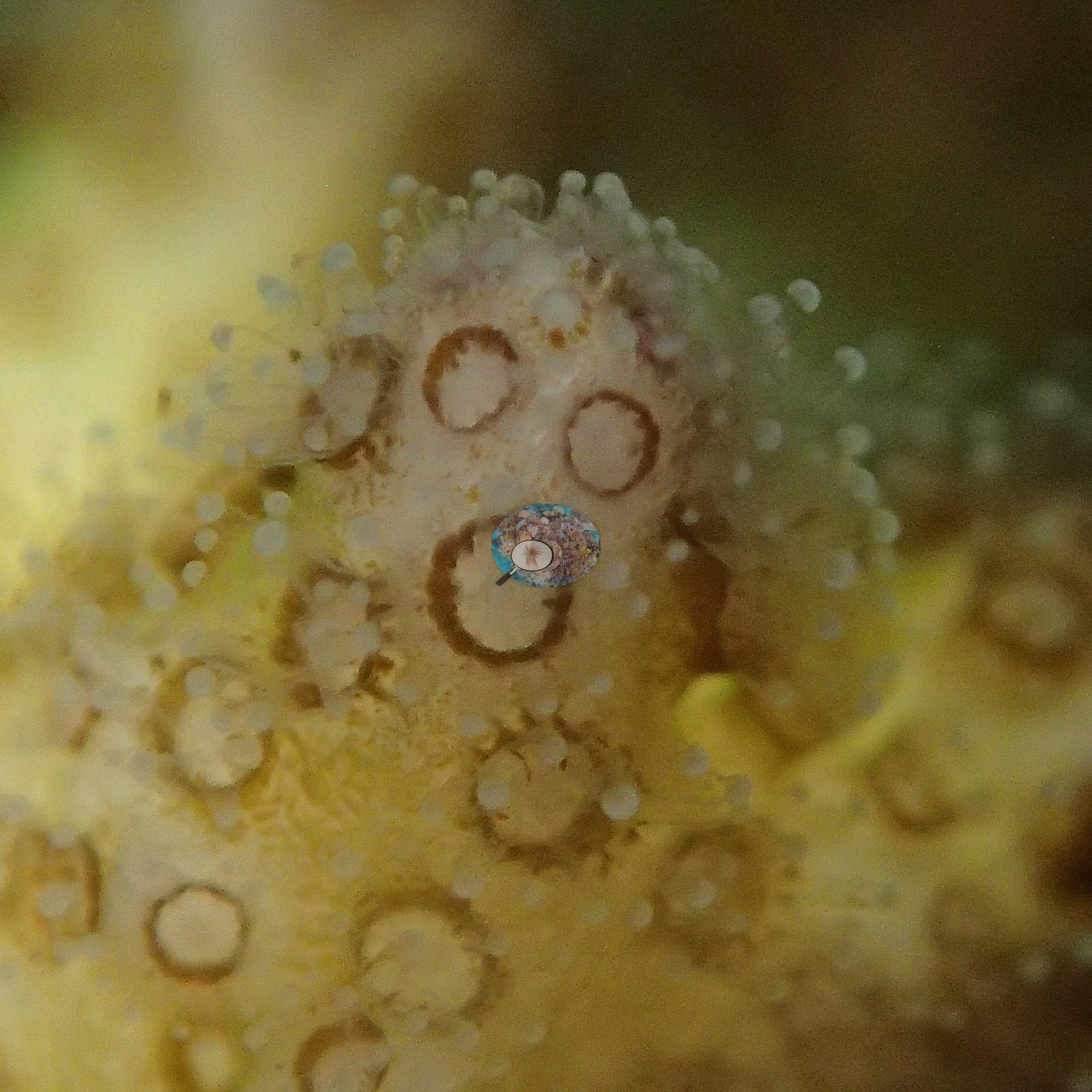
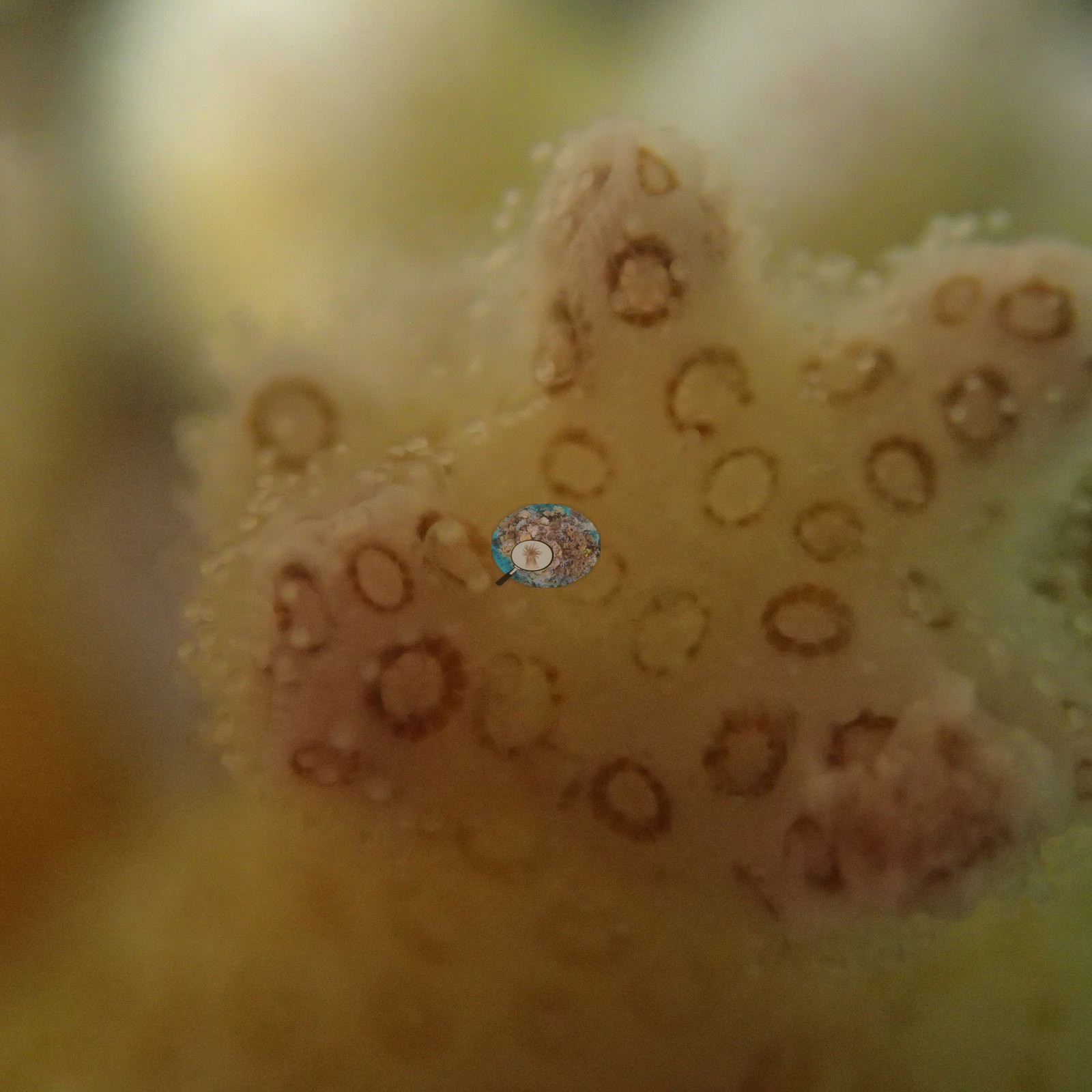
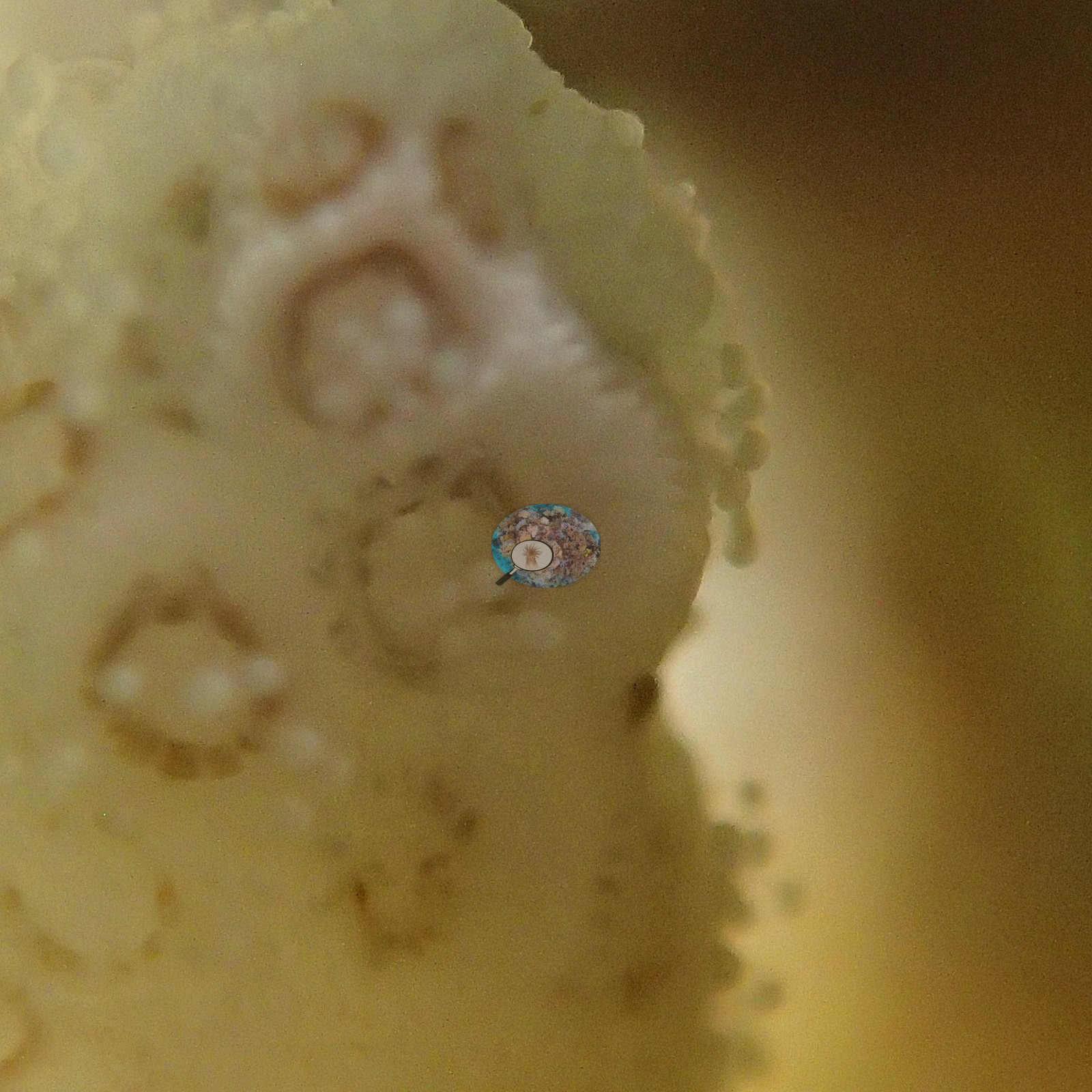
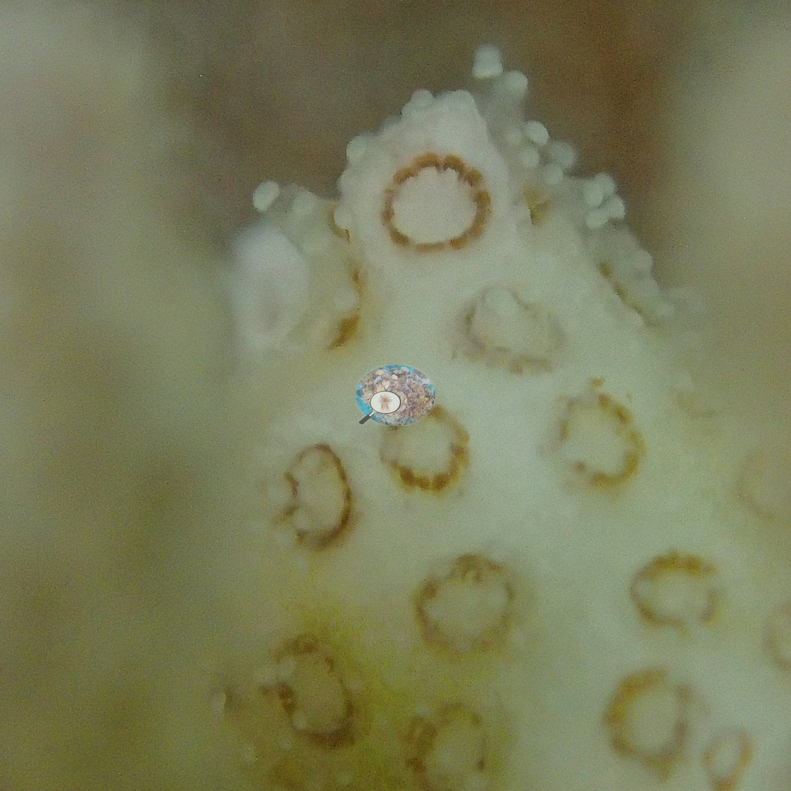
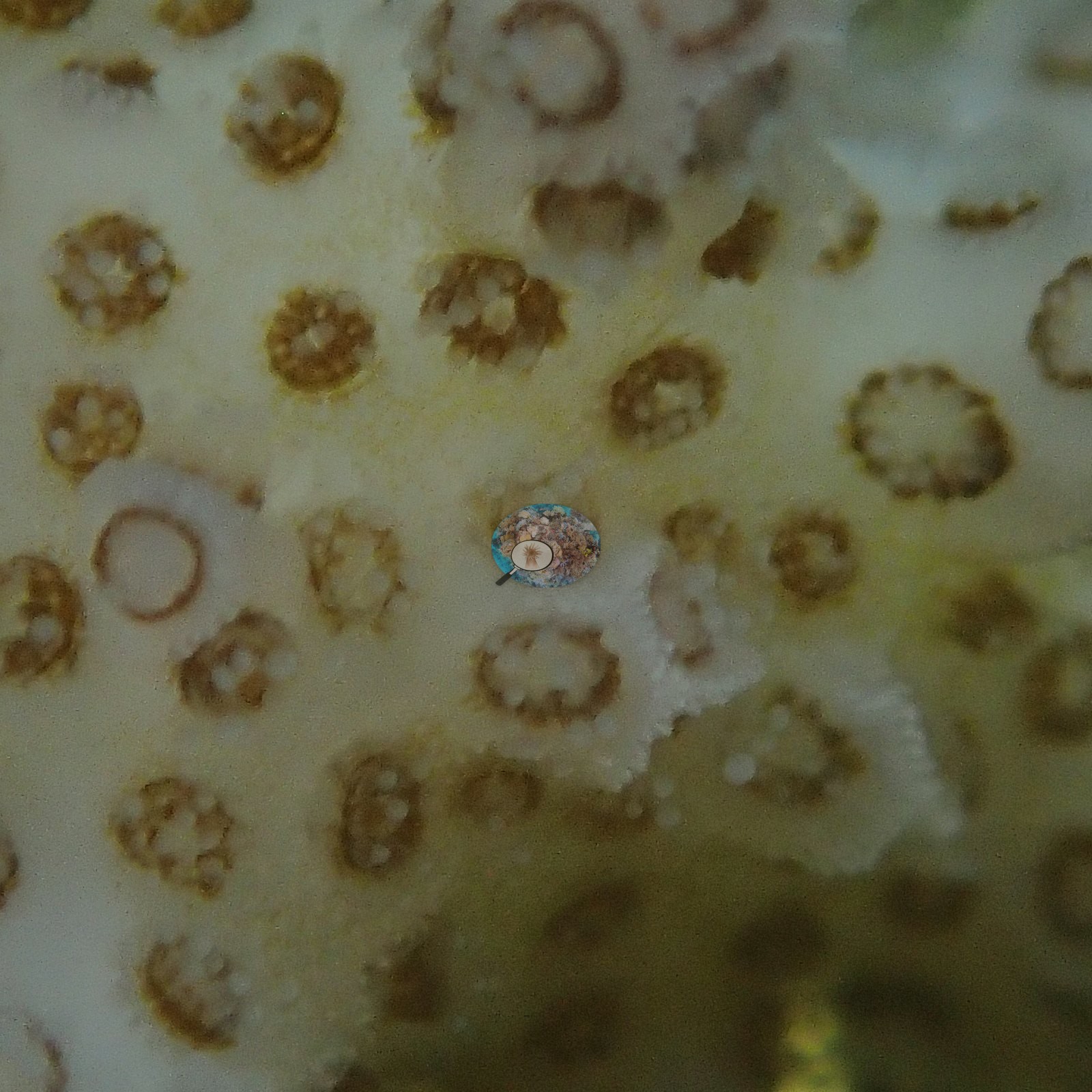
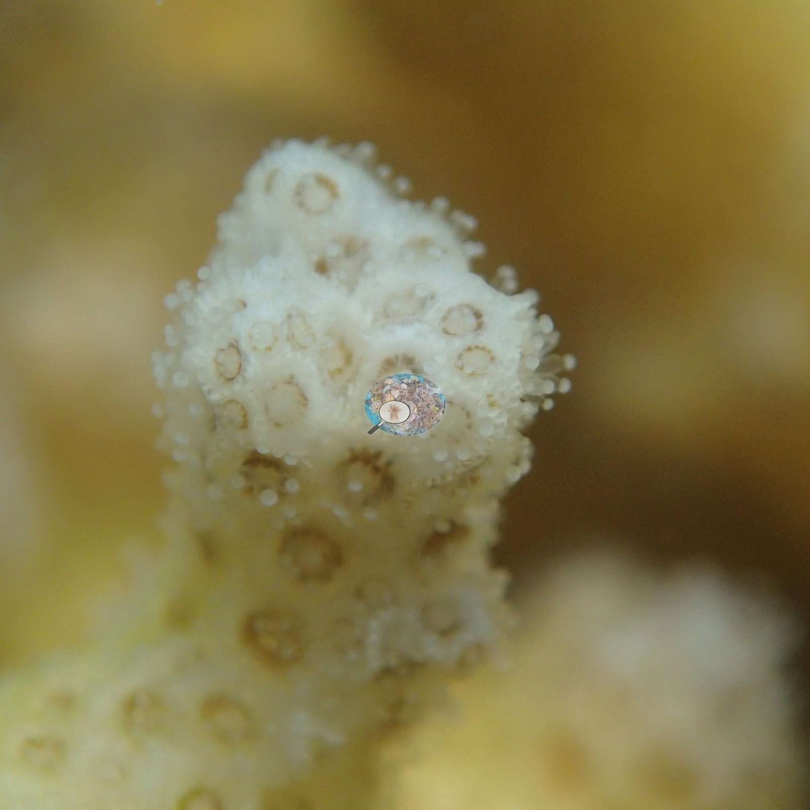
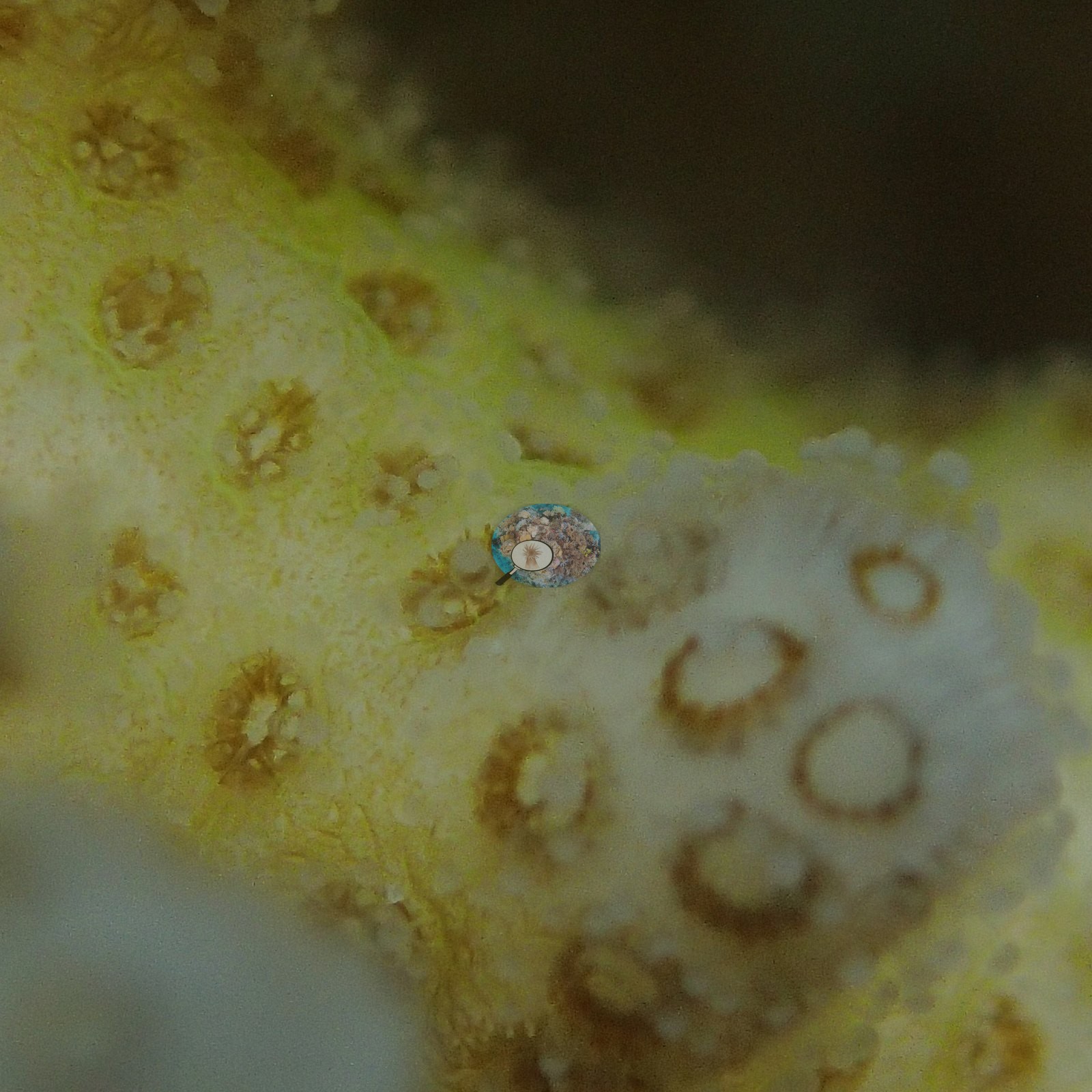
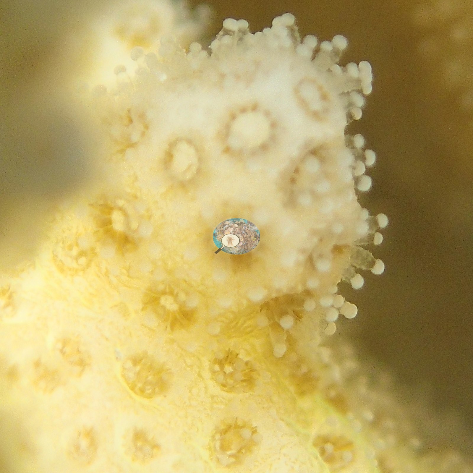
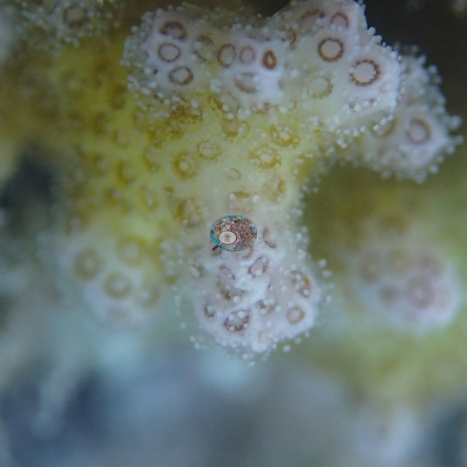
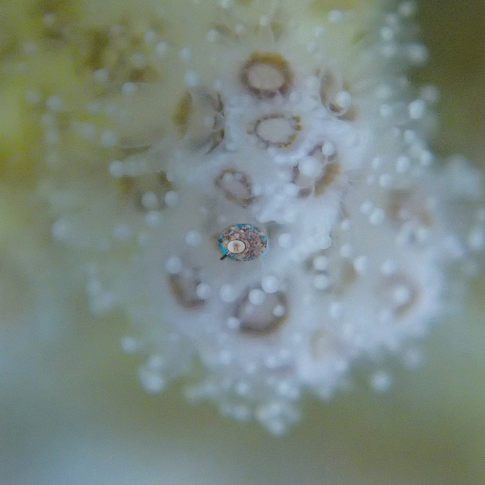
A long block of high-magnification, strobe-lit macro of individual small branching colonies runs through the middle of the 5 April dive (e.g., Image 67, Image 68, Image 69, Image 70, Image 71, Image 74, Image 75, Image 76, Image 77, Image 79, Image 80, Image 81, Image 82, Image 83, Image 84, Image 85, Image 90, Image 91, Image 92, Image 94, Image 95, Image 96, Image 97, Image 98, Image 100, Image 101, Image 102, Image 103, Image 104, Image 105, Image 106); it is the part of the photo set most likely to distort a photograph-derived cover figure, and the cover figure on this page does not exclude these frames.
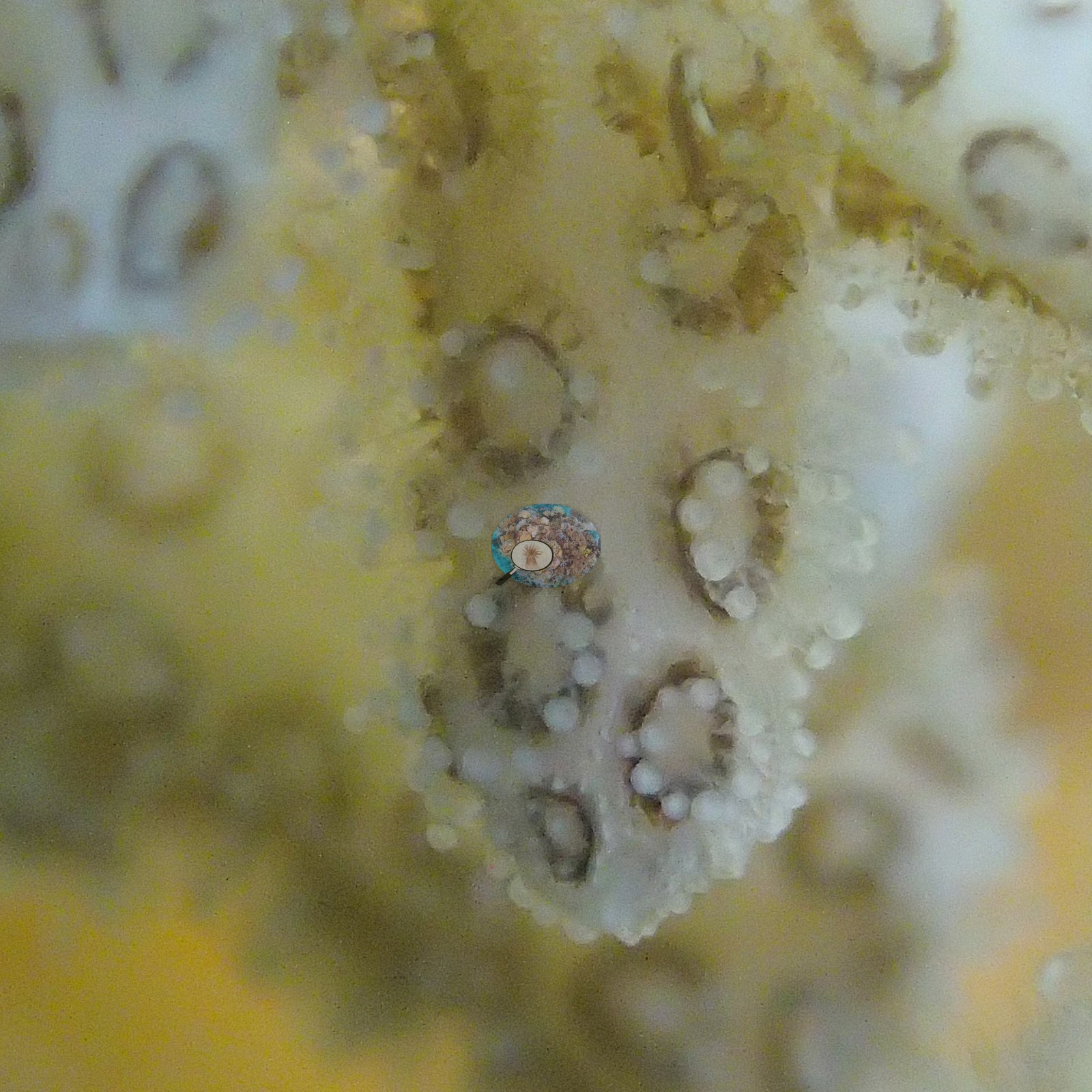
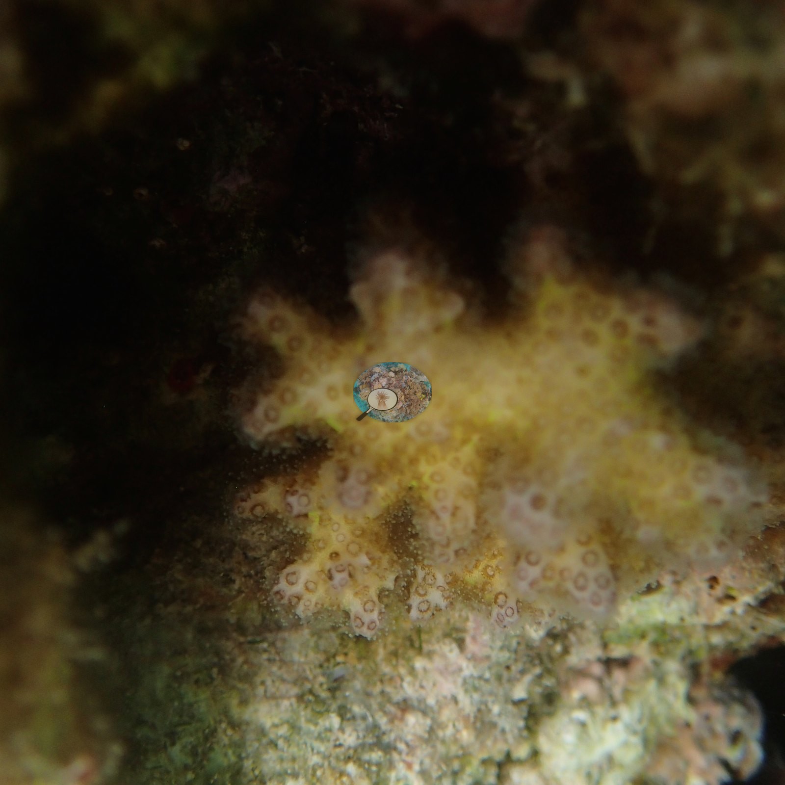
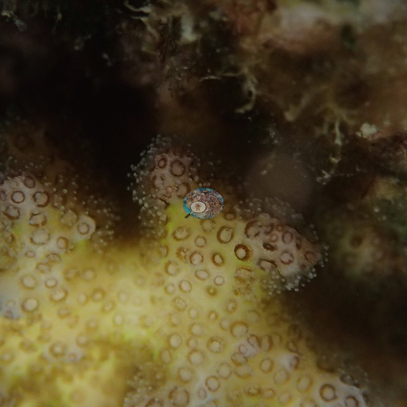
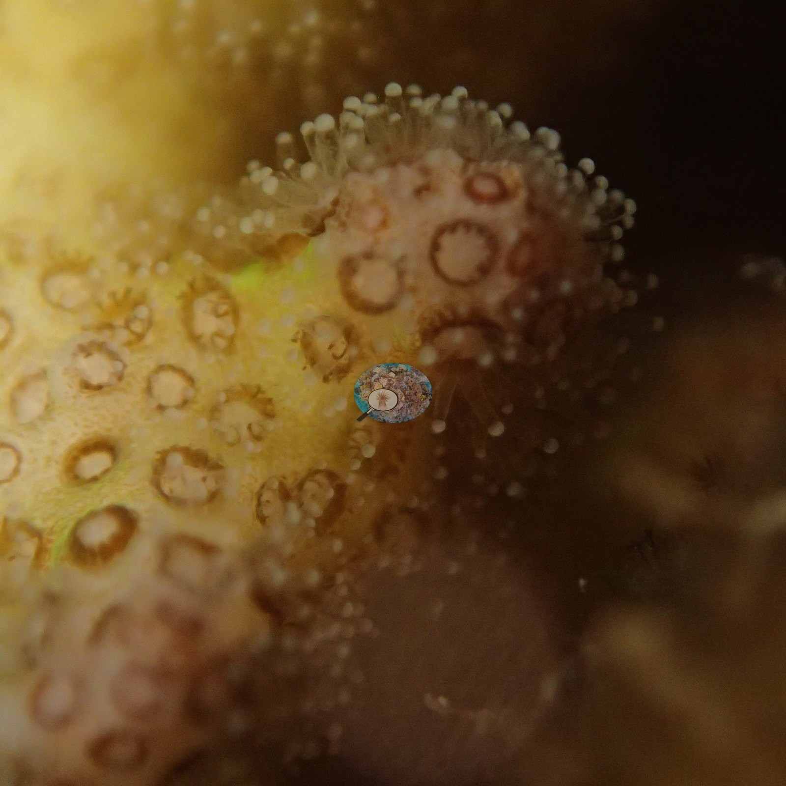
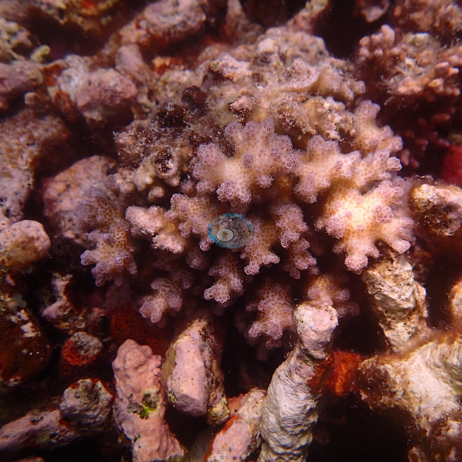
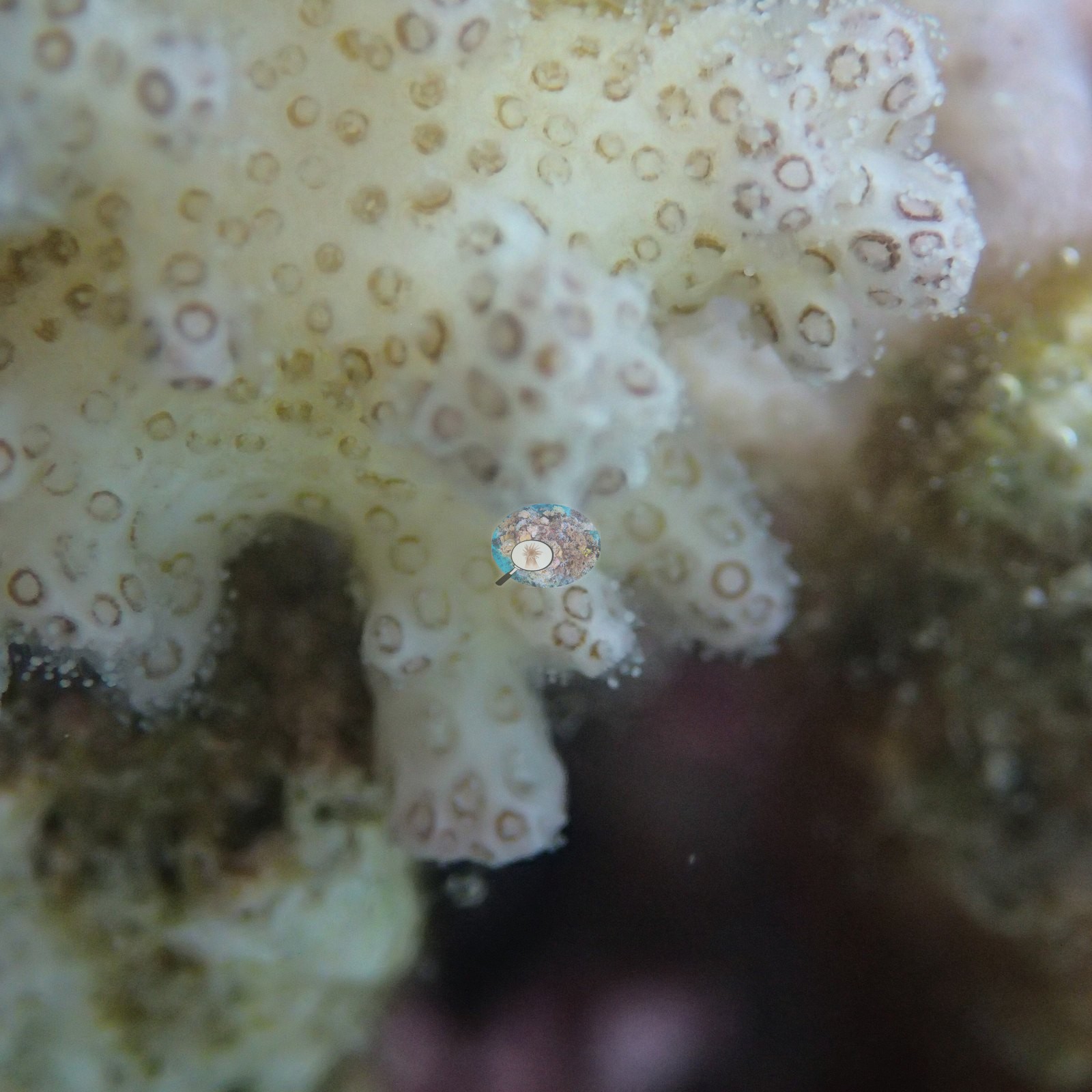
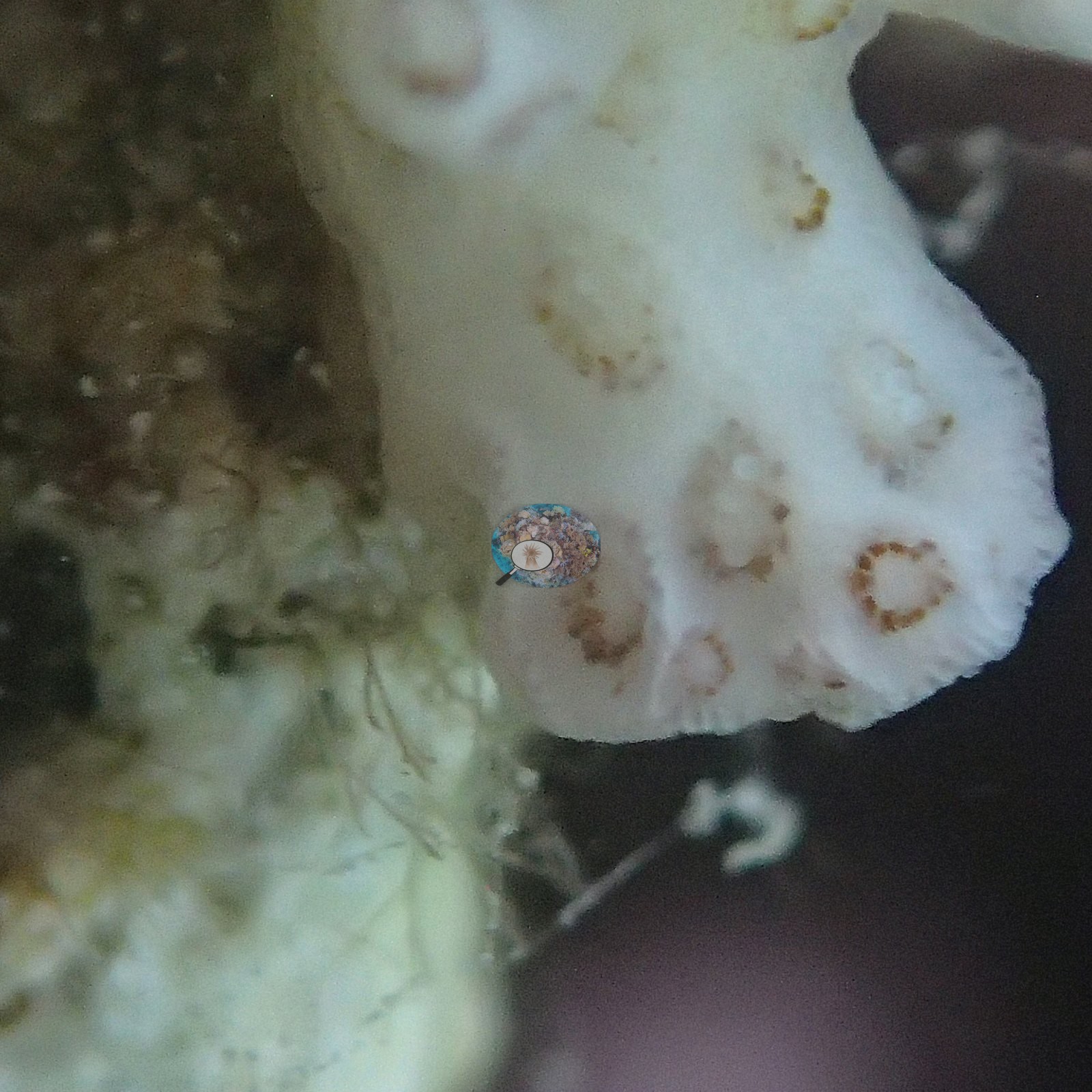
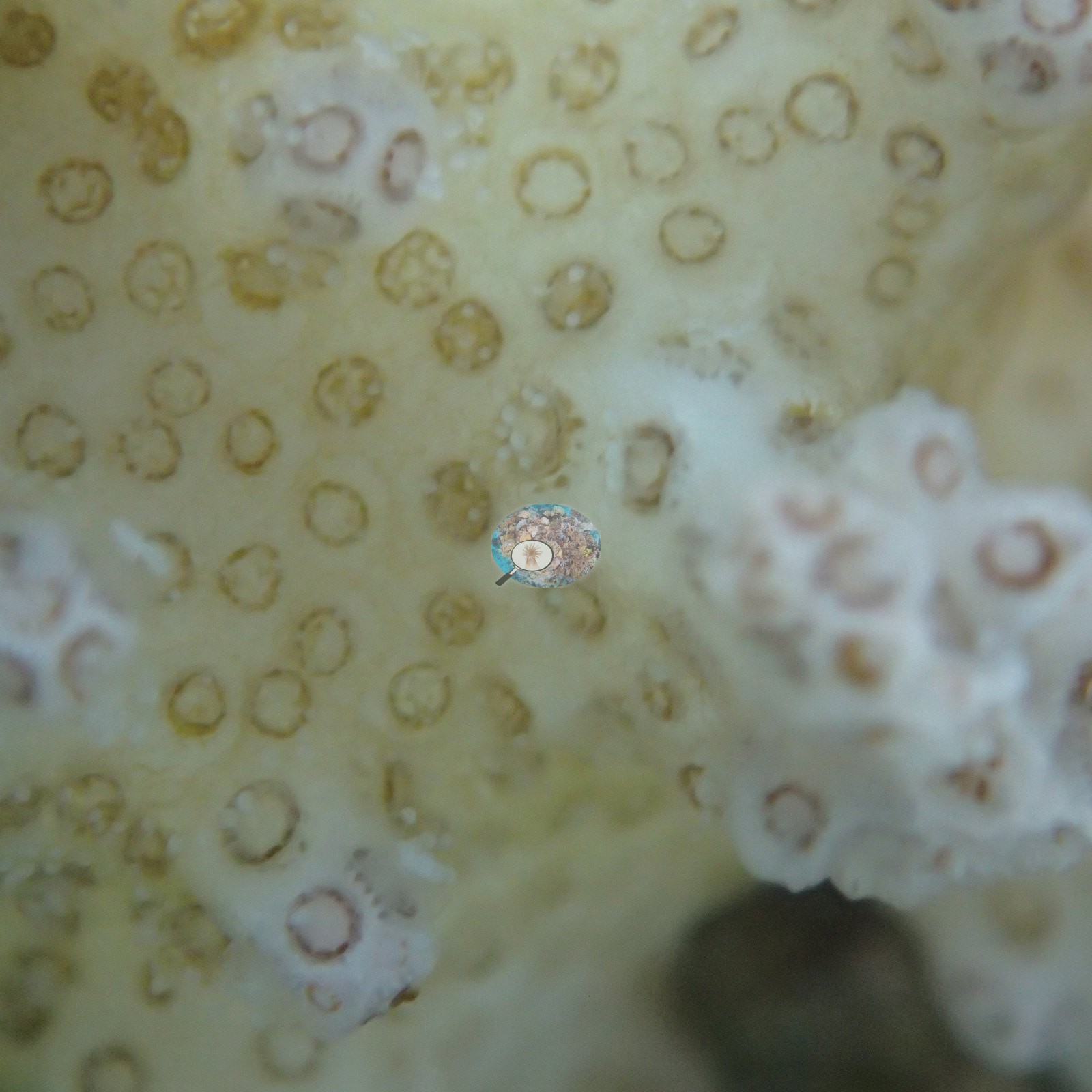
CV model comparison
More info
No computer-vision method yet estimates coral cover accurately on new images, so treat this as indicative rather than a measurement; it is revised as the methods improve.More info
Other rows are for comparison only and are never averaged. No diver transect exists for this trip, so agreement between rows is not accuracy.| Method | Hard coral cover (%) | Photos (n) ± SE |
|---|---|---|
| CoralSCOP edited | 39.2 | n = 88 · SE 2.2 |
| Claude | 56.5 | n = 115 · SE 2.2 |
CoralSCOP (edited photos) reads 39.2%, 17.3 pp below Claude's blind point count (56.5%); the headline stays Claude's blind point count because no transect or manual count exists here.
See a more detailed, stand-alone version of this table.
Data & links
All images from Coral Reef Diagnostics. Data are from Coral Reef Diagnostics.
© Coral Reef Diagnostics.