
Dongsha Atoll - Site 13
Part of the regional summaries: Taiwan - Dongsha and Taiwan
More info
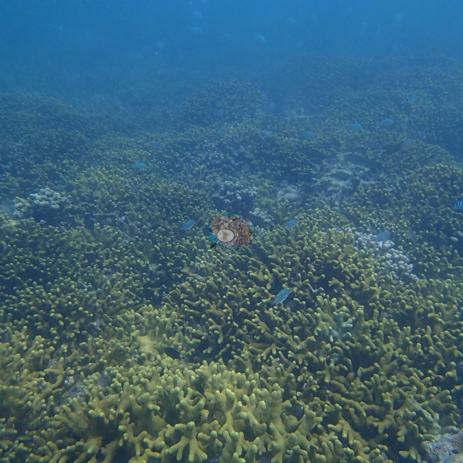
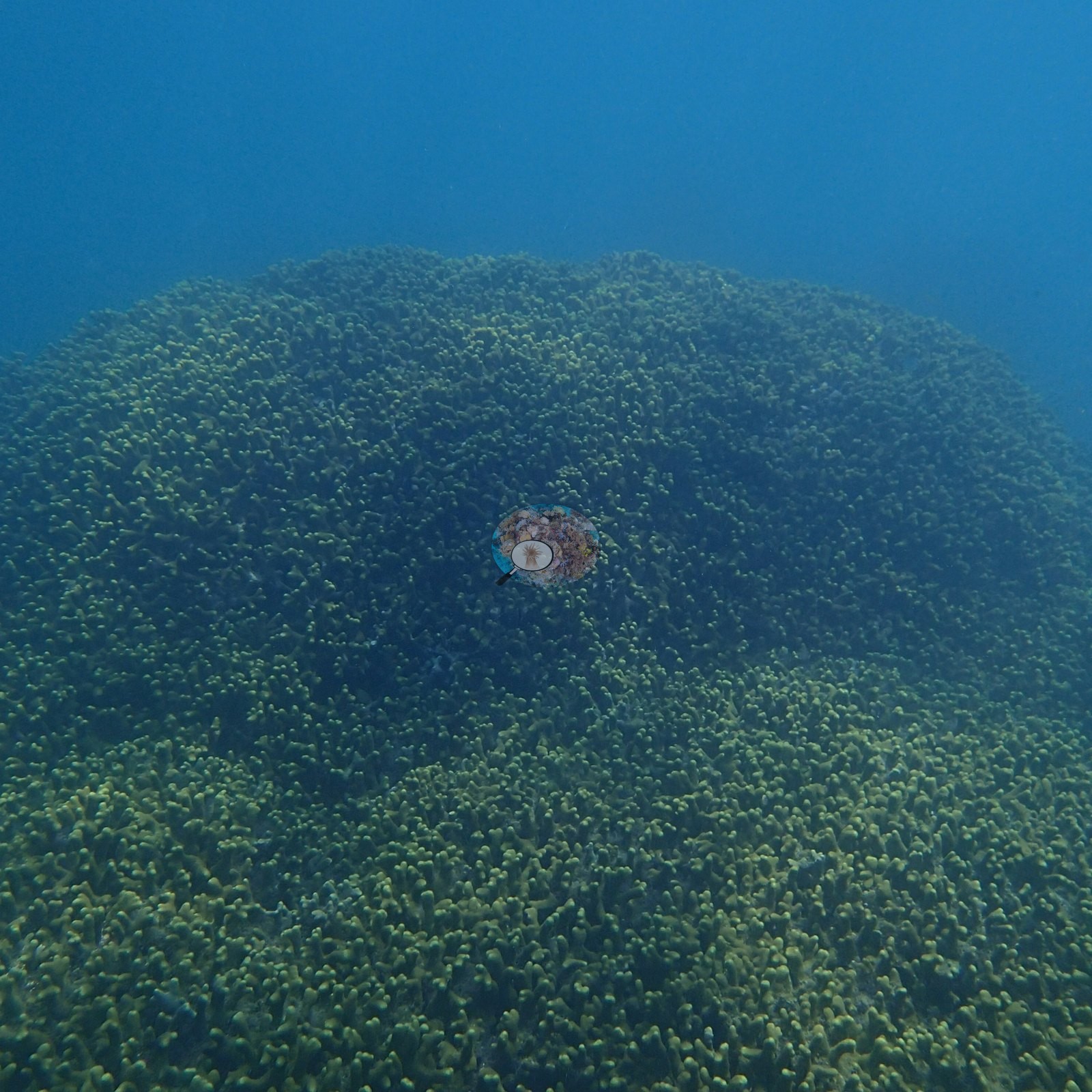
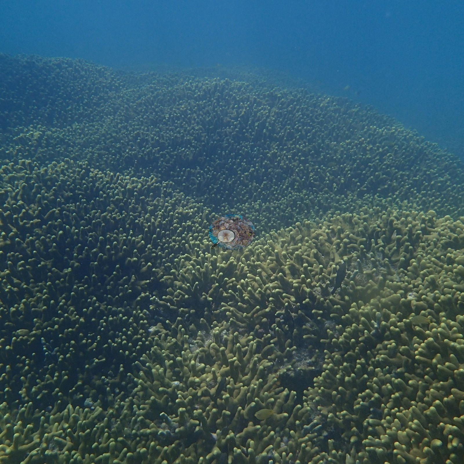
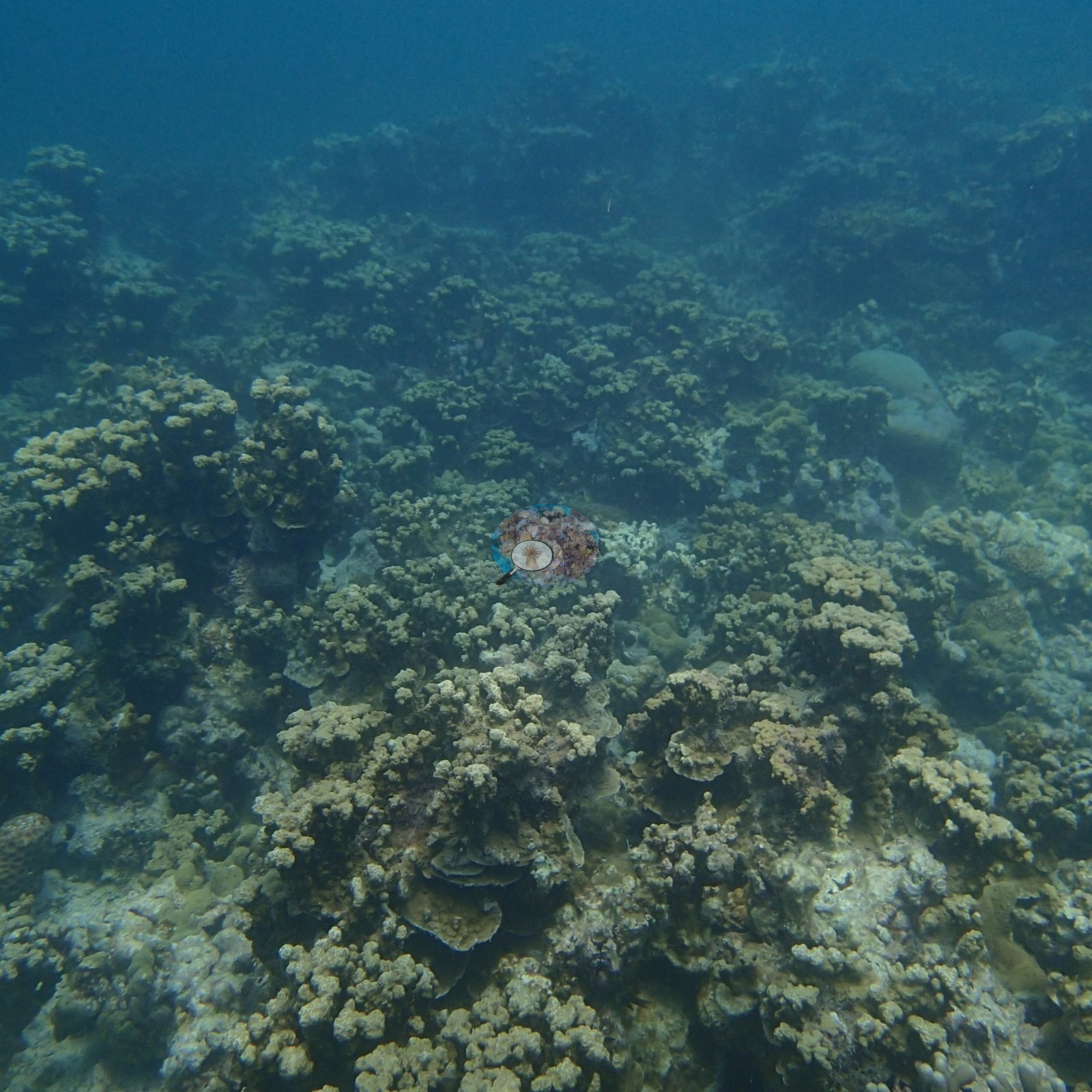
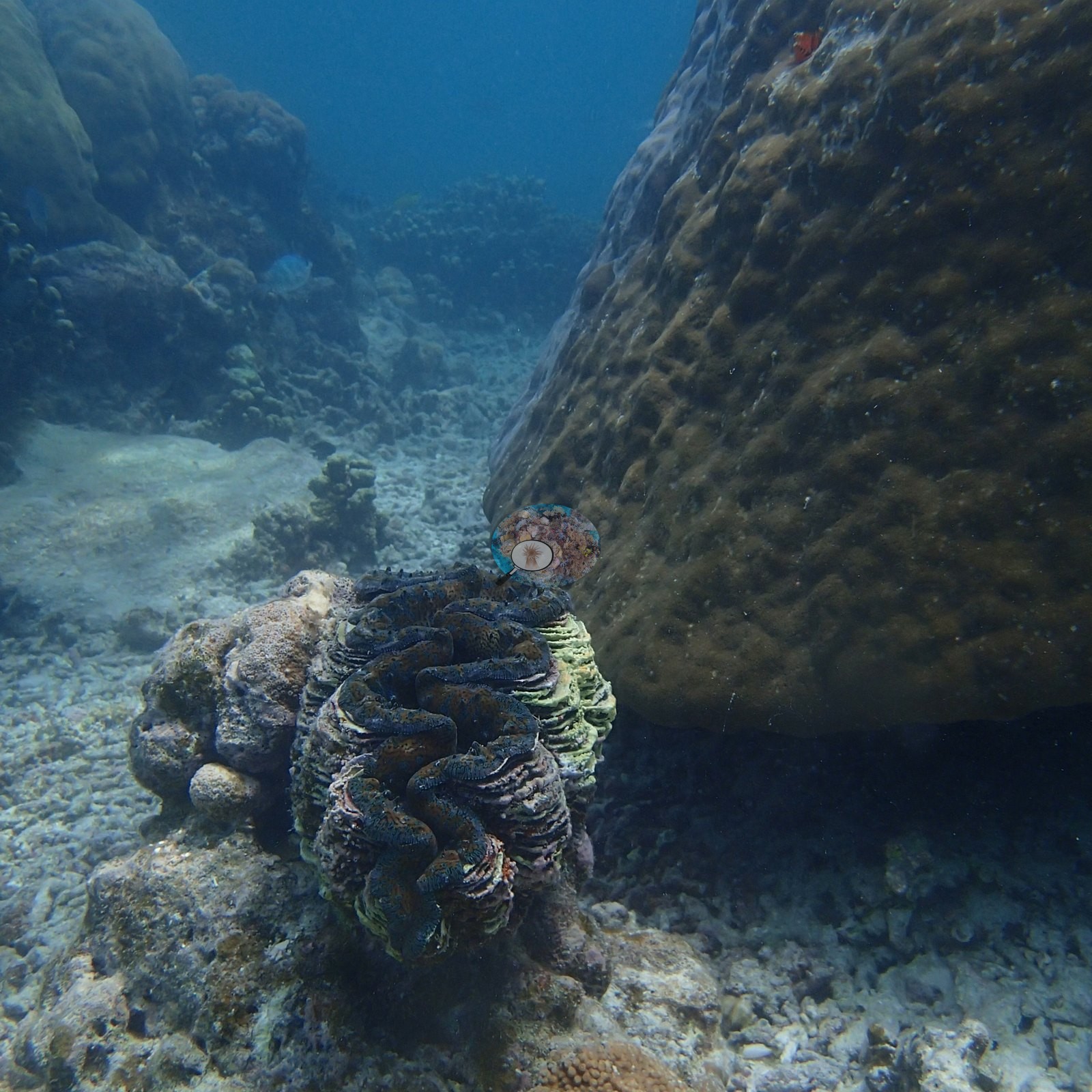
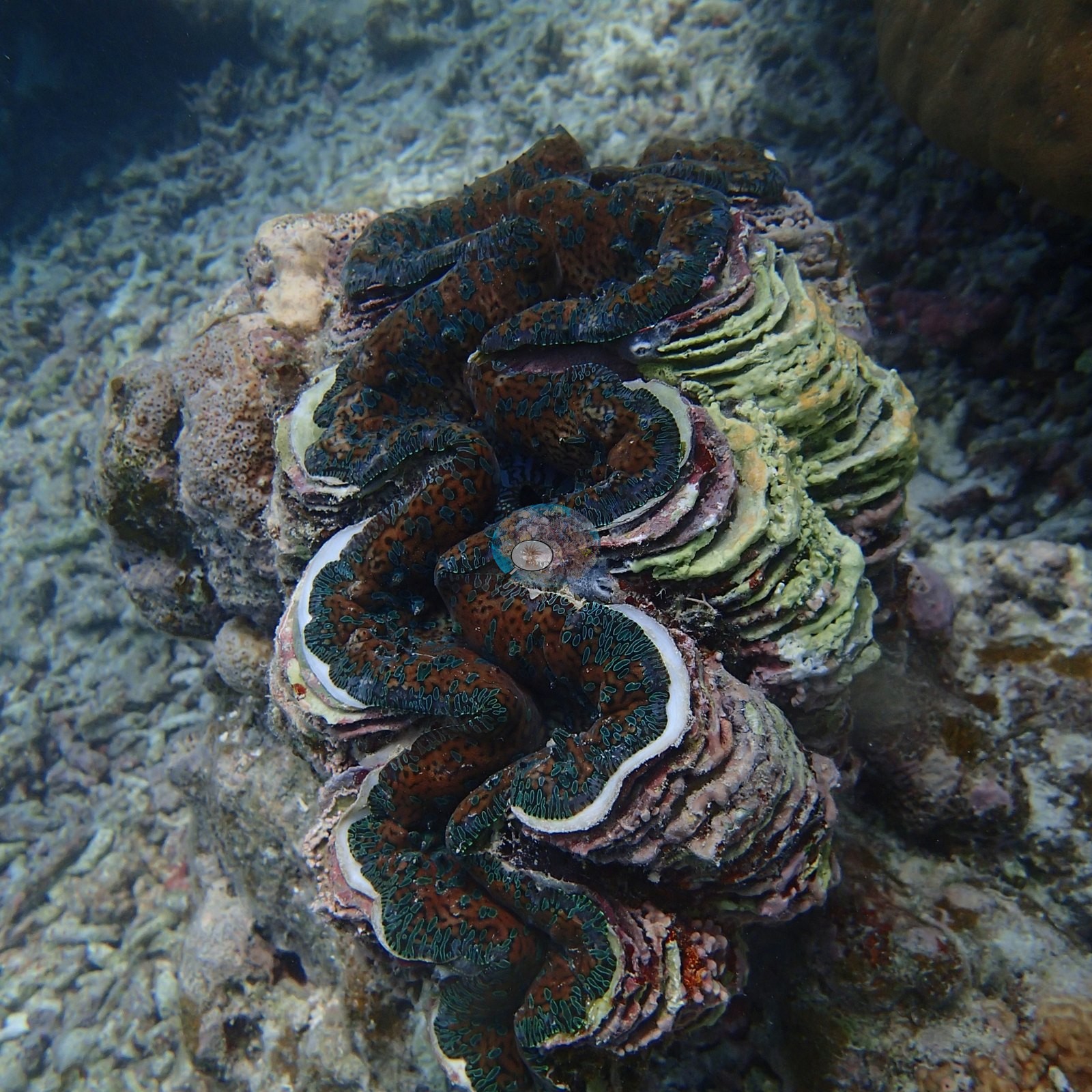
Dongsha, Taiwan (NOAA CRW virtual station)Site 13 at Dongsha Atoll is a vast bank of yellow-green finger corals studded with large massive Porites-type domes and striking tiered plate-coral pinnacles two to three meters tall, plus a giant clam. Anderson's sample sheet records one of the twelve branching corals sampled here as about 20% bleached or dead and another with recent partial mortality; derelict fishing line was draped across one massive coral.
東沙環礁 13 號測站是一大片黃綠色指形珊瑚構成的礁灘,其間點綴著巨大的塊狀 Porites 型圓丘,以及高達兩至三公尺、層次分明而醒目的片狀珊瑚塔,另有一顆巨硨磲。Anderson 的採樣紀錄表記載,在此採樣的十二株分枝狀珊瑚中,有一株約 20% 白化或死亡,另一株有近期的部分死亡;一顆塊狀珊瑚上披掛著廢棄漁線。
本摘要由 AI 翻譯;以英文版本為準。
One other CRD dive site with its own page within 5 km shown in orange — click a marker to open its page.
This page shows 50 of the 63 photographs from this site, including every one the text cites.
36 of the 50 photographs on this page are shown beside the text that discusses them under "Site description" below.
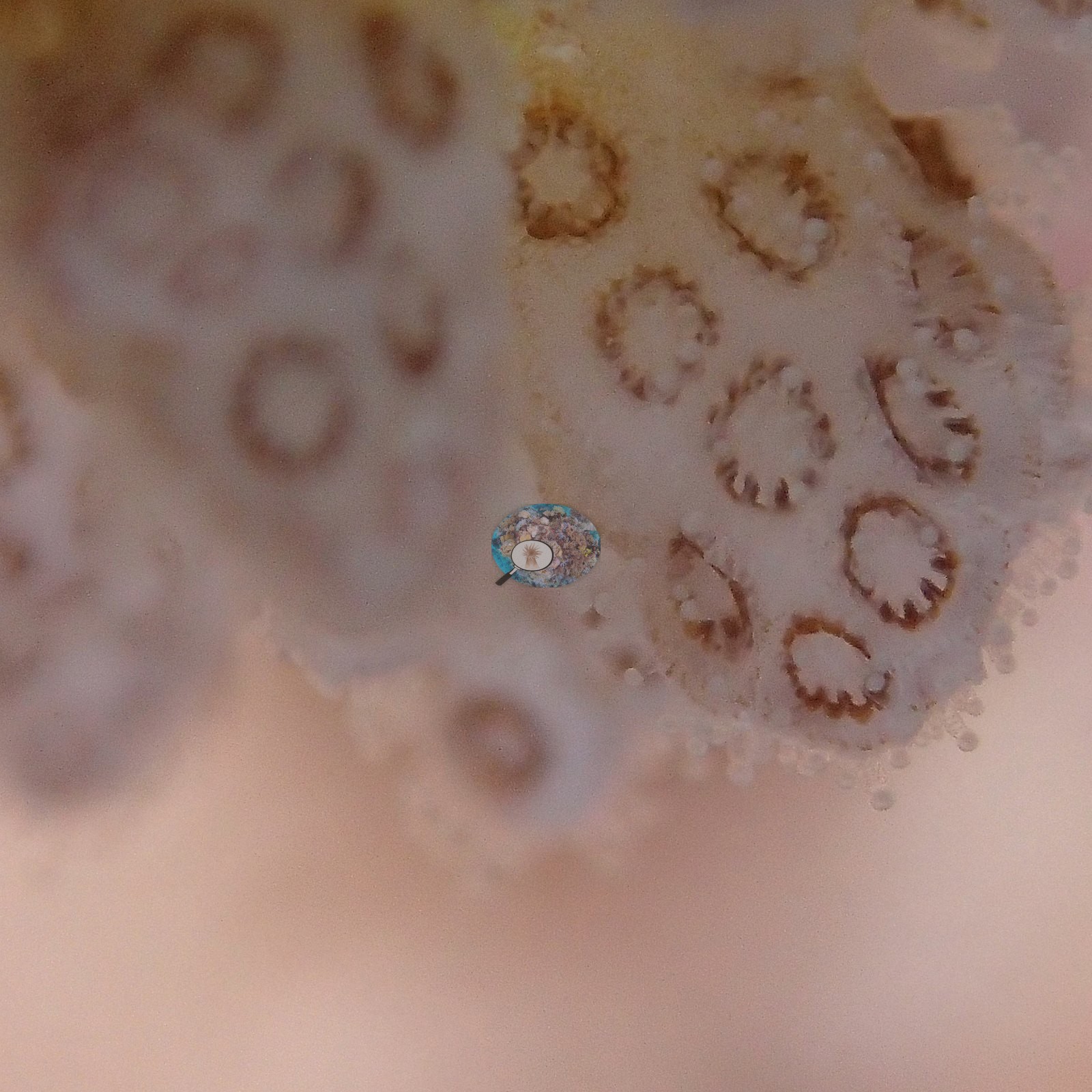
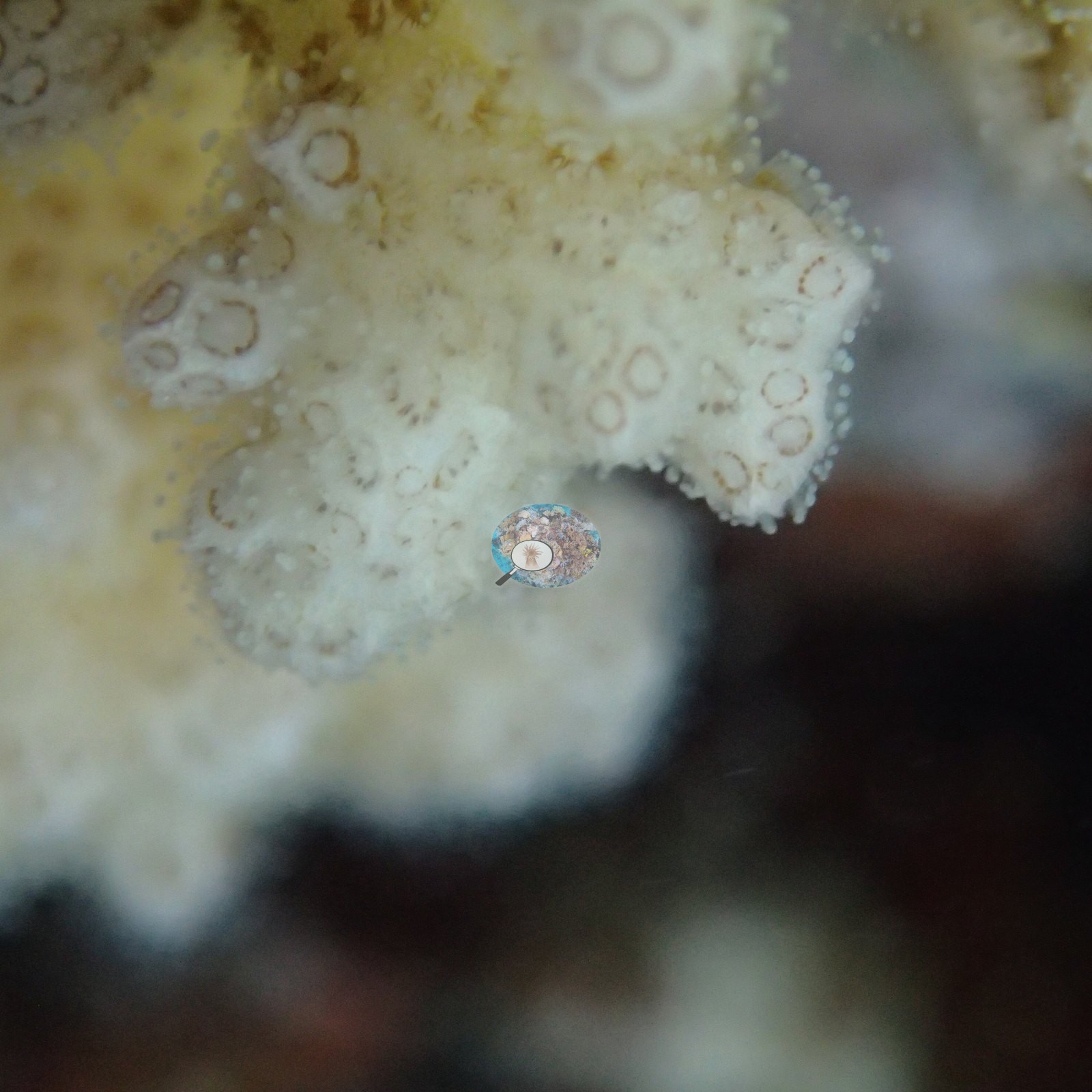
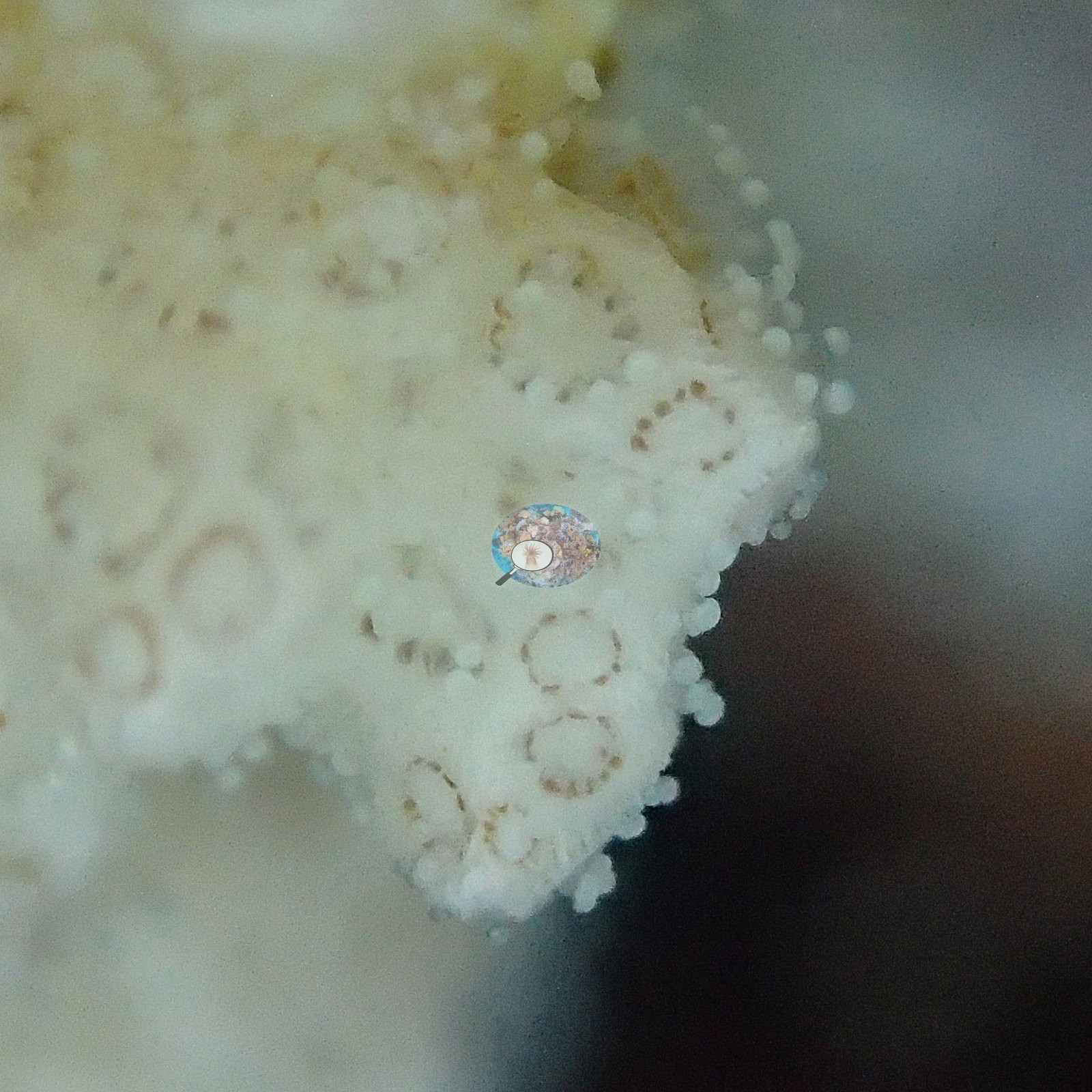
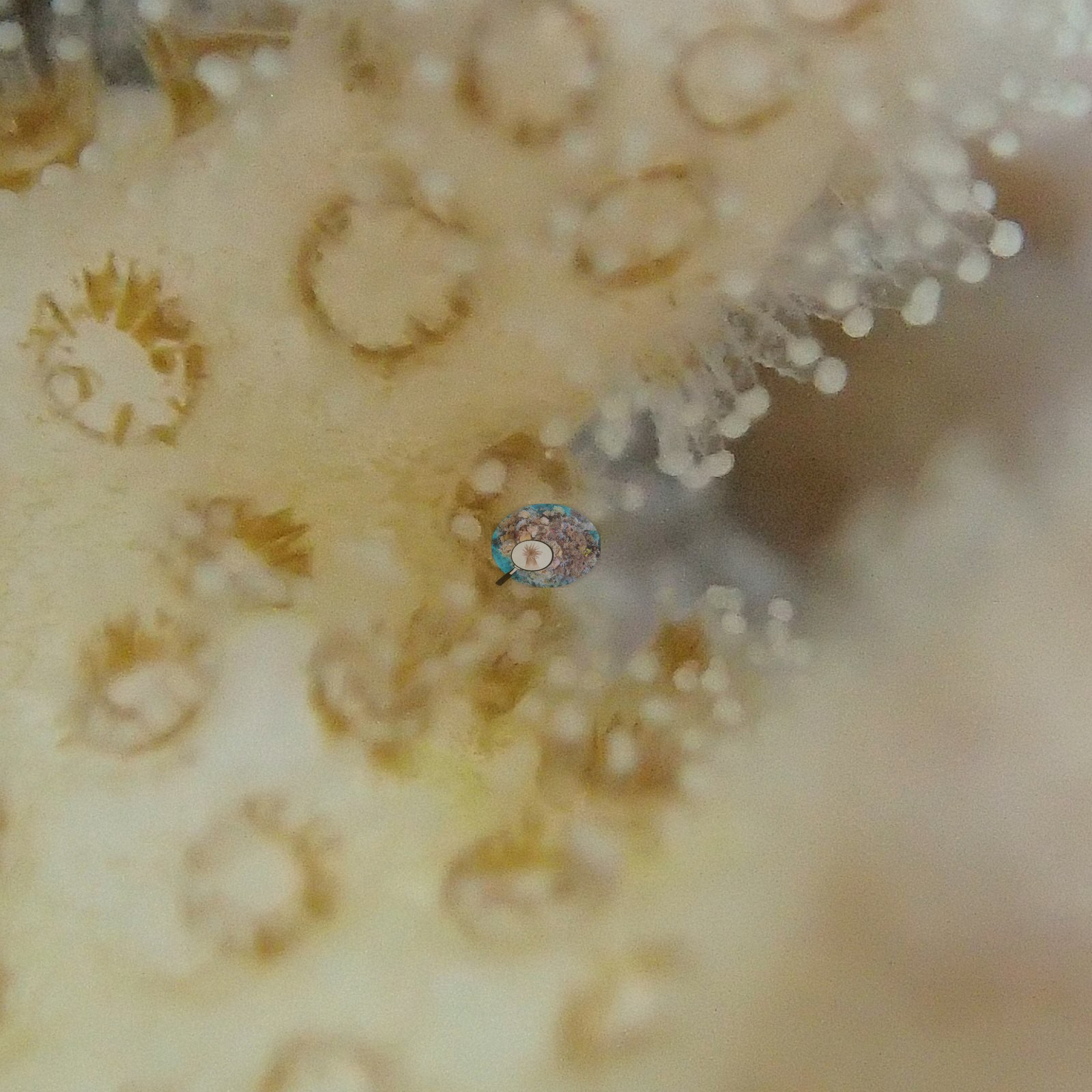
Site description
General benthic features
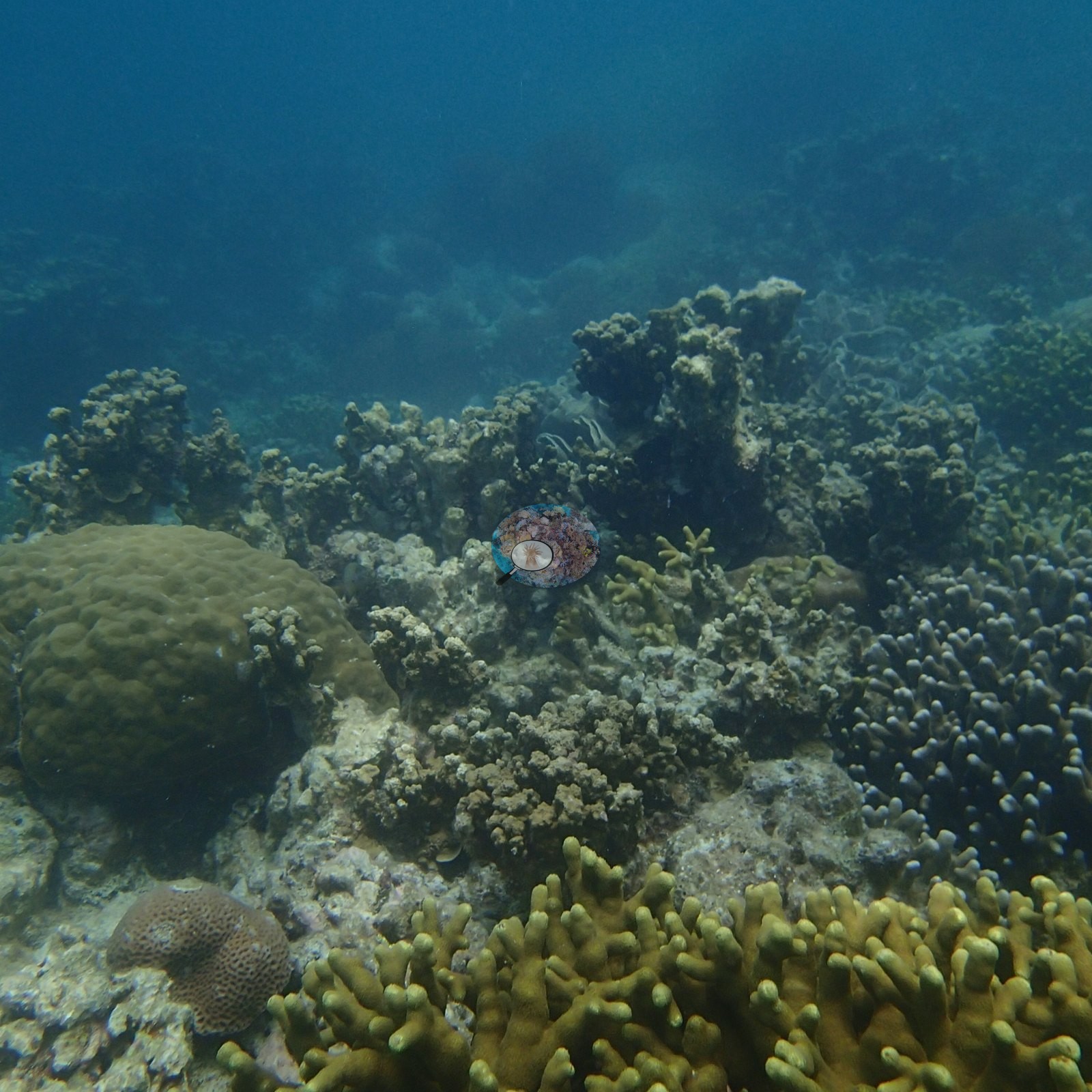
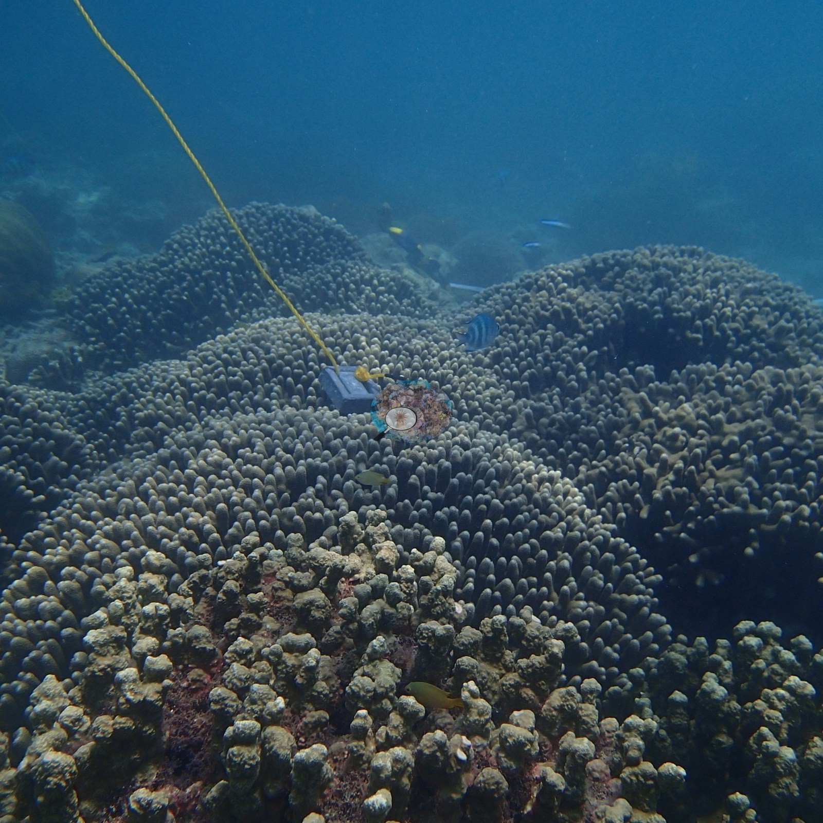
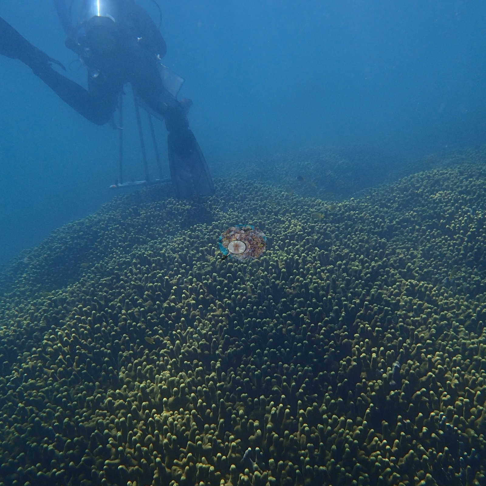
The full 63 frames show a big, structured reef. The station's dominant surface is a vast, low, monospecific-looking digitate coral field: yellow-green to olive finger-form colonies mounded over one another in a continuous bank running to the limit of visibility through six or more wide frames, with a diver descending over one of them for scale (e.g., Image 9, Image 18, Image 54, Image 55, Image 56).
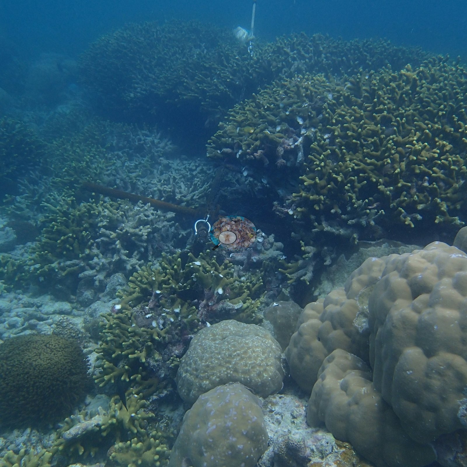
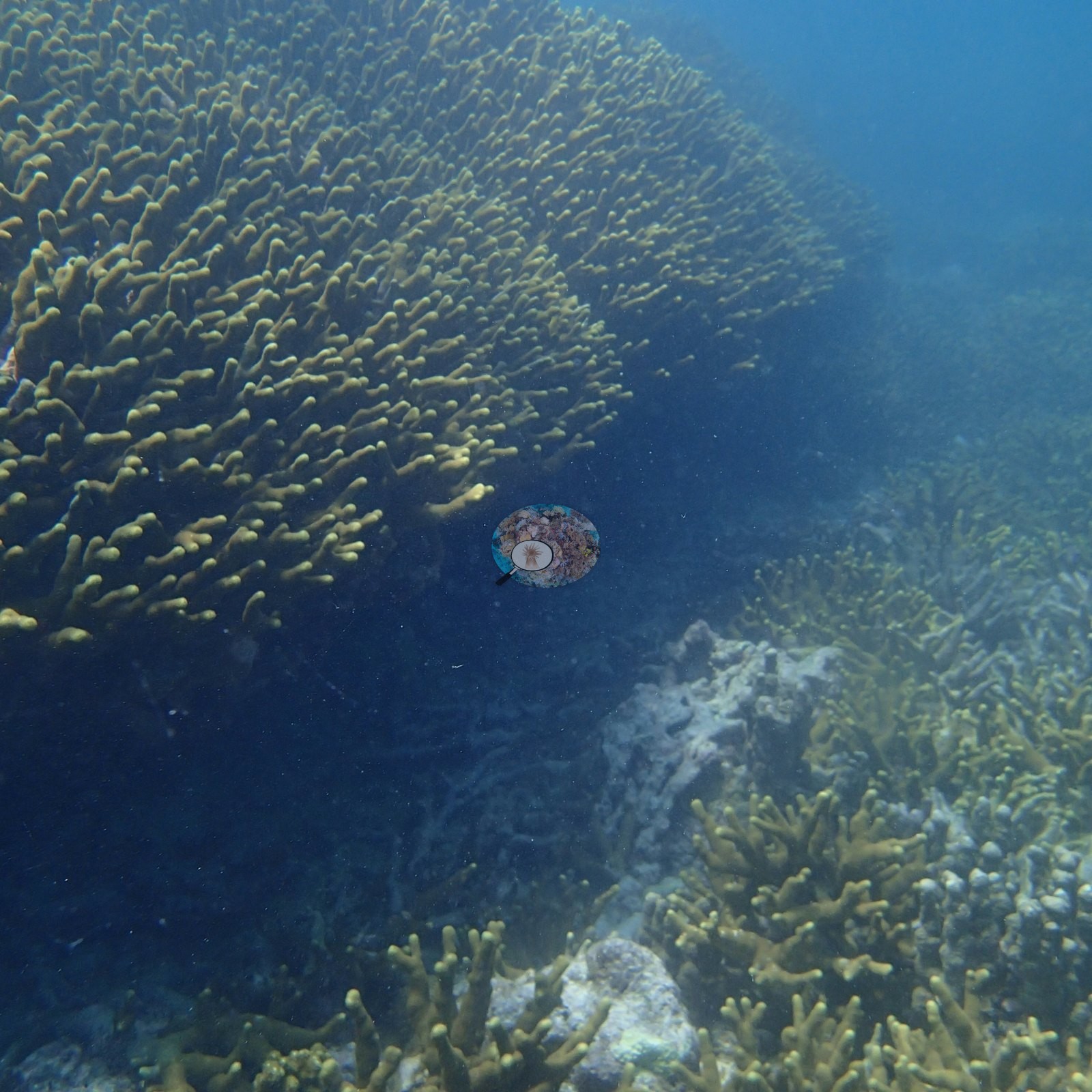
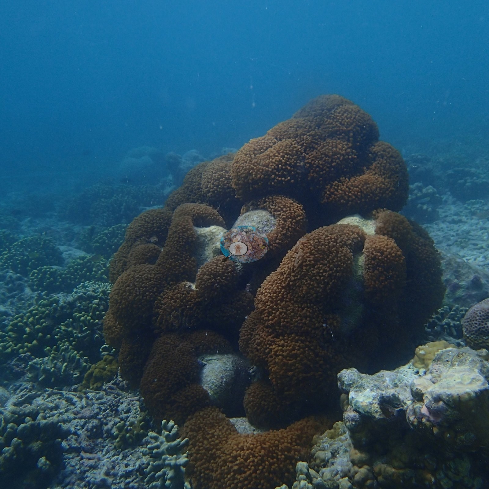
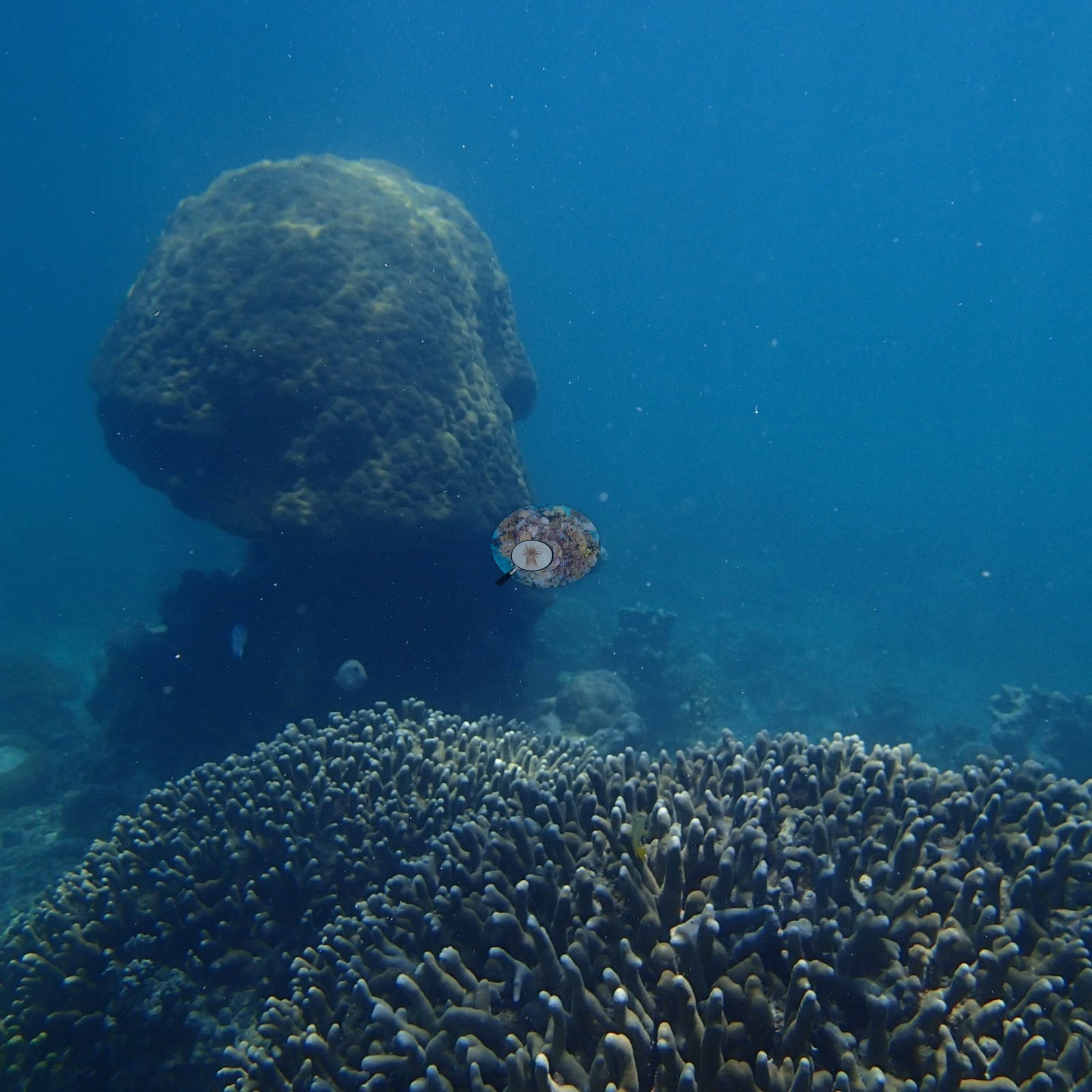
Standing above that bank are large massive Porites-type domes, one of them mushroom-shaped and deeply undercut at the base (e.g., Image 25, Image 52).
The photographs also show a set of tiered plate-coral pinnacles: columns of shingled tabulate and foliose plates stacked in overlapping tiers, two to three meters of relief, standing clear of the surrounding reef with damselfish and sergeant majors swirling around them (e.g., Image 42, Image 43, Image 44, Image 45, Image 46).
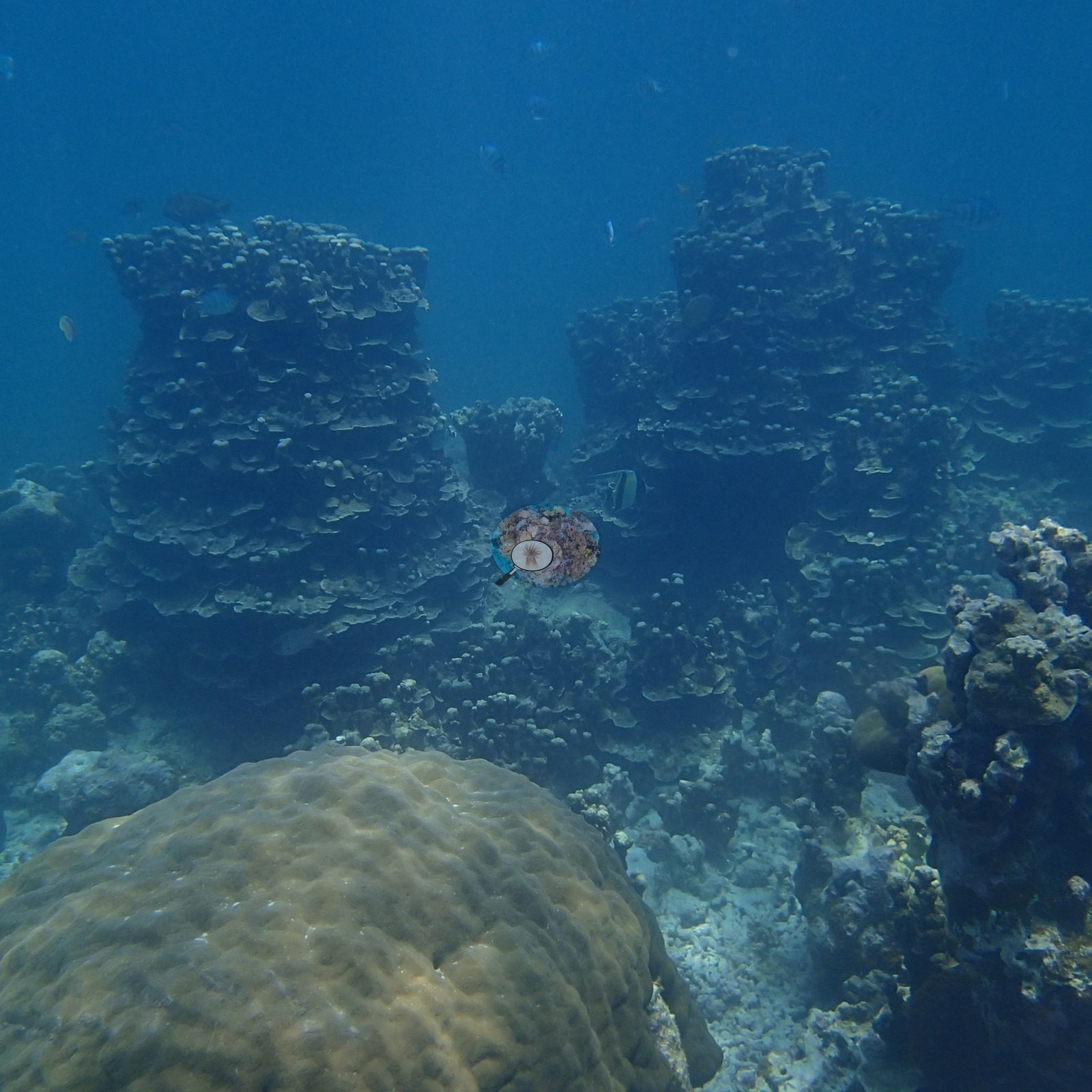
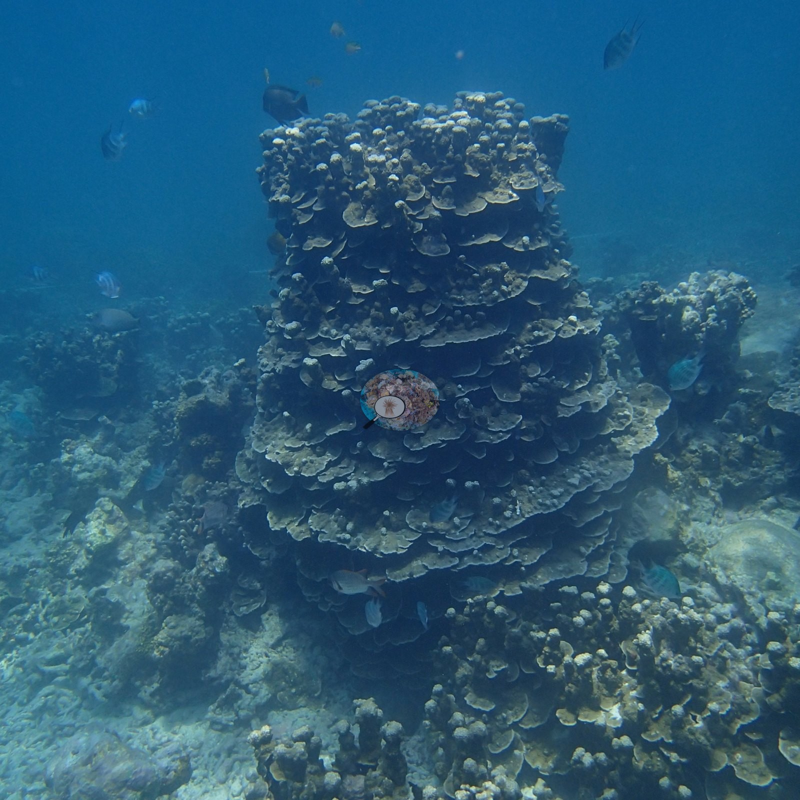
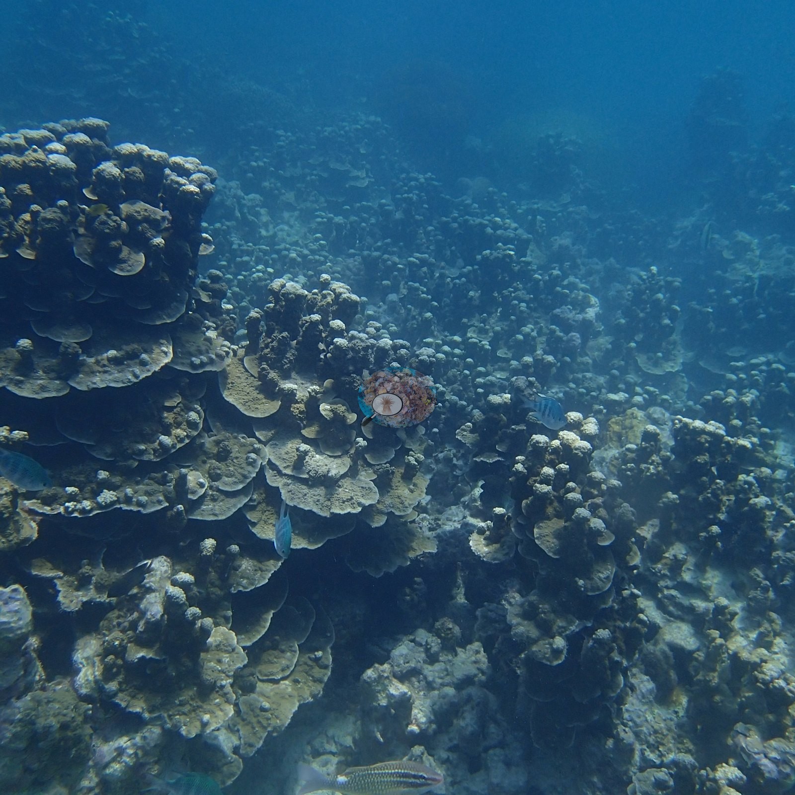
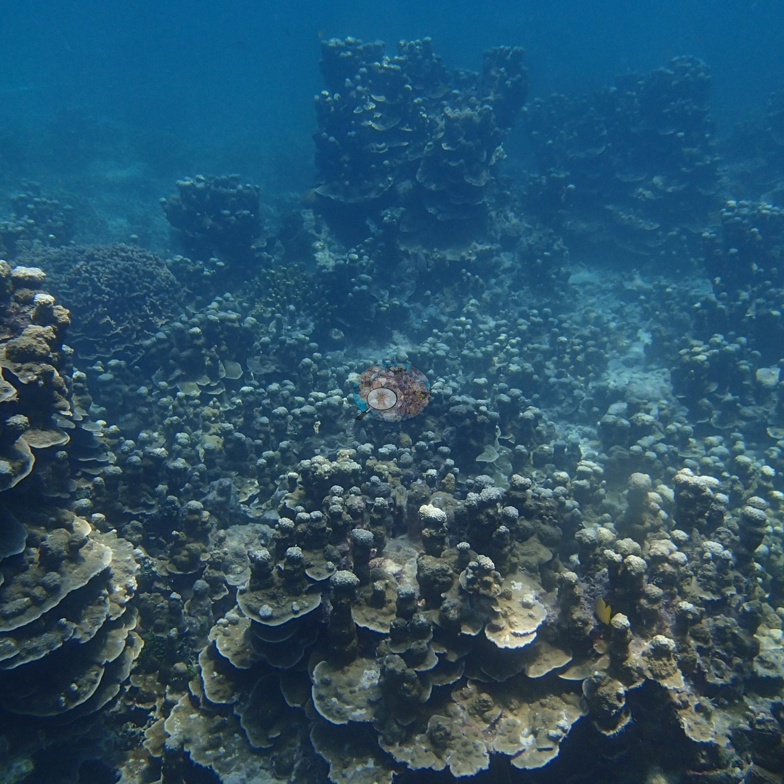
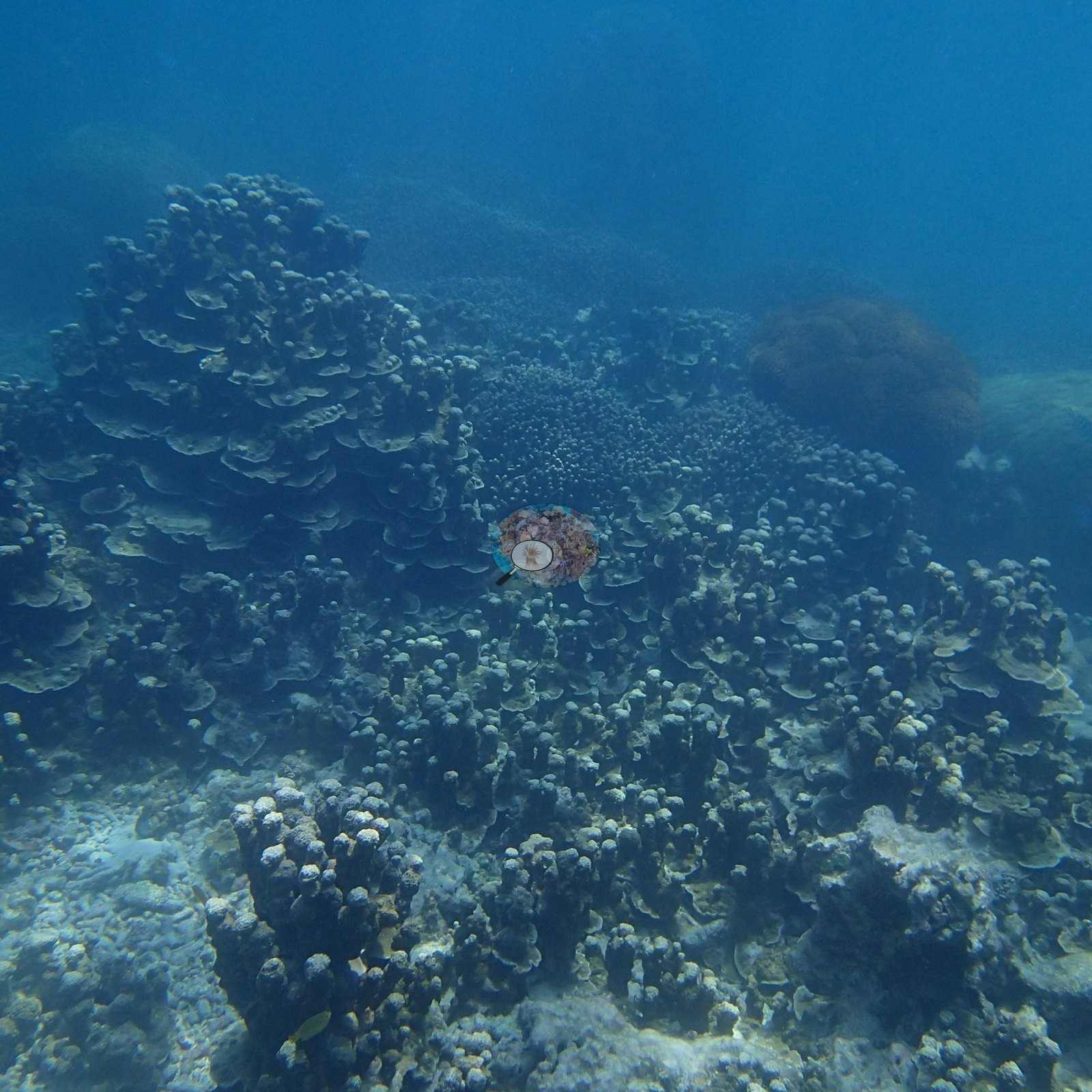
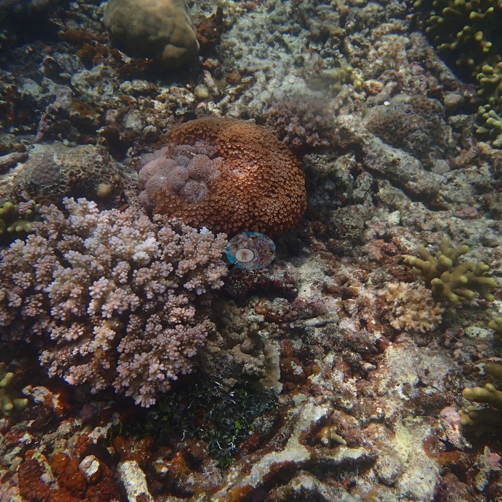
Five frames are given to them, and they are the most striking structure at the station. Elsewhere the frames show dense fields of brown hummocky colonies, blue-tipped digitate Acropora, foliose plates, and coralline-crusted rubble filling in between (e.g., Image 26, Image 27).
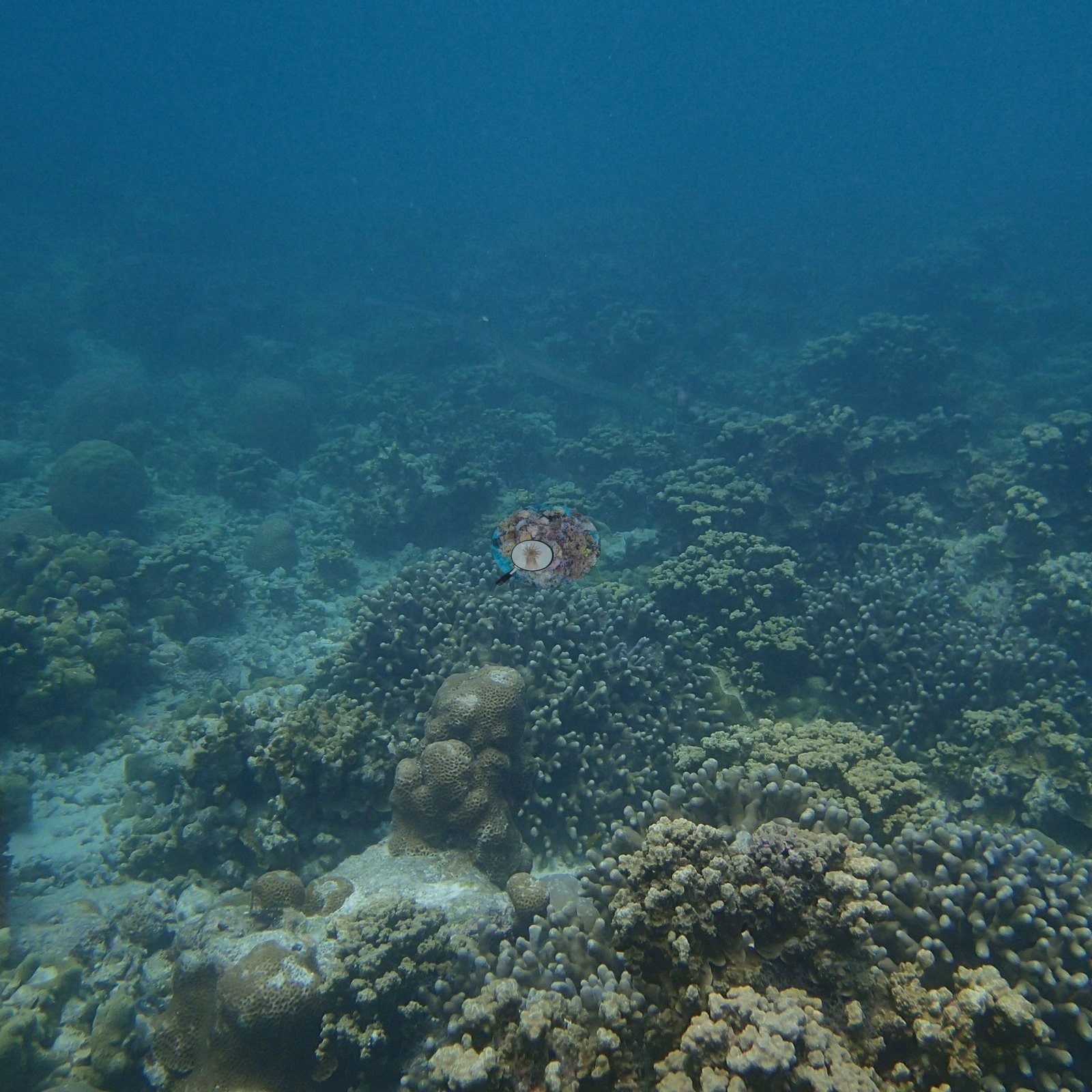
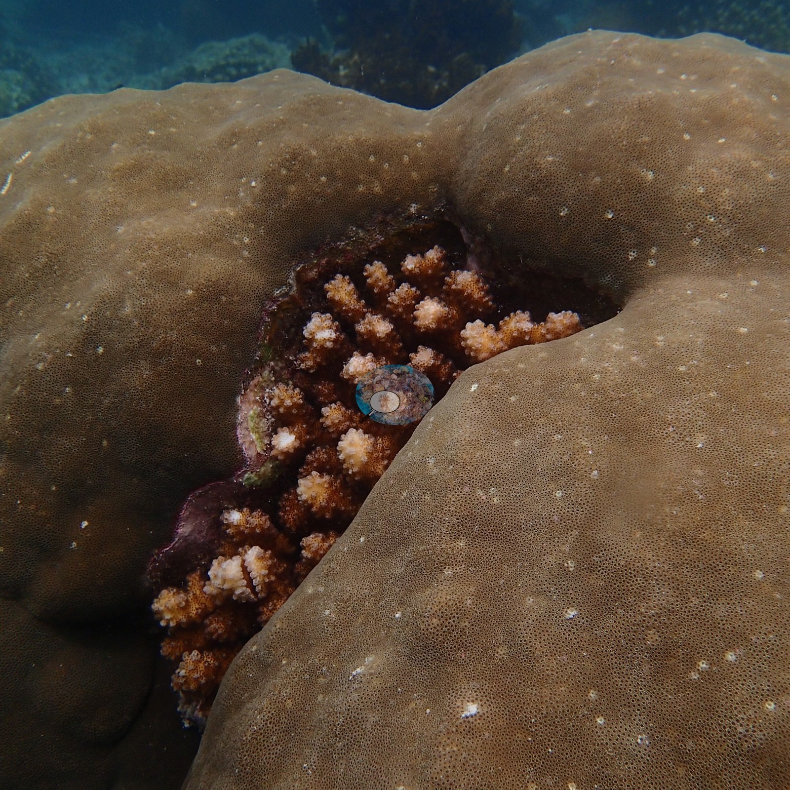
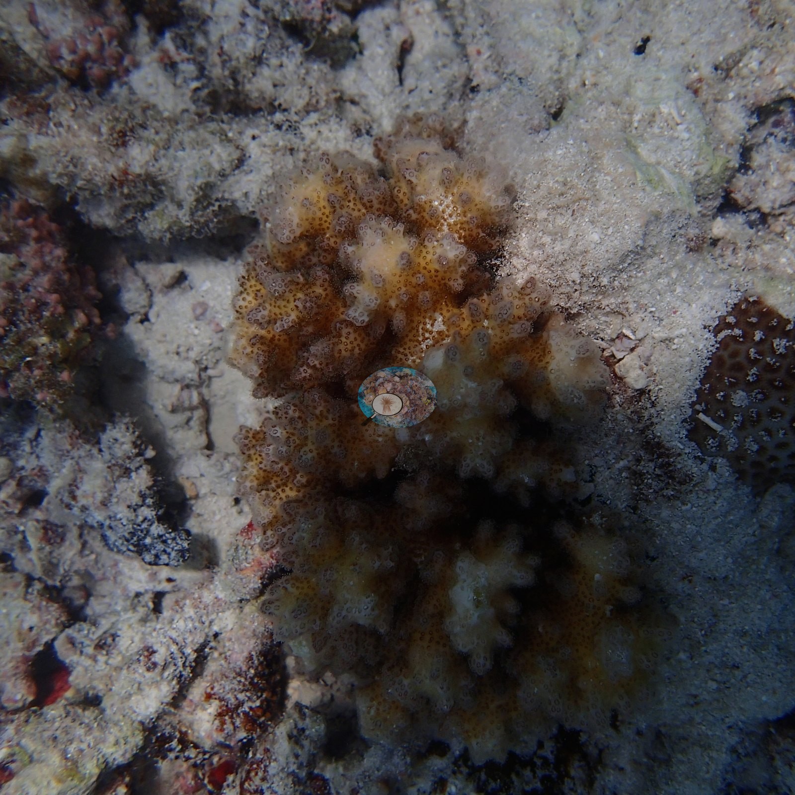
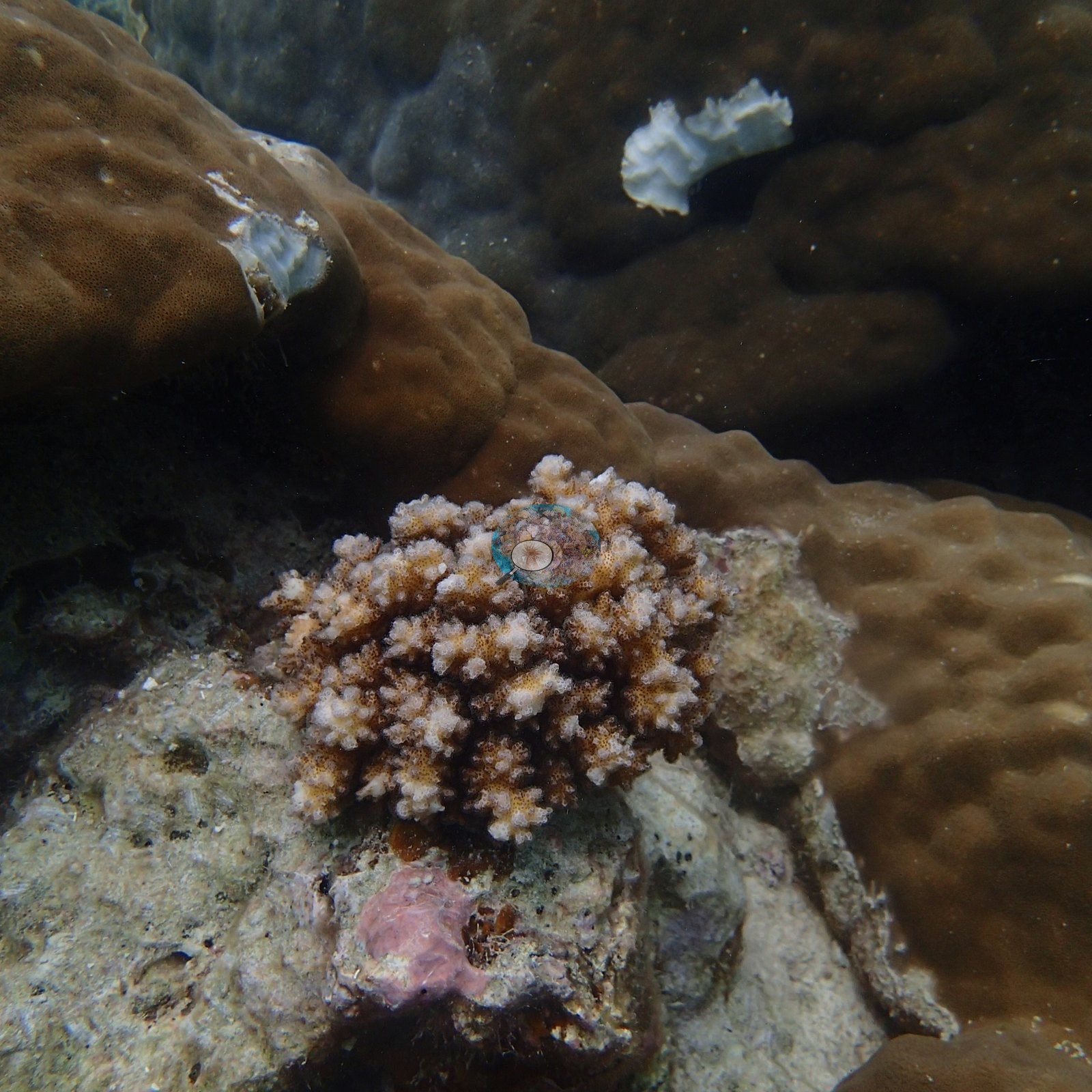
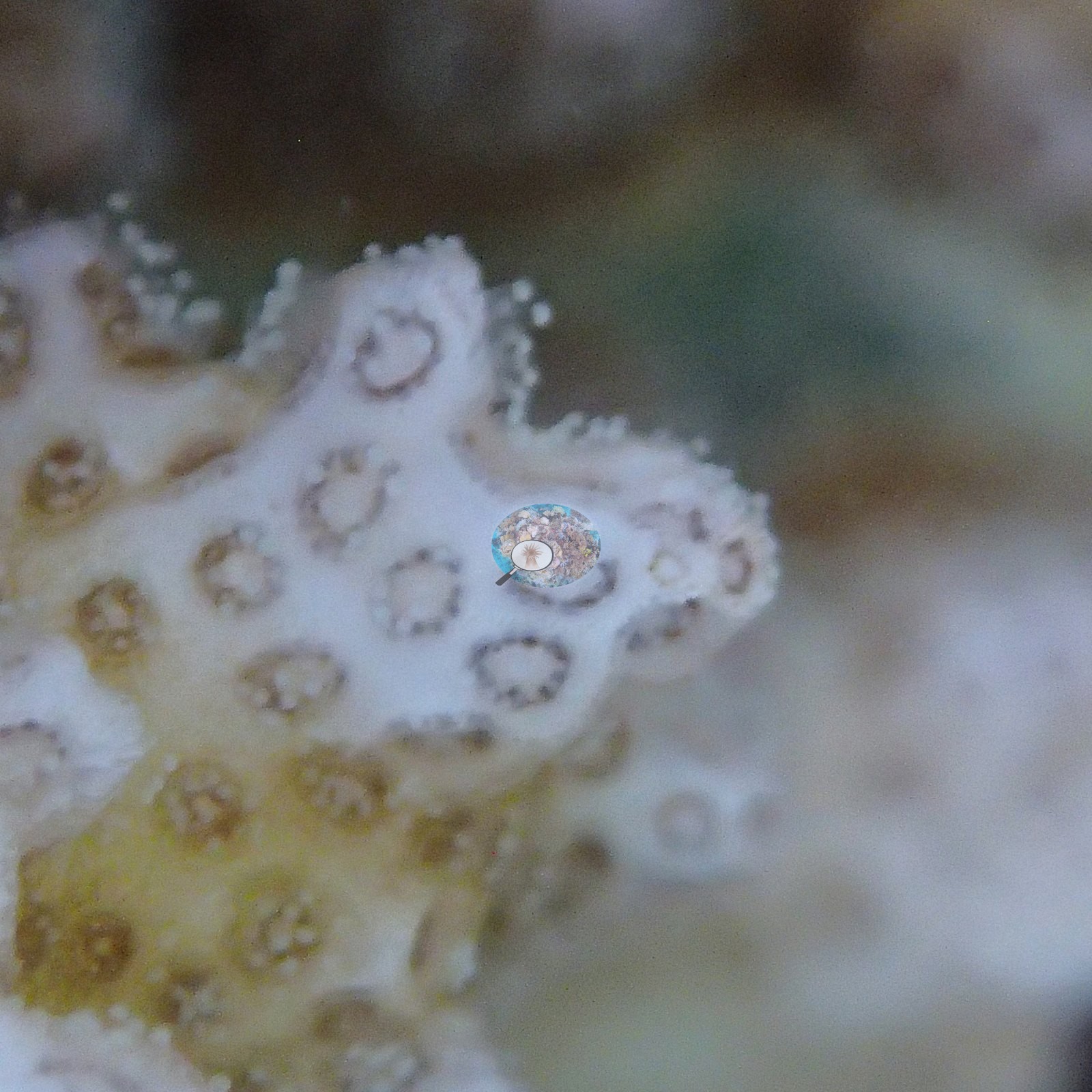
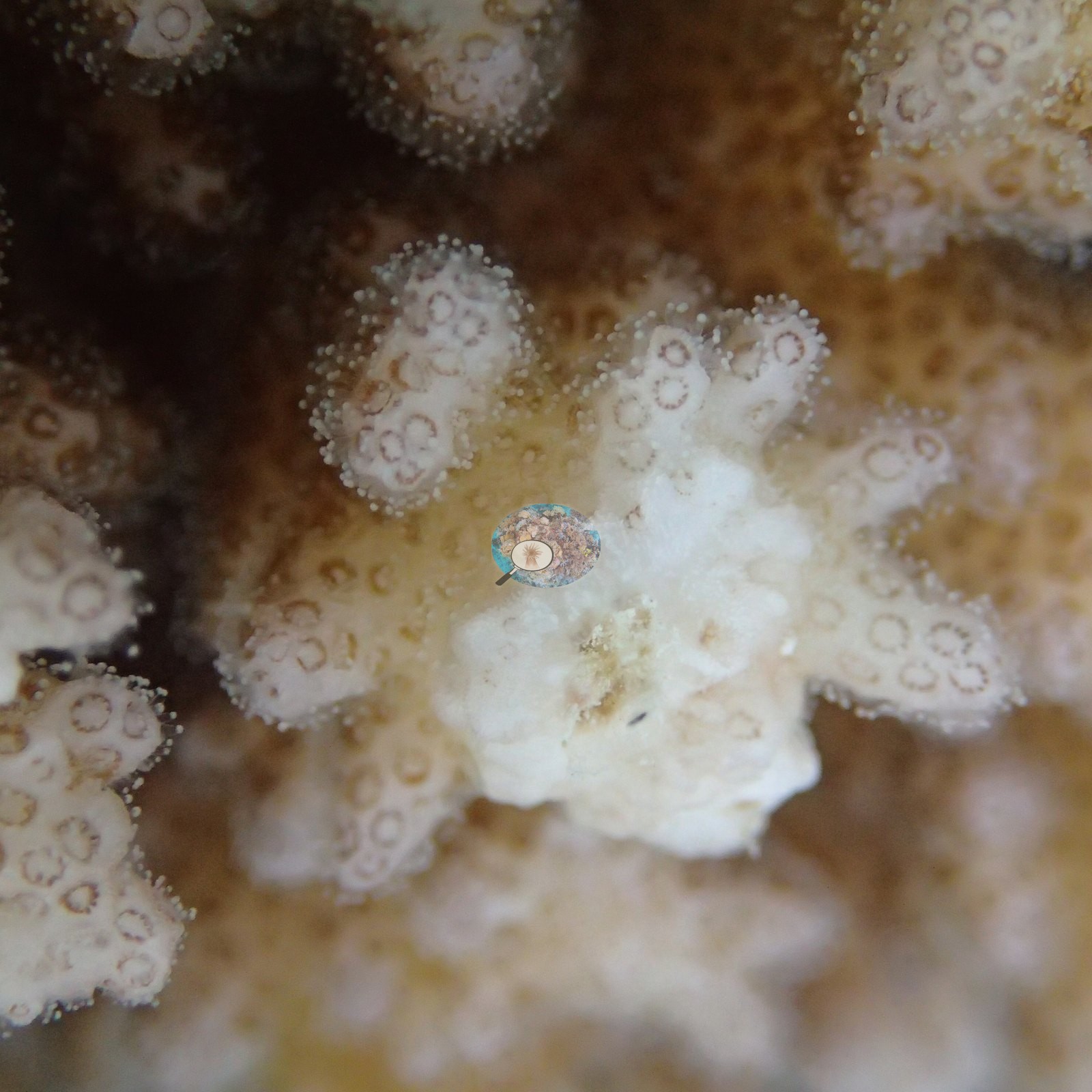
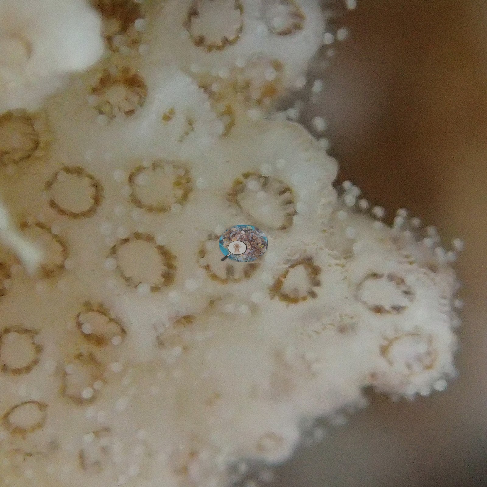
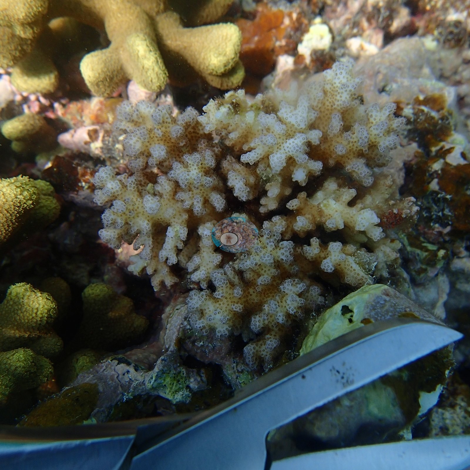
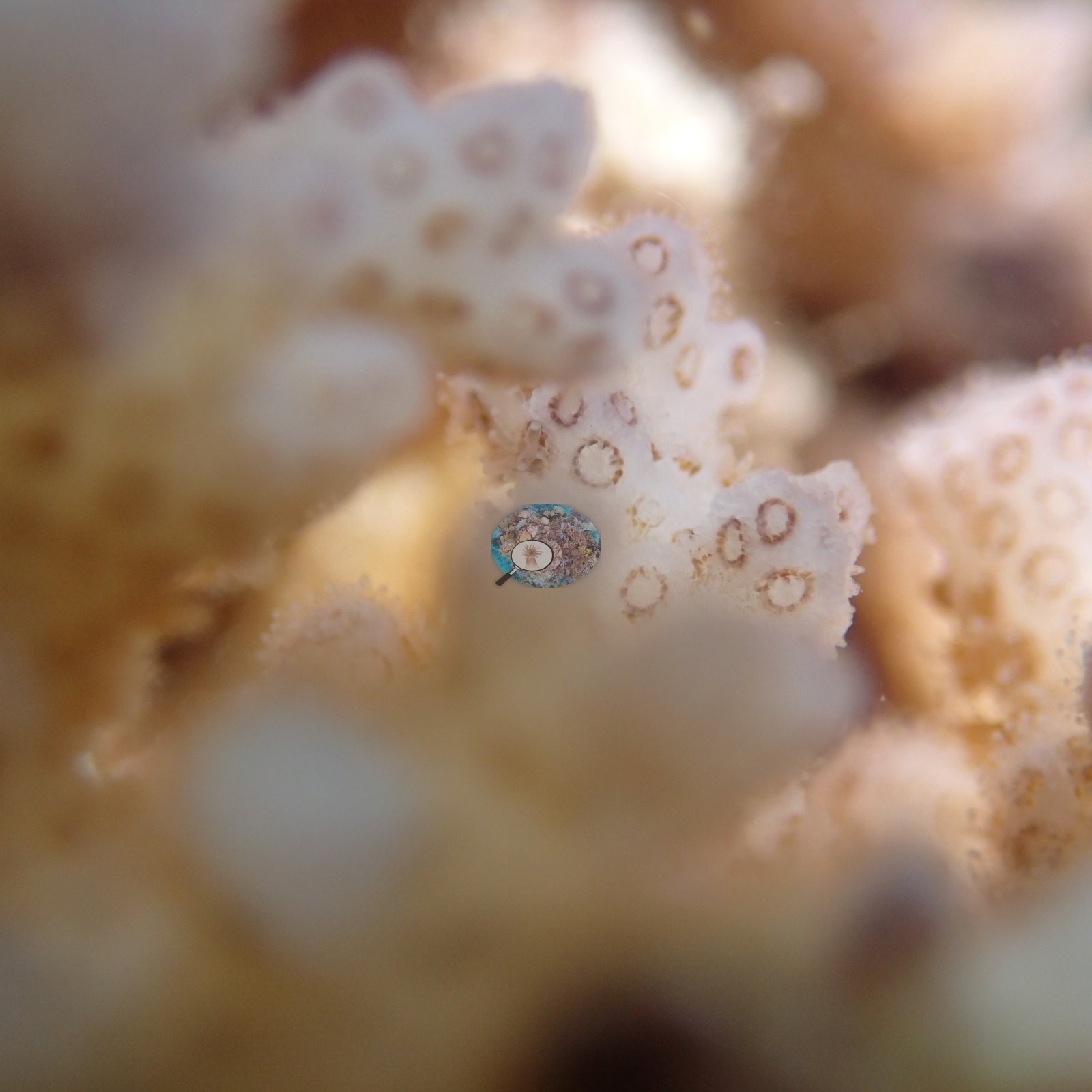
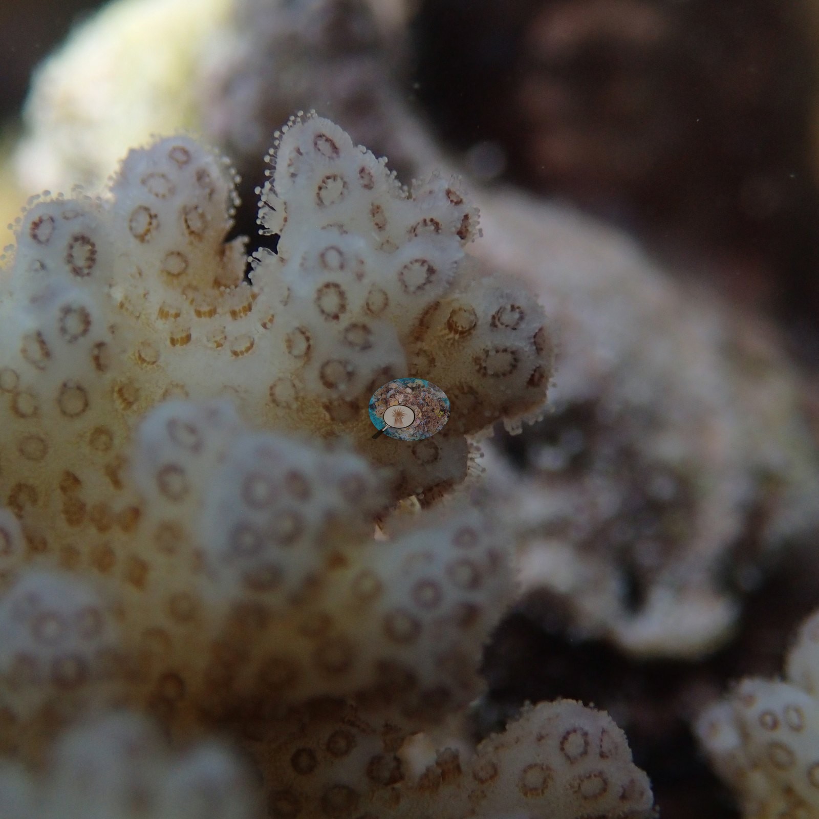
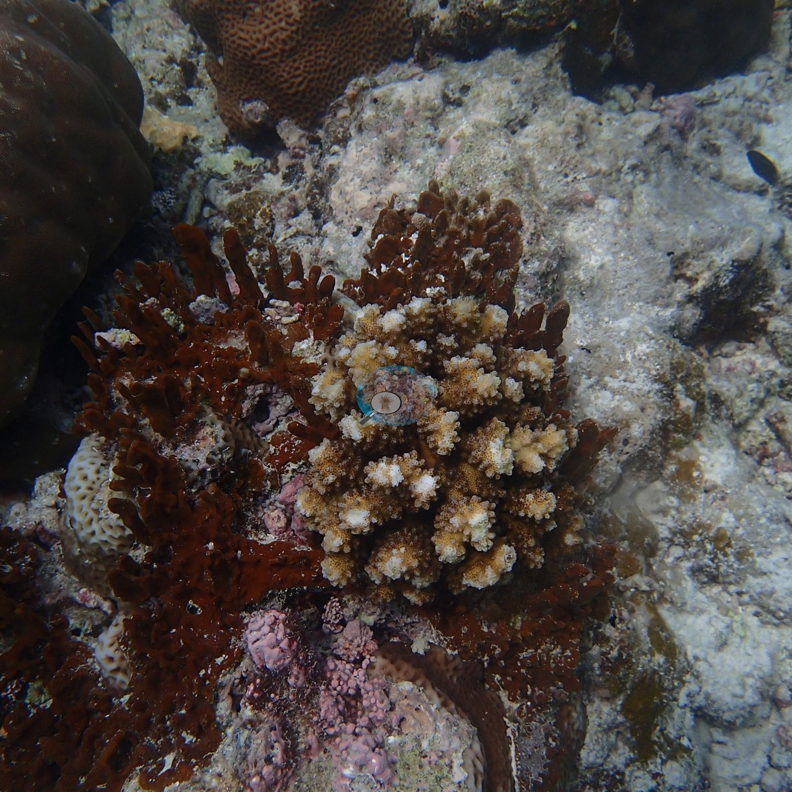
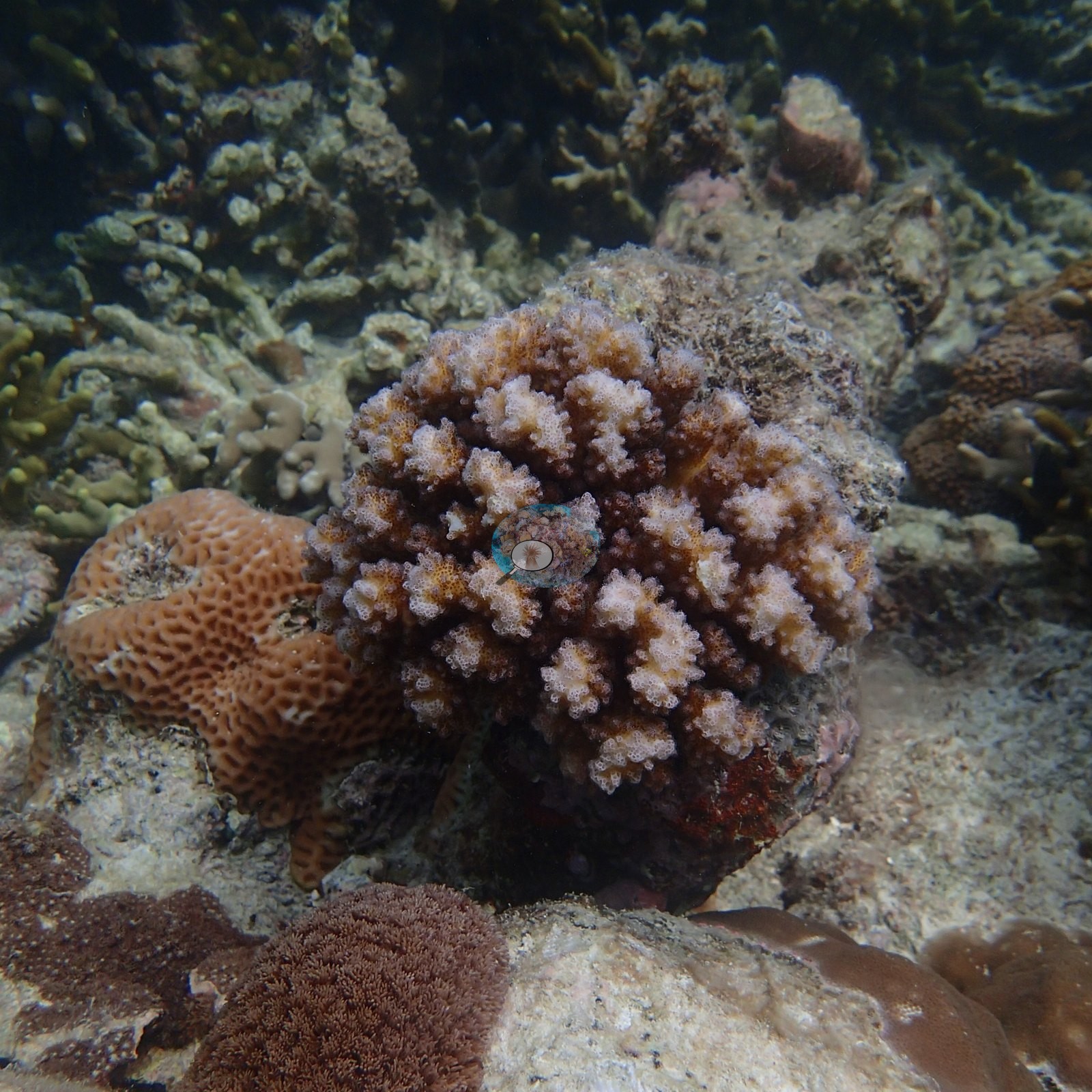
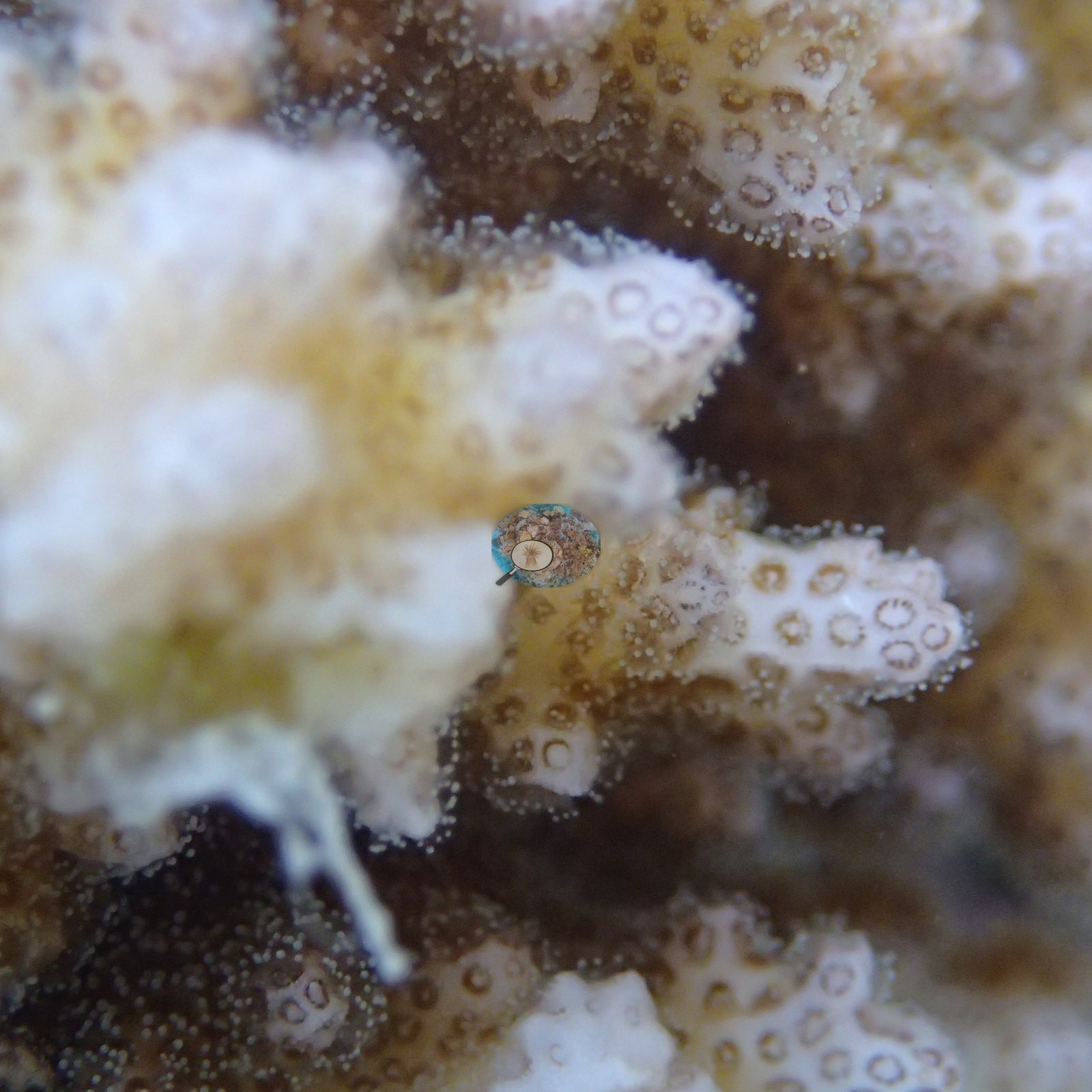
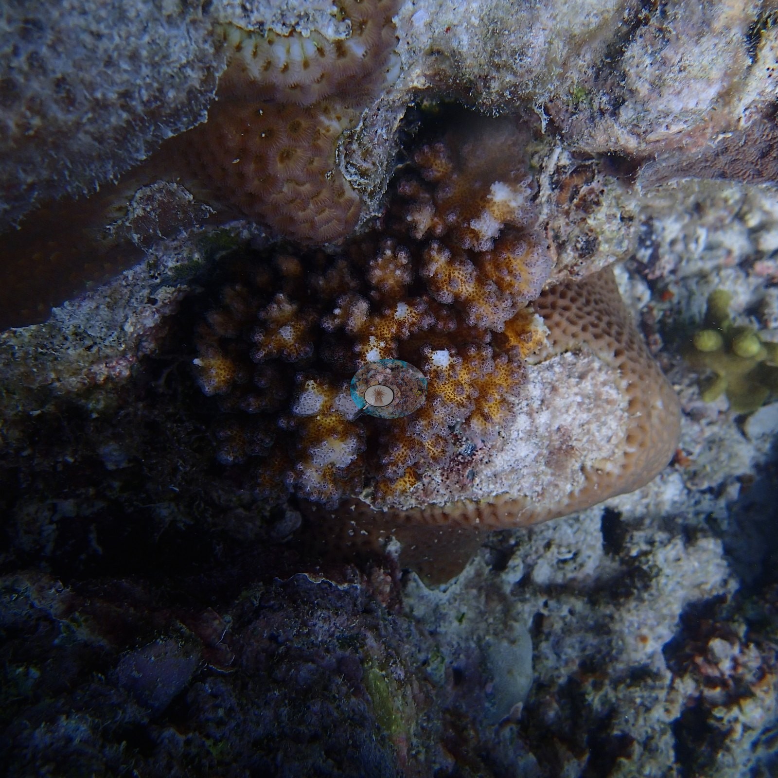
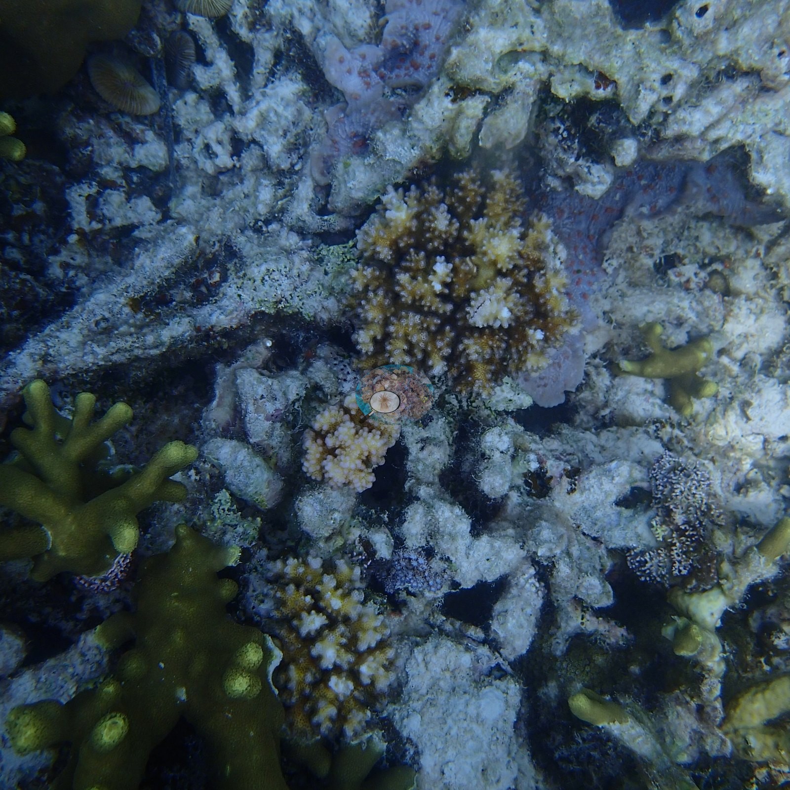
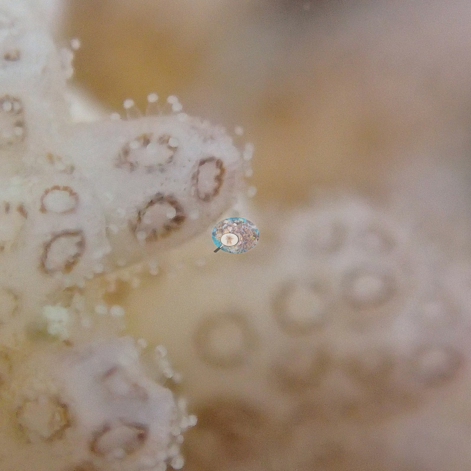
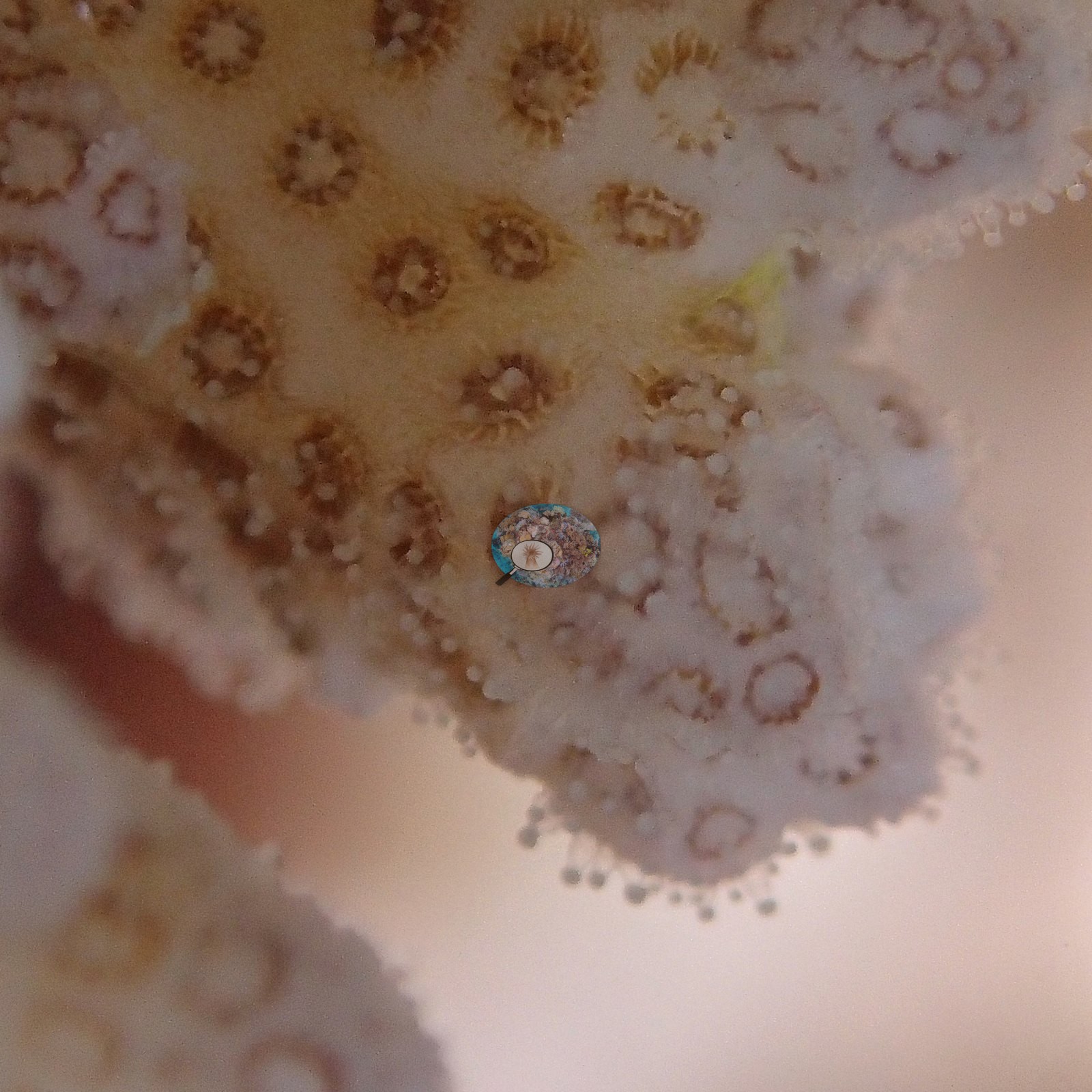
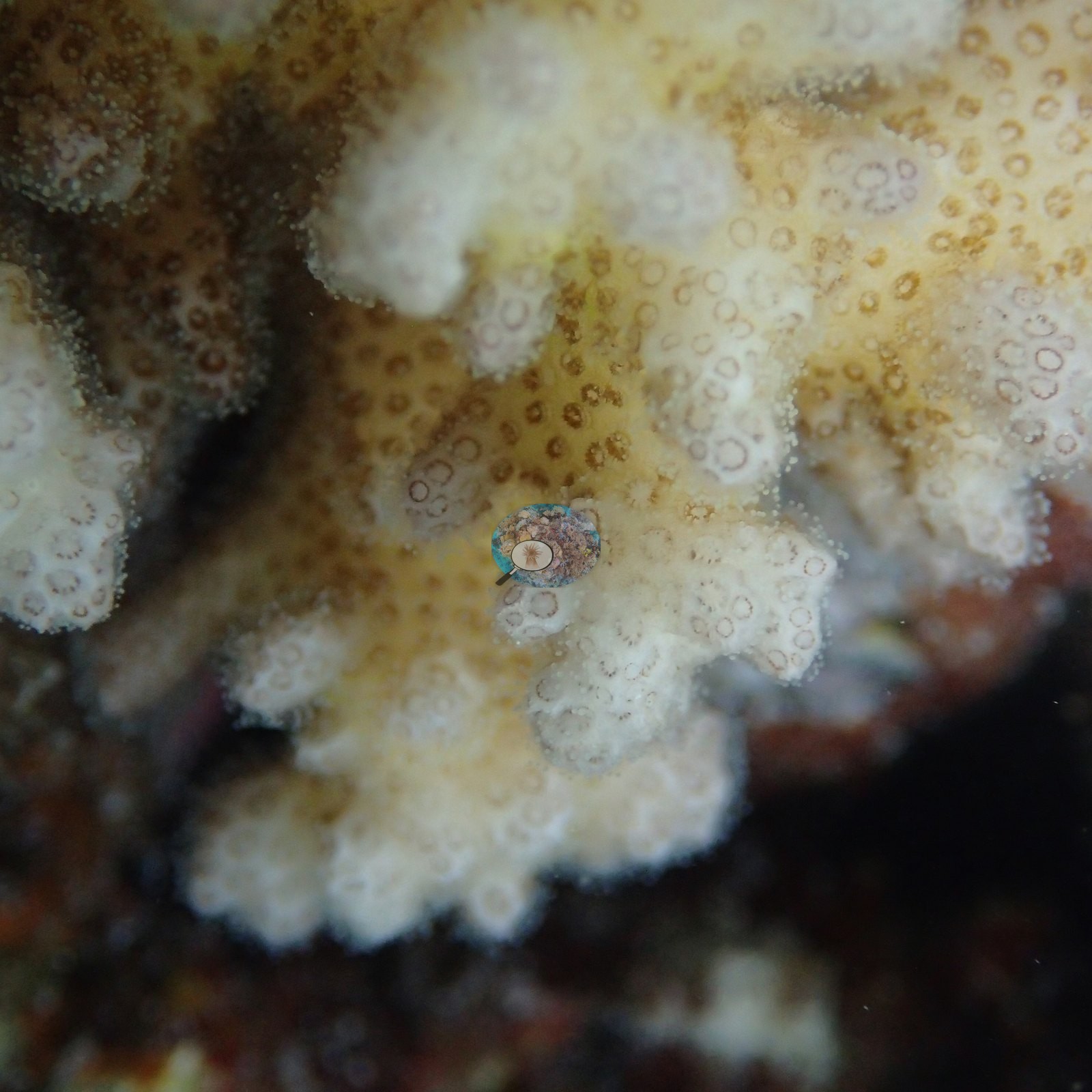
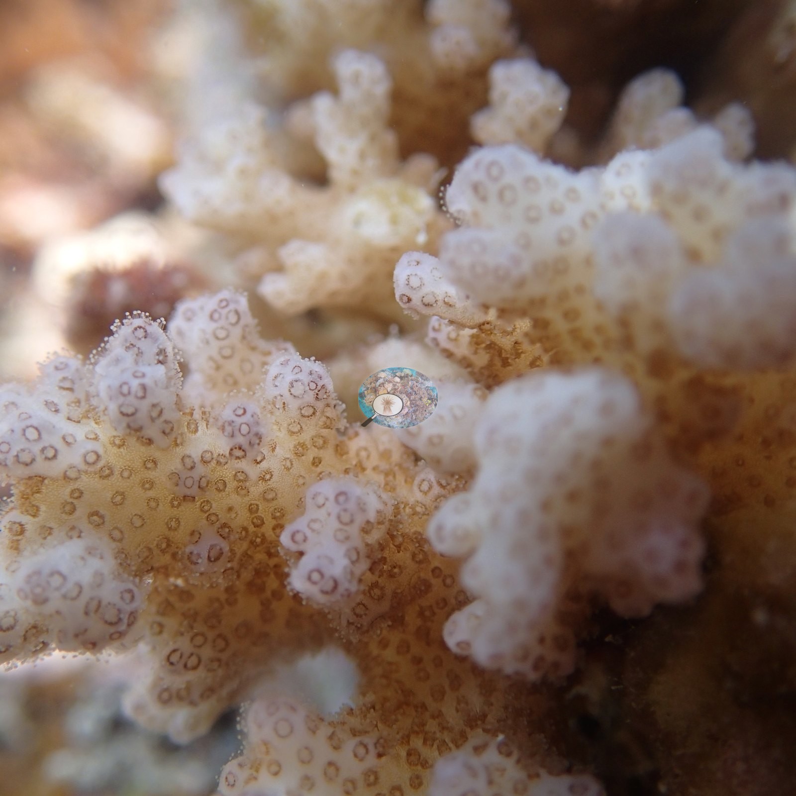
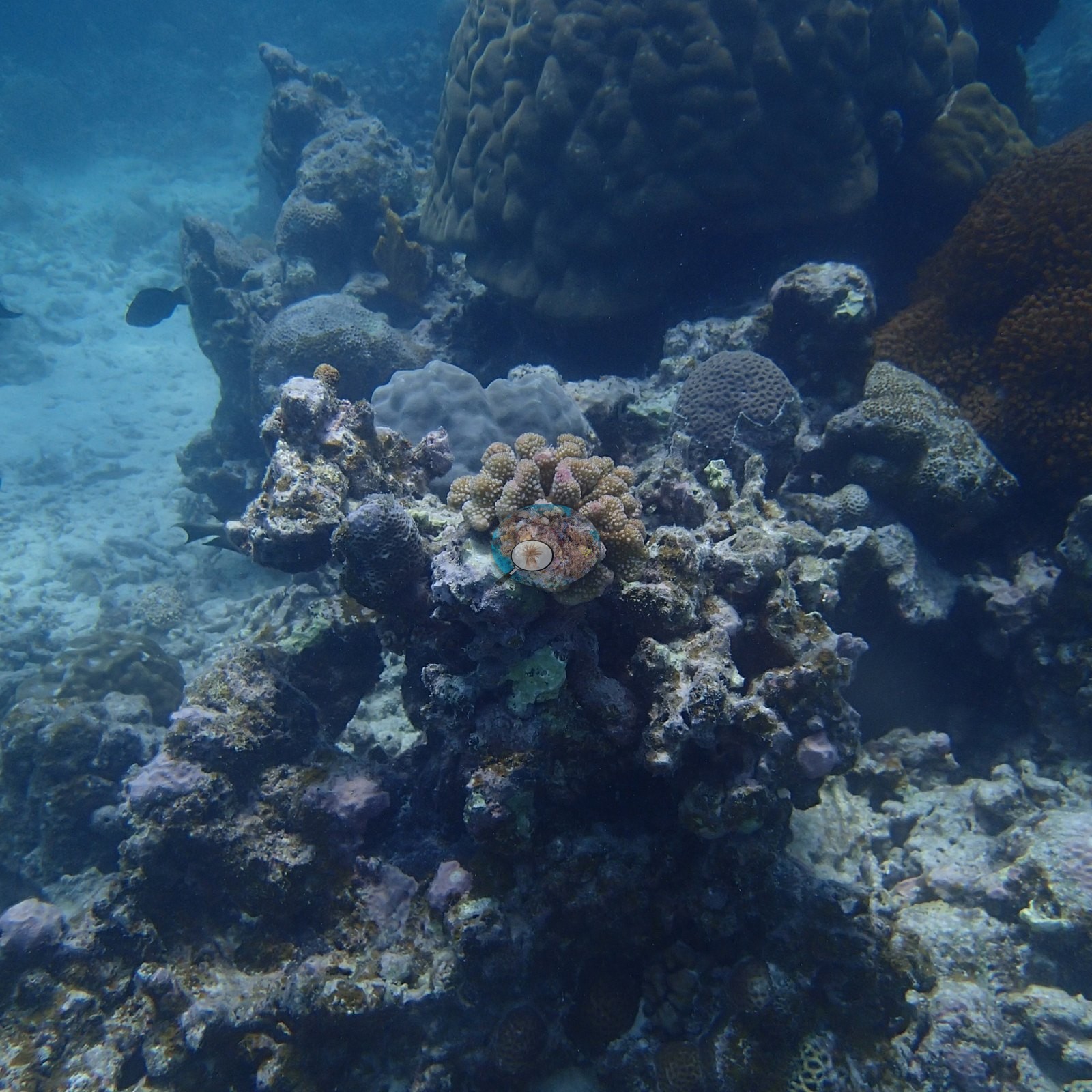
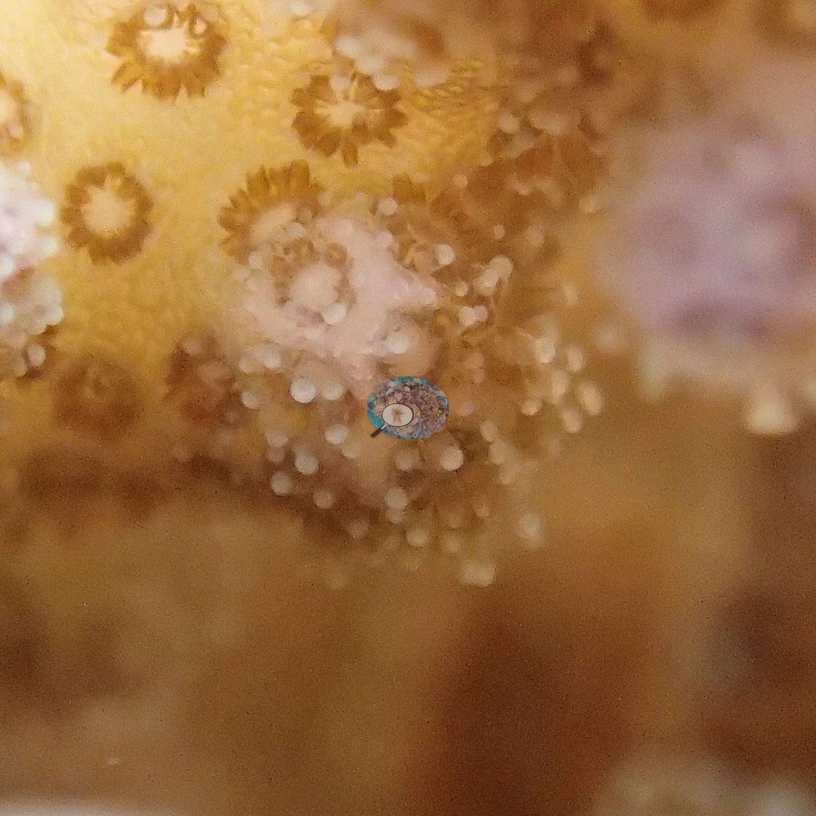
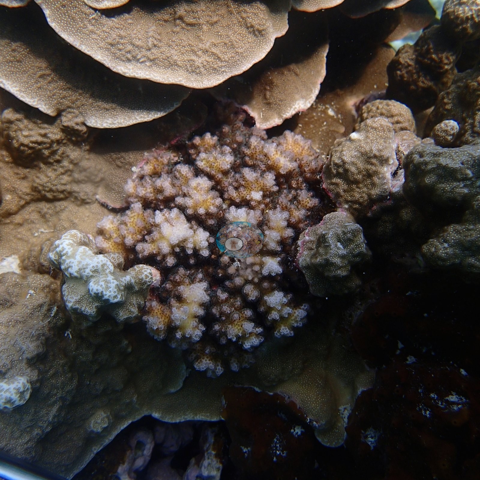
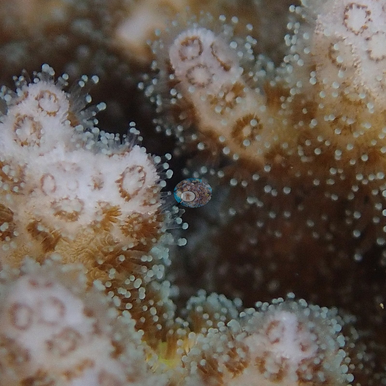
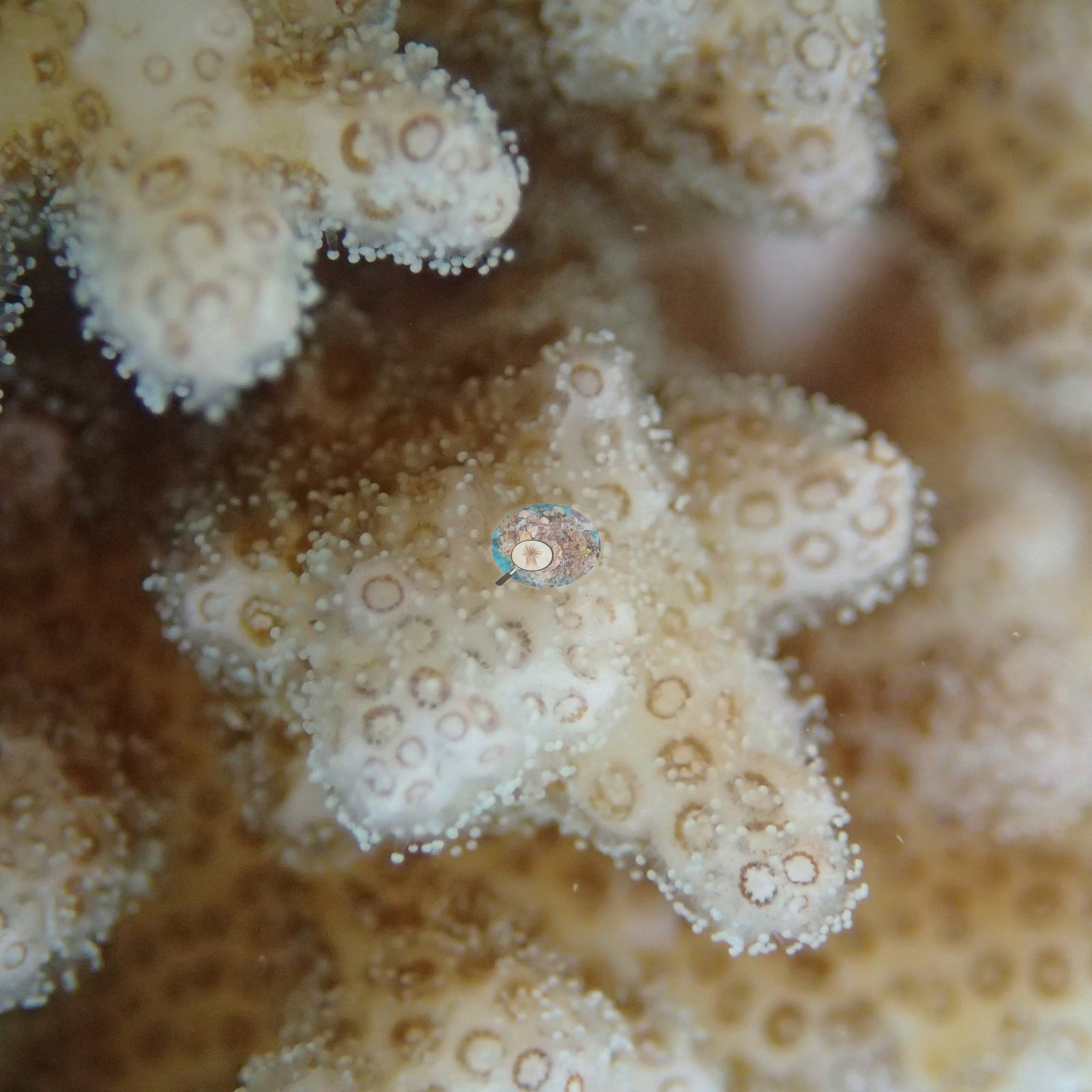
The pocilloporid colonies that were the point of the visit recur throughout, growing out of clefts between smooth tan massive boulders and on coralline-crusted rubble (e.g., Image 1, Image 10, Image 12, Image 14, Image 17, Image 19, Image 22, Image 24, Image 28, Image 33, Image 37, Image 41, Image 47, Image 51, Image 58).
Coral cover
- Reference: Claude CV point count (this site's photos)
- Coral cover: 54% — a blind Claude computer-vision point count of 37 of Anderson's unedited photographs (20 points per photo; water, fish, and obstructions excluded). Other computer-vision methods are listed in the method table.
Stressors
Anderson's sample sheet records two of the twelve branching colonies sampled here with partial loss: one at 2.5 m scored 20% bleached or dead, and one at 3.2 m had 20% recent mortality (40% in the sheet's combined bleached-or-dead score). The other ten score 0%, and most are noted as healthy, two as darkly pigmented. The digitate banks, the plate pinnacles, and the massive Porites-type domes are normally pigmented in the photographs. Two further things stand out in the full photo set. One frame shows thin monofilament line draped across a knobby Porites-type colony — derelict fishing gear on the reef (e.g., Image 16).
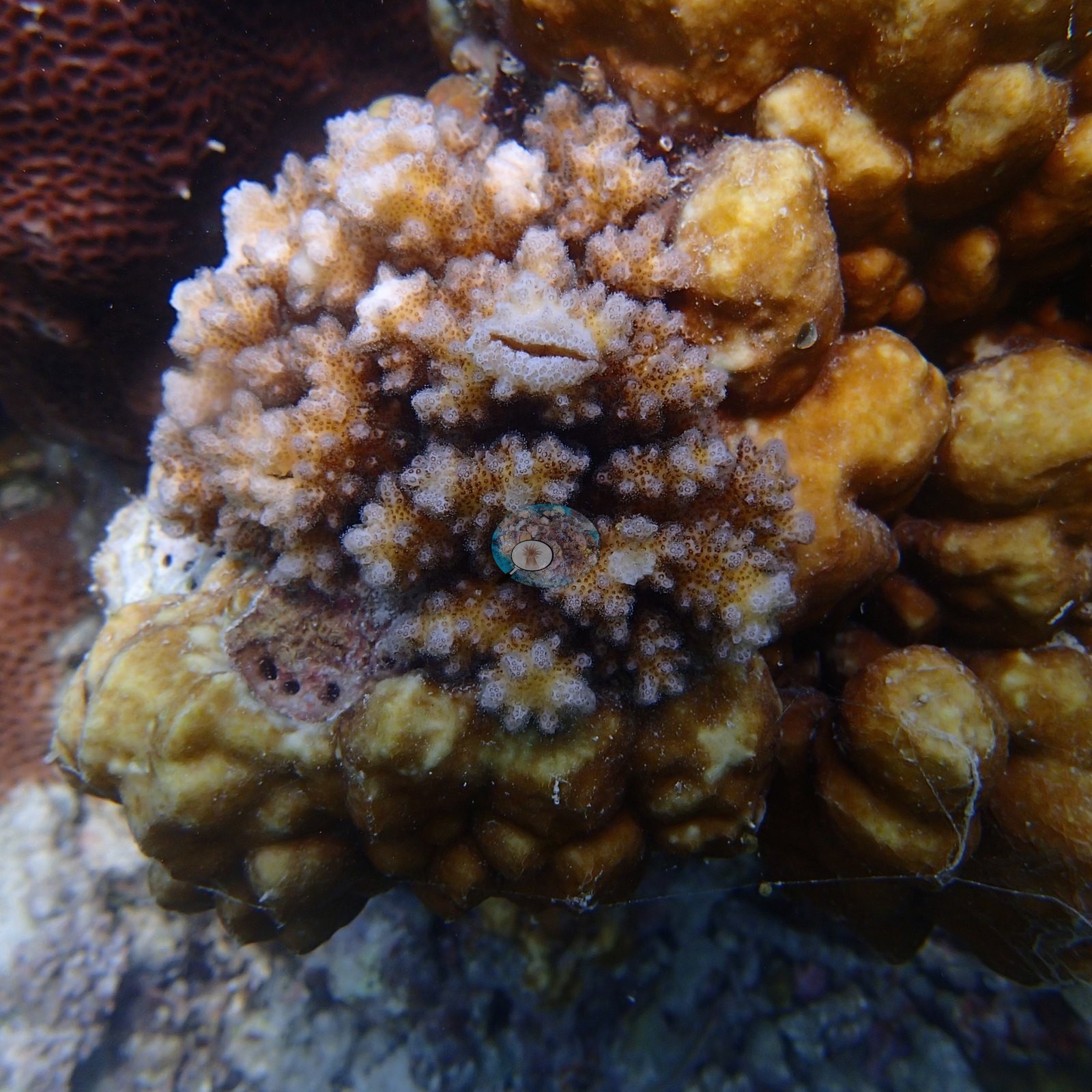
Another shows a clean patch of bare white skeleton with a sharp tissue margin on the shoulder of a massive dome: recent partial tissue loss, cause not determinable from a photograph. No crown-of-thorns starfish (COTS) or other corallivores were evident.
Fish and other fauna
Two frames hold a large fluted giant clam (Tridacna) with a mottled brown, orange, and iridescent green mantle (e.g., Image 7, Image 8).
Comparisons with other data
We found no other surveys confirmed at this site as of 2026. If you would like to share yours, please email us.
Dive record
Site 13 was worked in two dives on the morning of 4 April 2016, logged on Anderson's sample sheet as in at 10:11 (45 minutes, maximum depth 4.0 m) and in at 11:26 (45 minutes, maximum depth 4.6 m); the camera's frames run 10:44–12:10 across both dives.
Other notes
The station is also where the science is most visible. A small gray instrument housing on a yellow line — a deployed logger — sits on top of a digitate colony in one frame (e.g., Image 4).
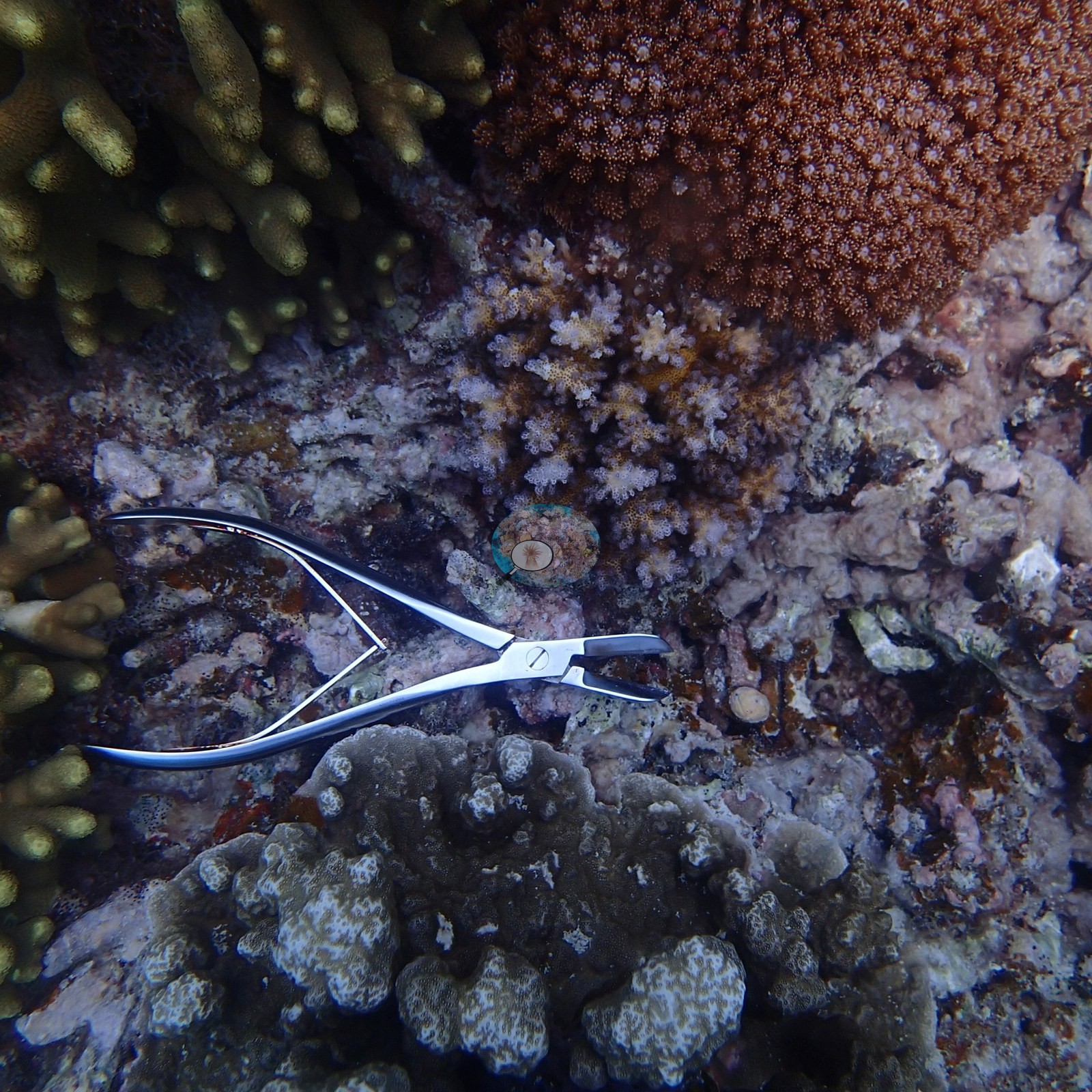
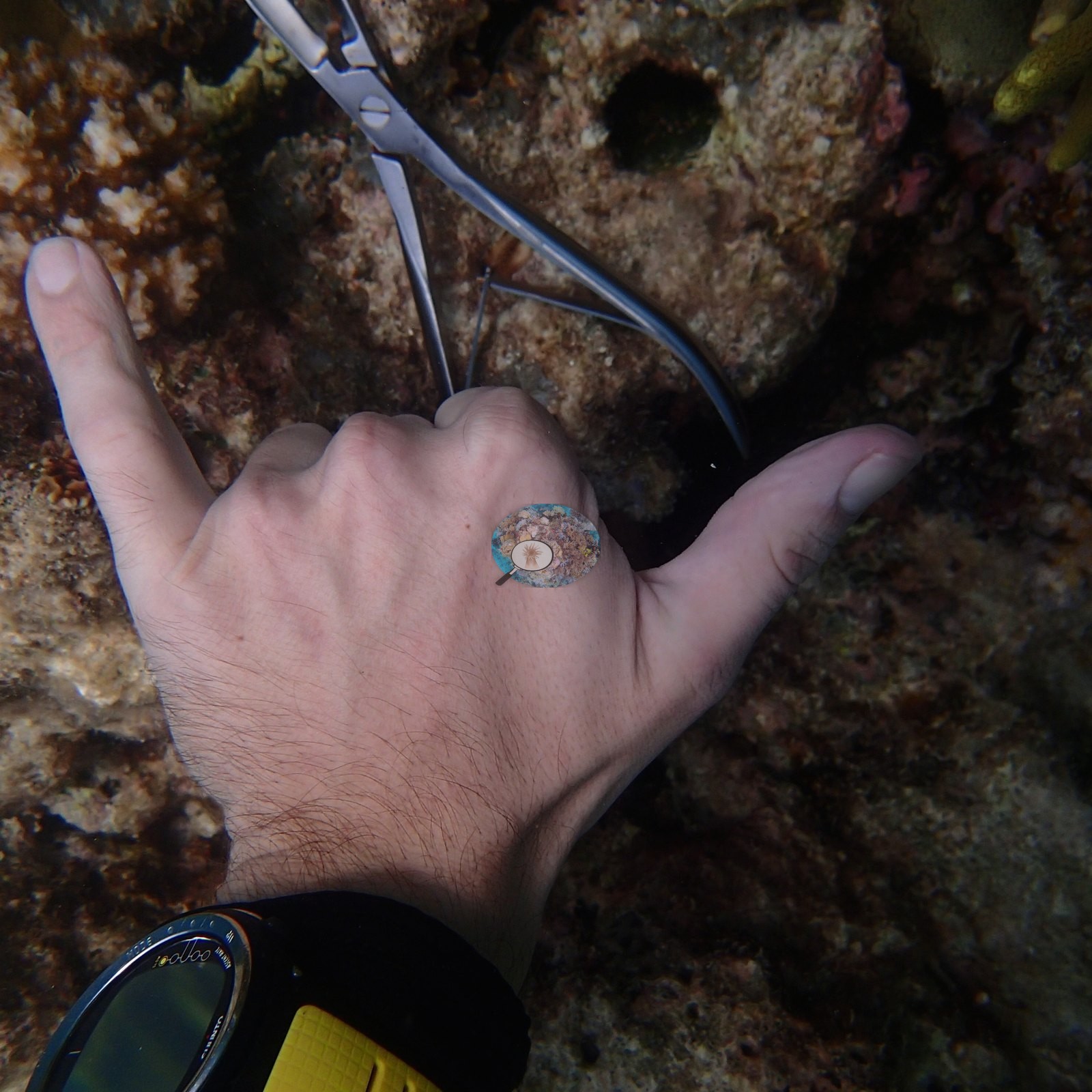
Anderson's own bare hand, with a dive computer on the wrist, reaches into another beside a pair of open stainless bone-cutting forceps; the forceps appear on the substrate again in a second frame (e.g., Image 3, Image 13).
A diver descends carrying a metal frame over the digitate field (e.g., Image 57).
CV model comparison
More info
A few low-cover photos (sand, rubble, or open bottom) pull the mean down; the median is the typical photo. No computer-vision method yet estimates coral cover accurately on new images, so treat this as indicative rather than a measurement; it is revised as the methods improve.More info
Other rows are for comparison only and are never averaged. No diver transect exists for this trip, so agreement between rows is not accuracy.| Method | Hard coral cover (%) | Photos (n) ± SE |
|---|---|---|
| CoralSCOP unedited | 35.2 | n = 37 · SE 3.0 |
| Claude | 53.9 | n = 37 · SE 4.2 |
CoralSCOP (unedited photos) reads 35.2%, 18.7 pp below Claude's blind point count (53.9%); the headline stays Claude's blind point count because no transect or manual count exists here.
See a more detailed, stand-alone version of this table.
Data & links
All images from Coral Reef Diagnostics. Data are from Coral Reef Diagnostics.
© Coral Reef Diagnostics.