
Turks and Caicos - Shark Hotel

Part of the regional summary: Turks and Caicos Islands
More info
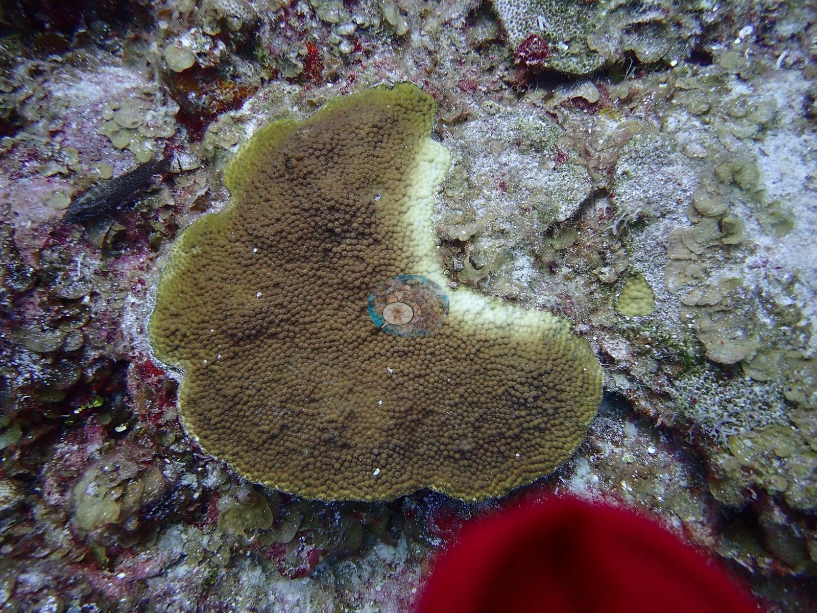
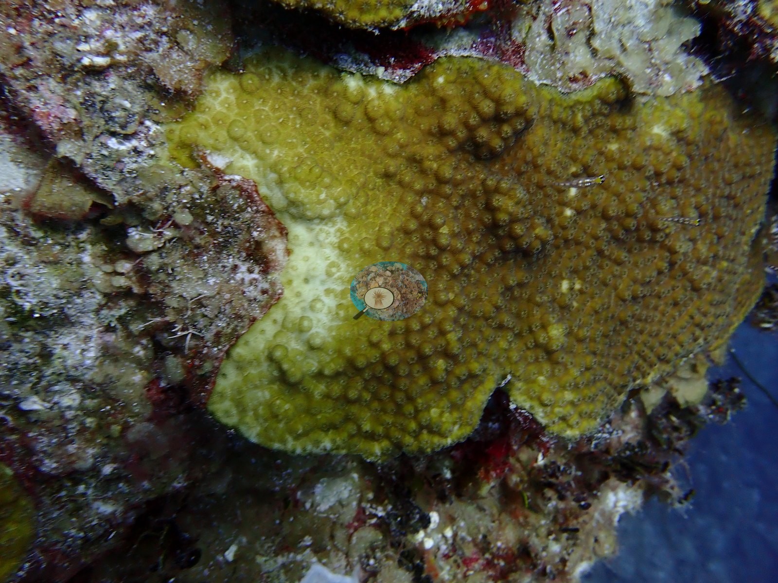
Turks and Caicos (NOAA CRW virtual station) · Bahamian ecoregion mean: 9.1 degree heating weeksA wall dive with a chimney swim-through from about 40 ft to 130 ft, with plate corals on the upper wall and a sand terrace below where Caribbean reef sharks, southern stingrays, and barracuda cruise. Active tissue loss was widespread, with about a dozen frames of fresh white lesions on star corals and one shot of ten or more small colonies each carrying its own lesion.
Five independent survey stations shown with the program’s logo — click one to see how far it is from this site.
Four other CRD dive sites within 5 km shown in orange — click a marker to open its page.
This page shows 50 of the 55 photographs from this site, including every one the text cites.
46 of the 50 photographs on this page are shown beside the text that discusses them under "Site description" below.
Site description
Setting
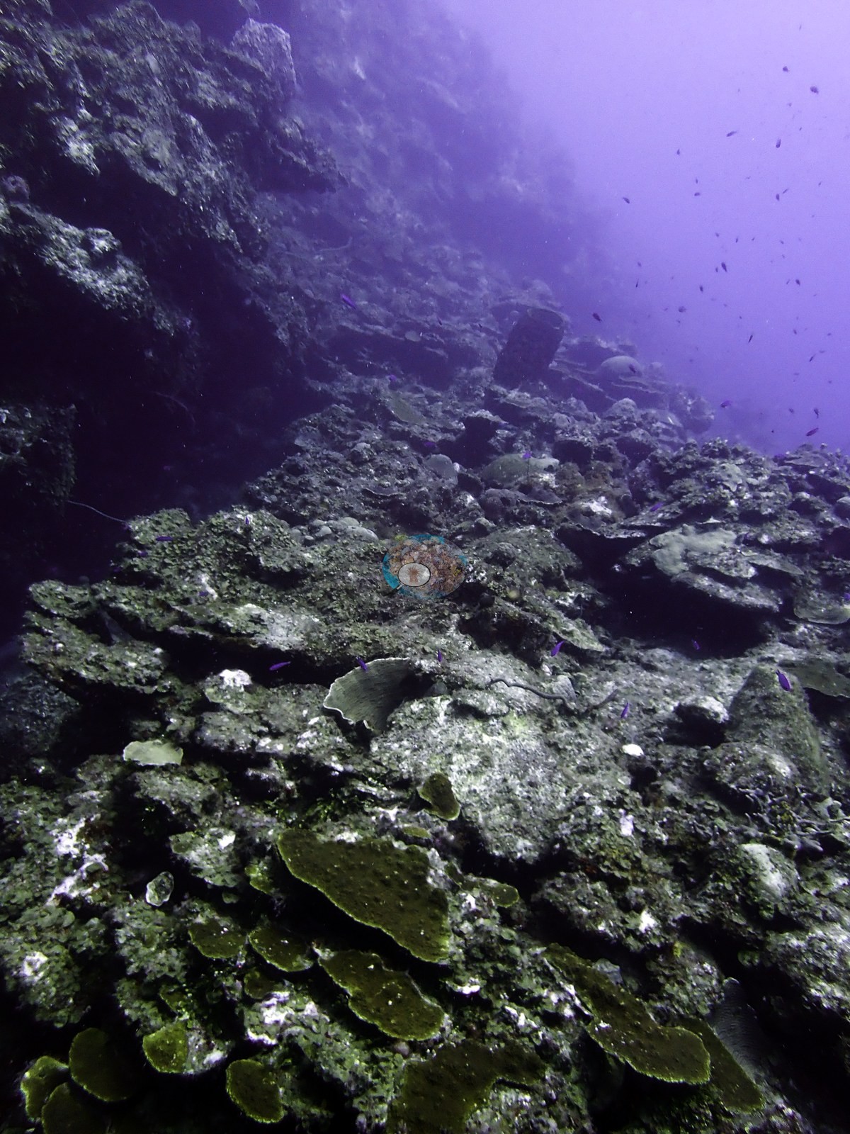
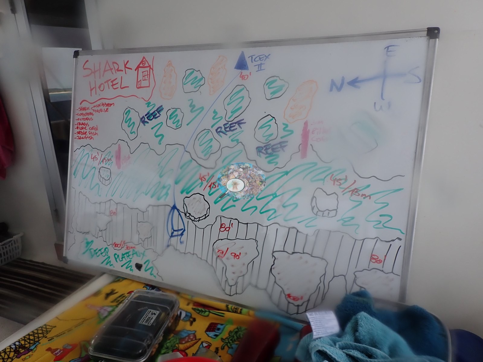
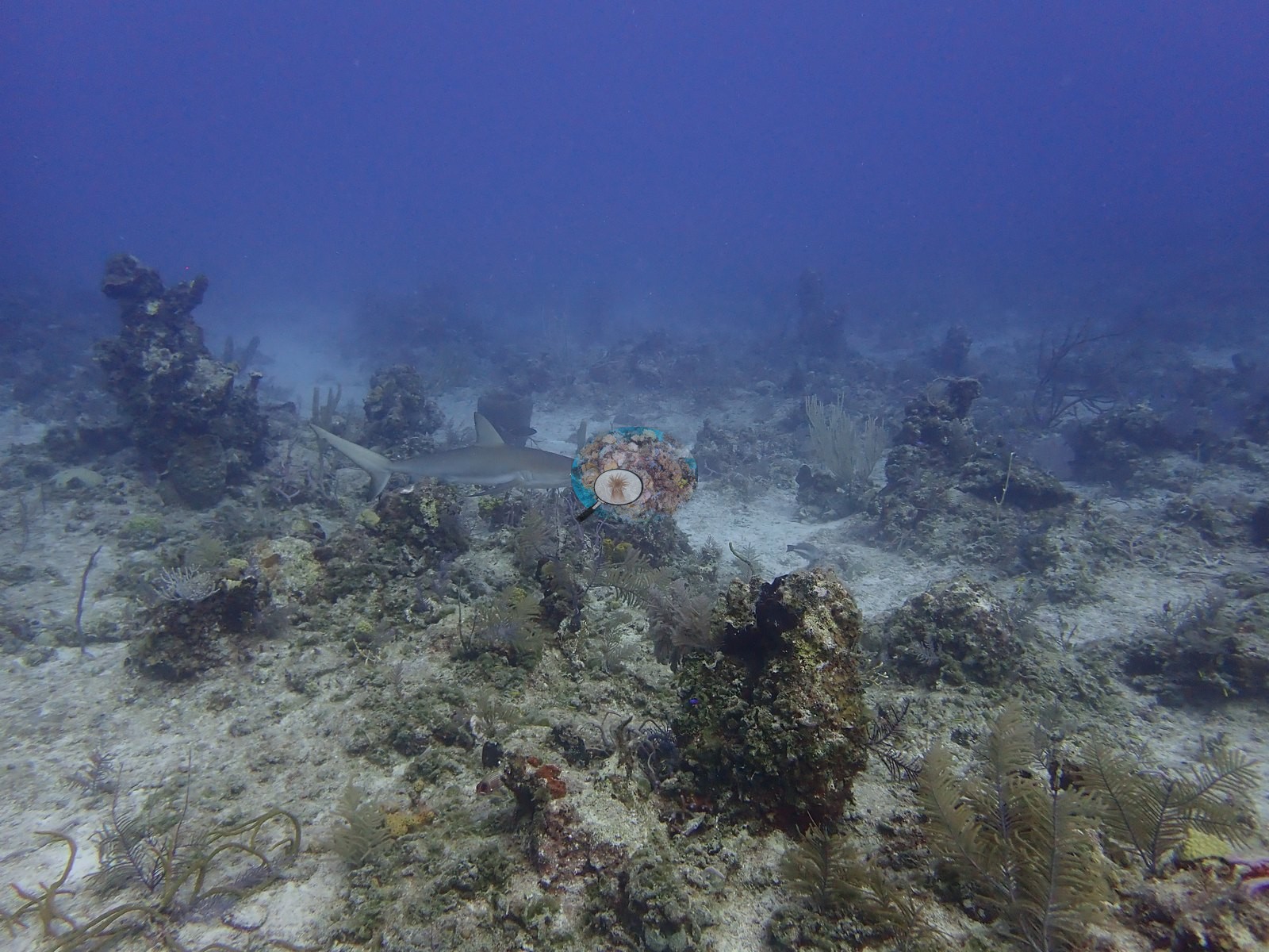
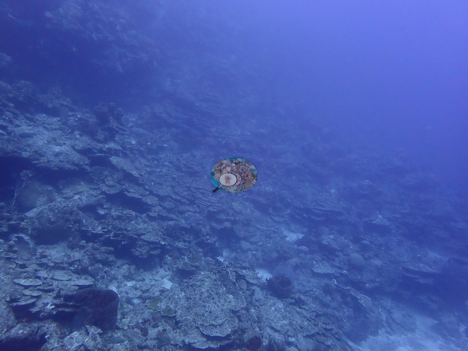
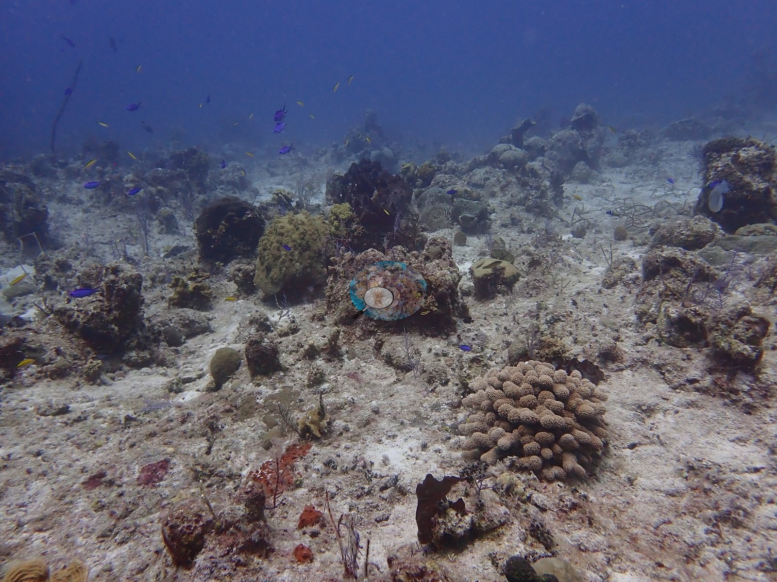
Shark Hotel is the wall dive of this Northwest Point group, and the frames separate cleanly into four zones (e.g., Image 5, Image 11, Image 19, Image 55).
General benthic features
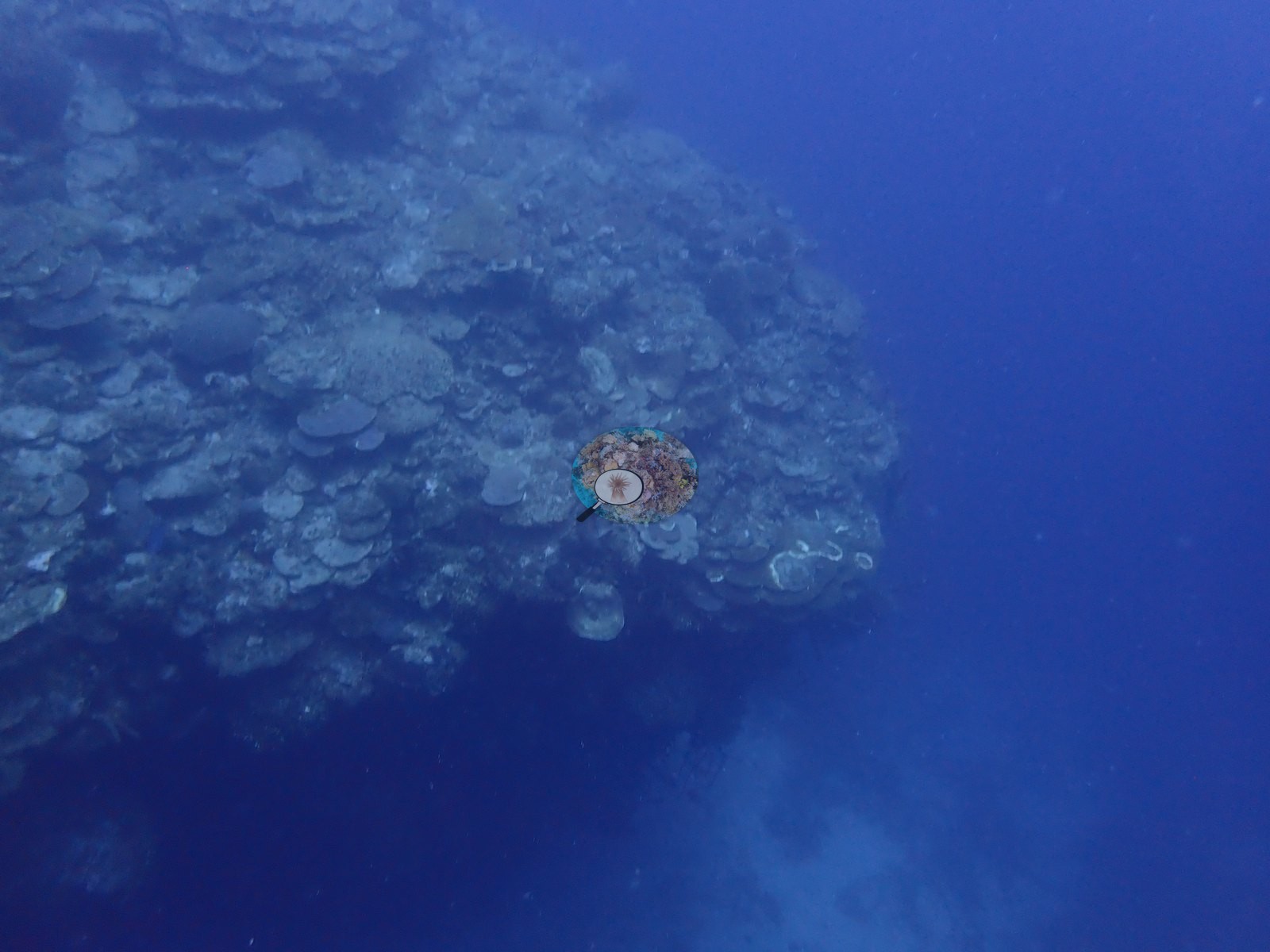
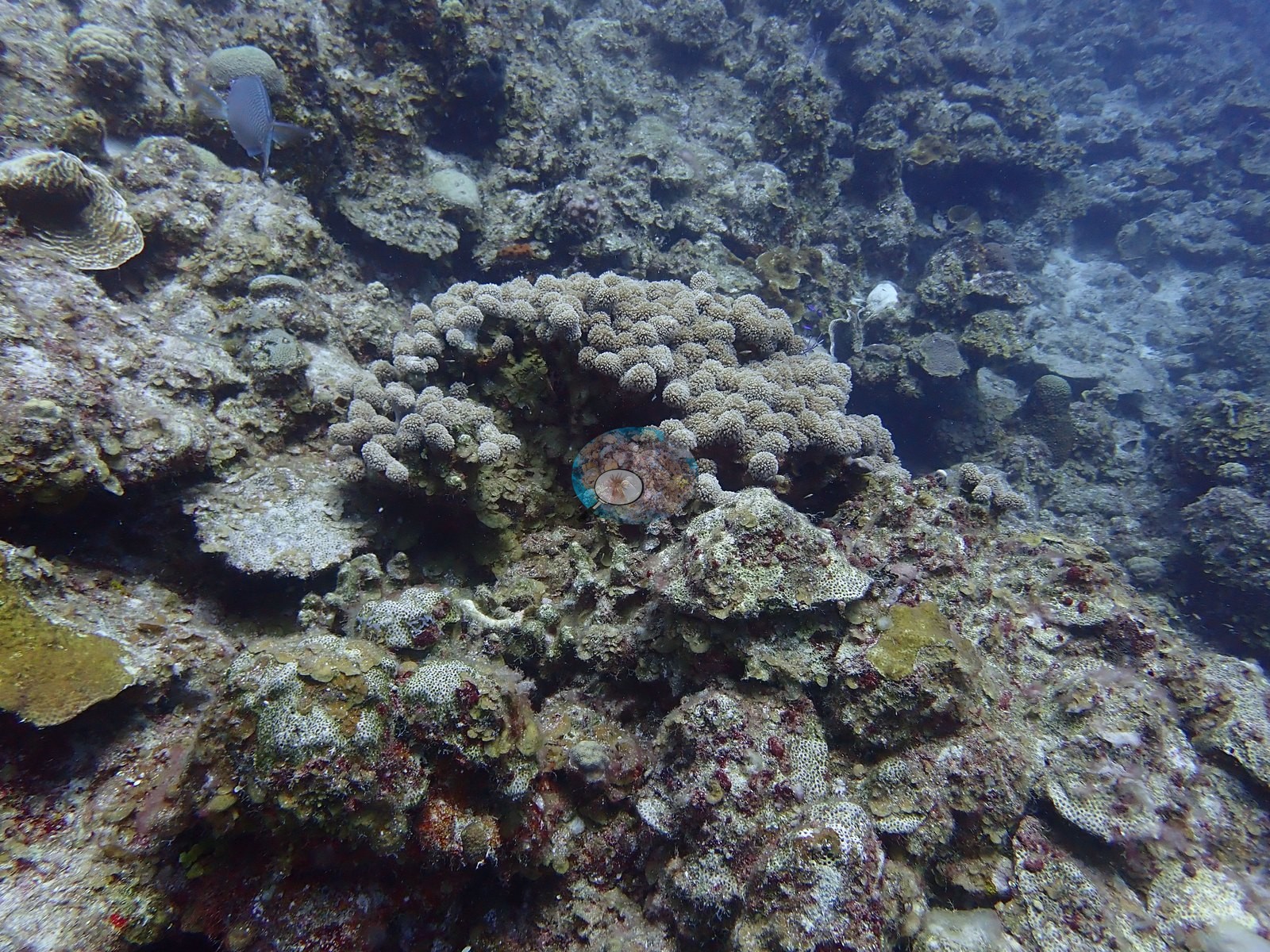
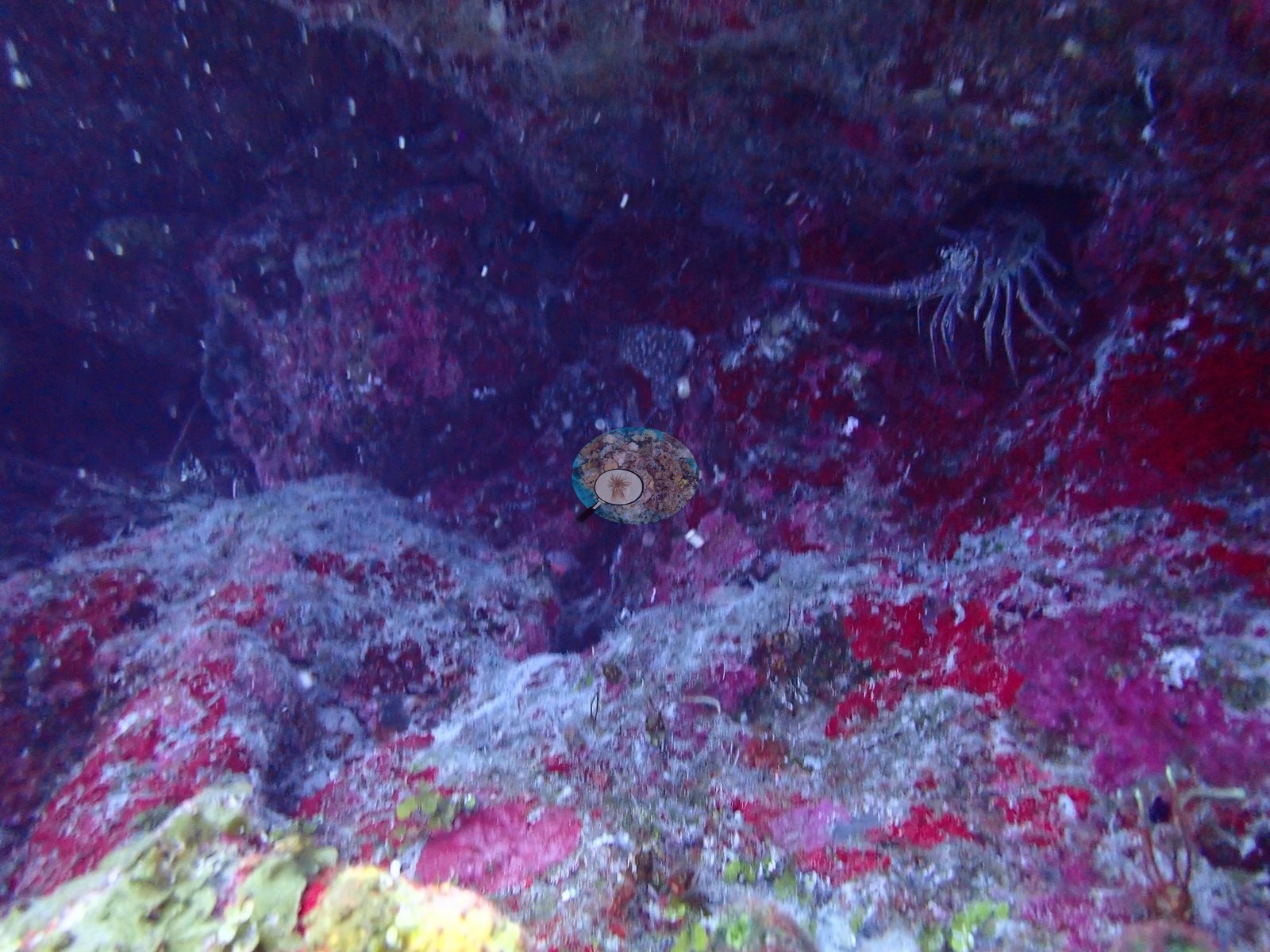
The wall itself is near-vertical and mostly austere: turf- and coralline-crusted rock with sparse coral (e.g., Image 36, Image 41), a dark vertical slot where the chimney breaks the face, and overhangs whose ceilings are painted pink with crustose coralline algae (e.g., Image 49, Image 53) and red rope sponges (e.g., Image 14).
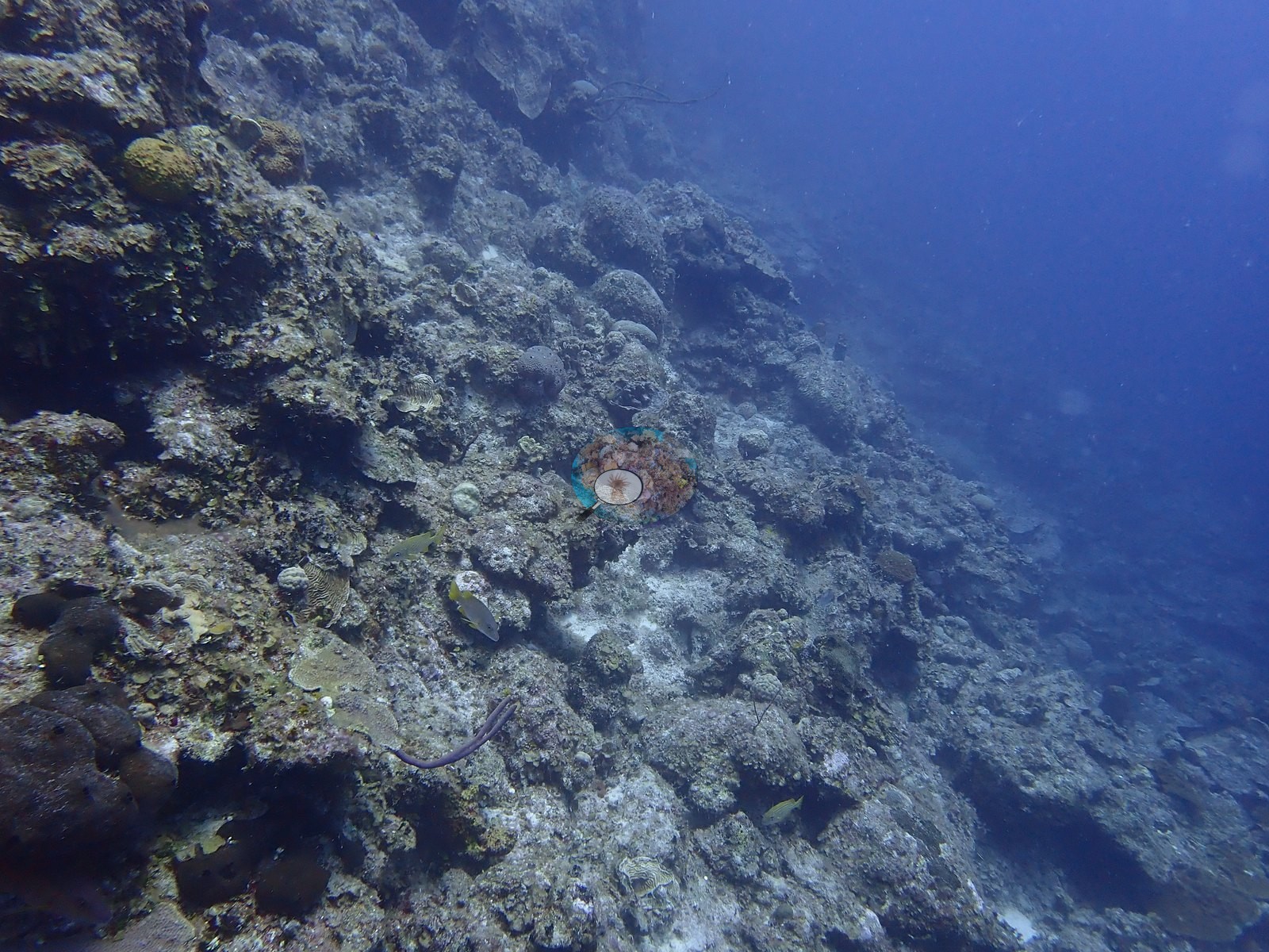
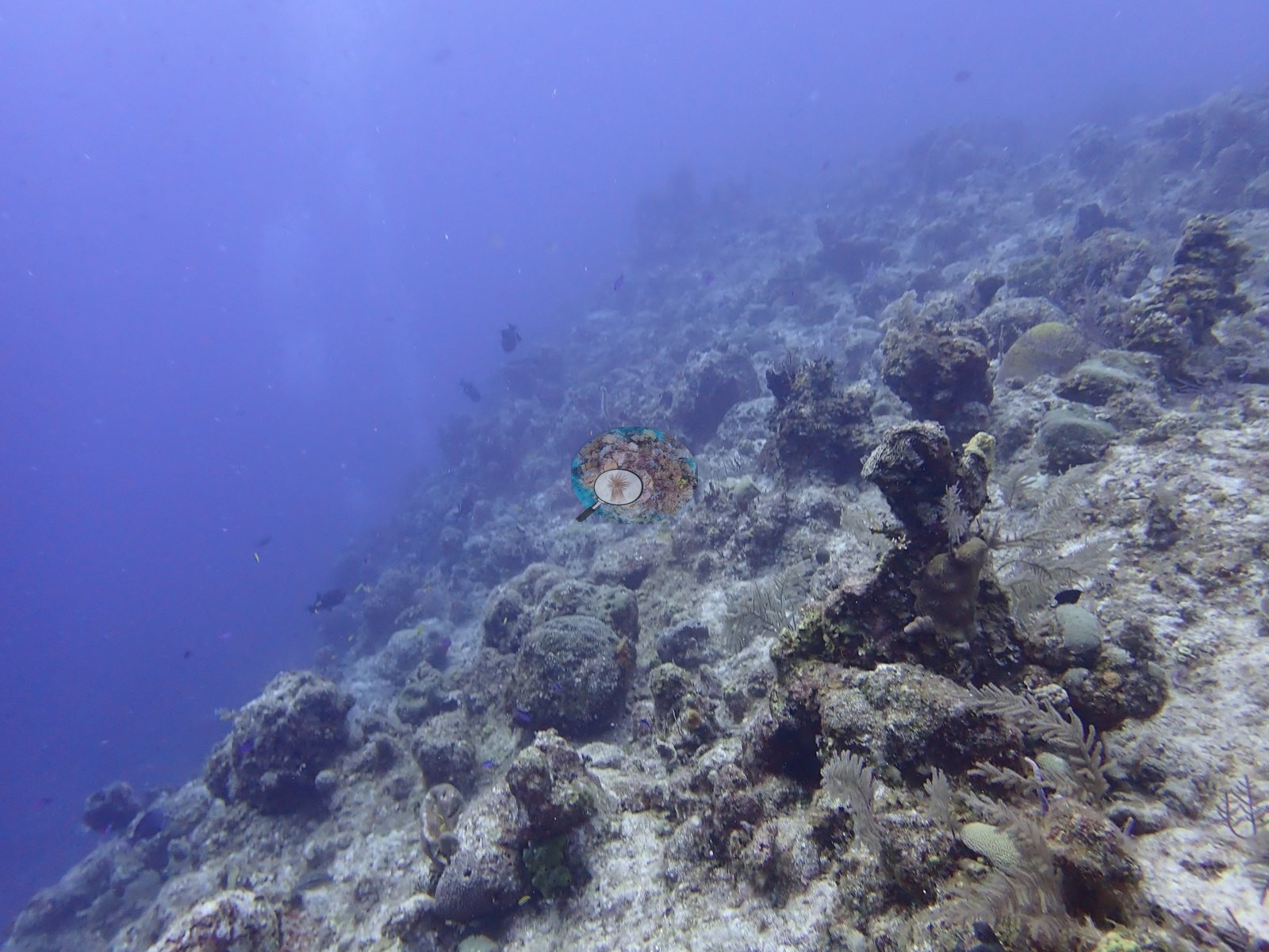
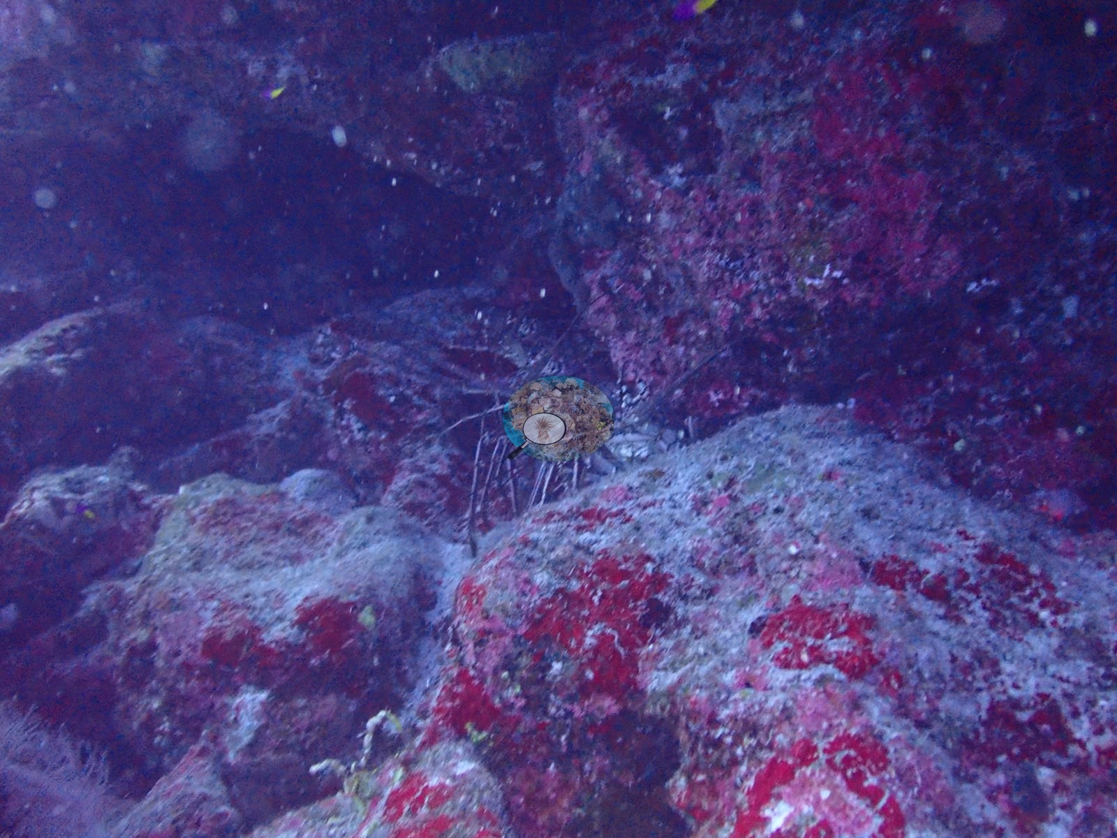
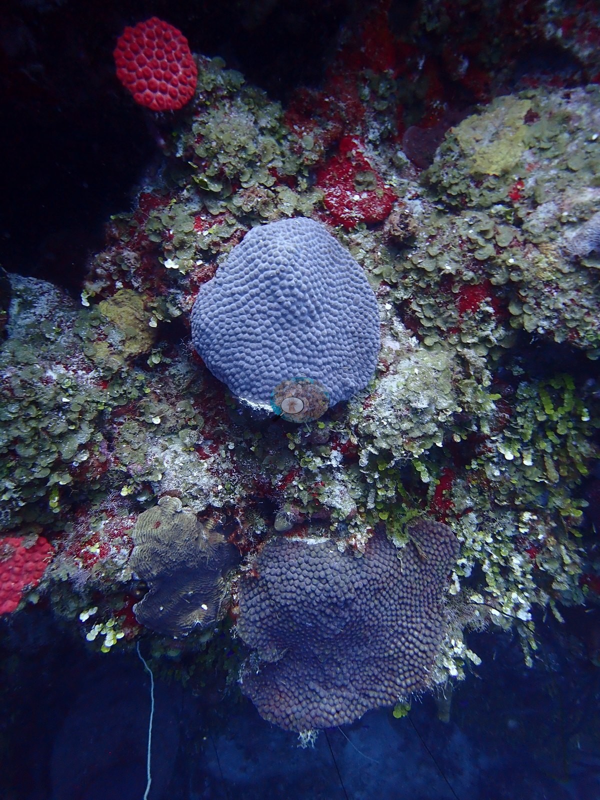
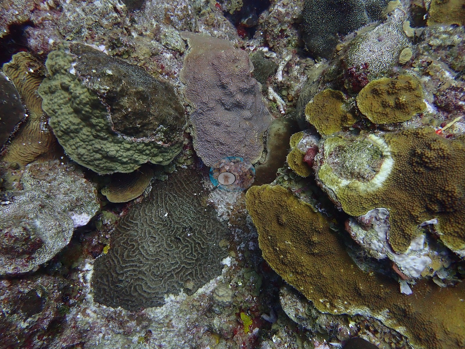
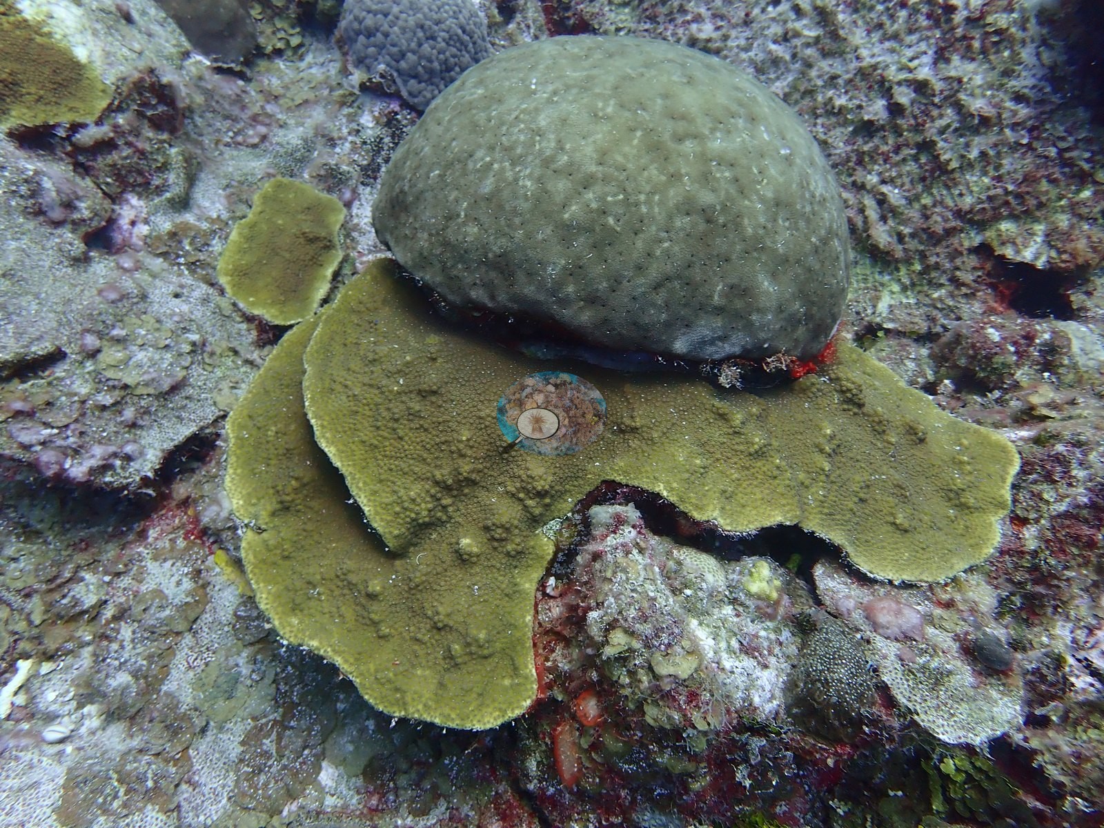
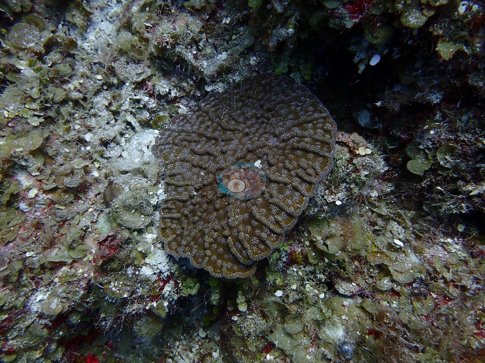
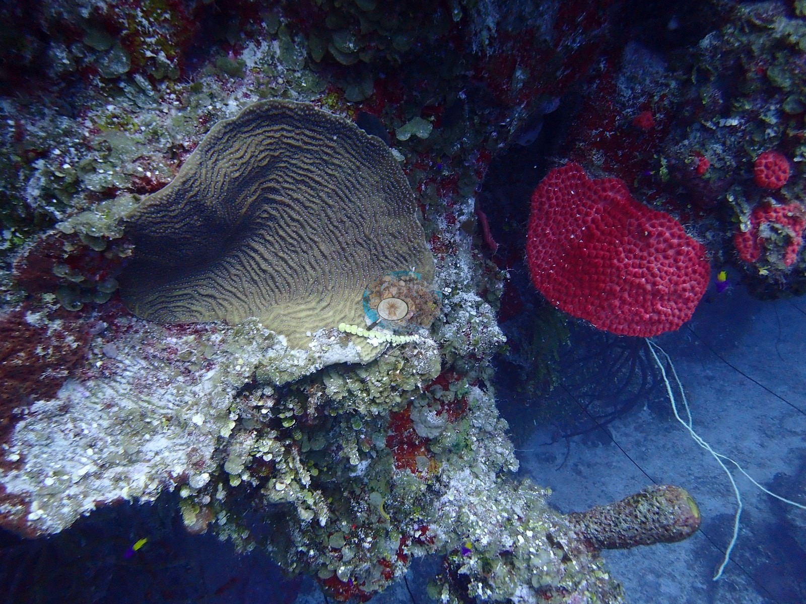
The upper wall carries the best coral - broad fields of stacked Agaricia-type plates and mauve Montastraea cavernosa plates, tier on tier (e.g., Image 3, Image 22, Image 27, Image 30, Image 43).
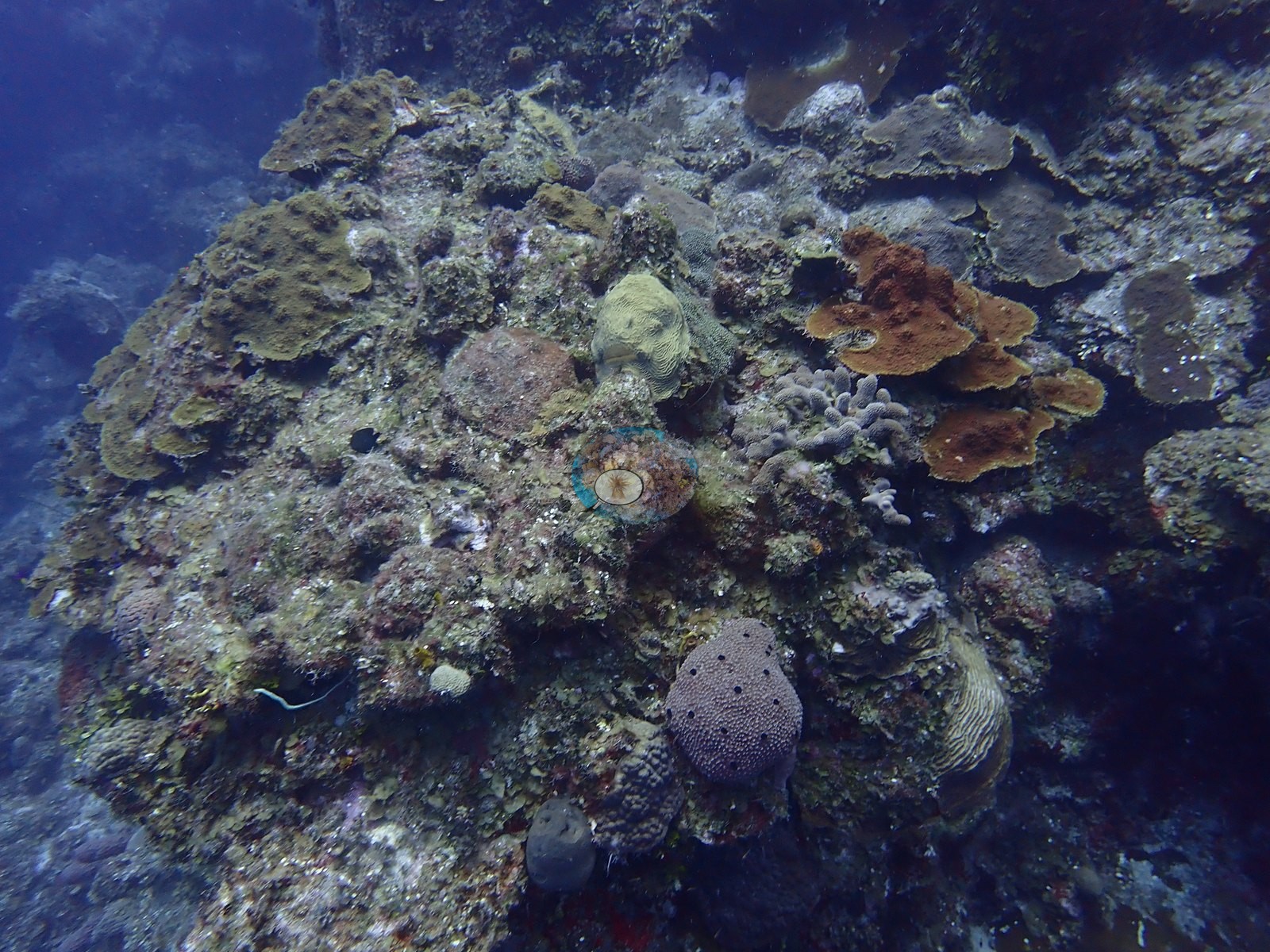
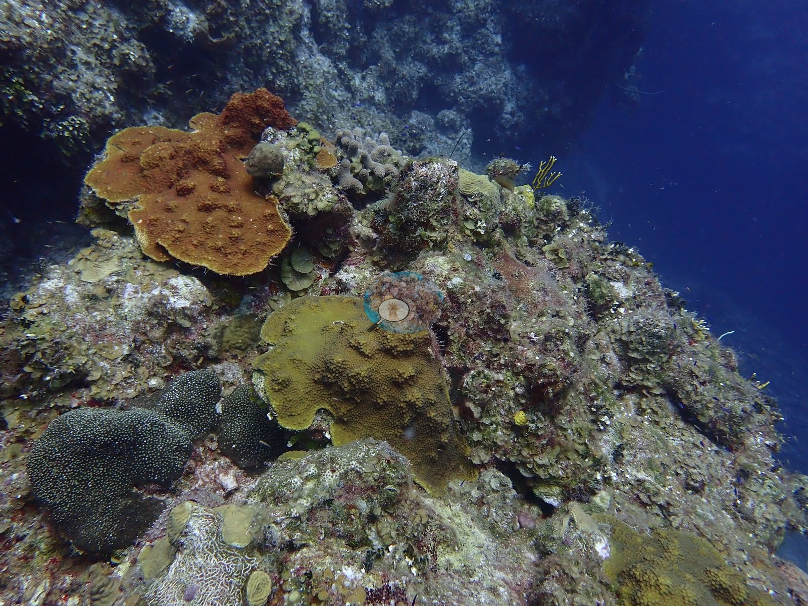
The reef top above is the opposite: wide, low-relief pavement, largely dead, and crusted, in places carpeted in green turf (e.g., Image 4, Image 10, Image 24).
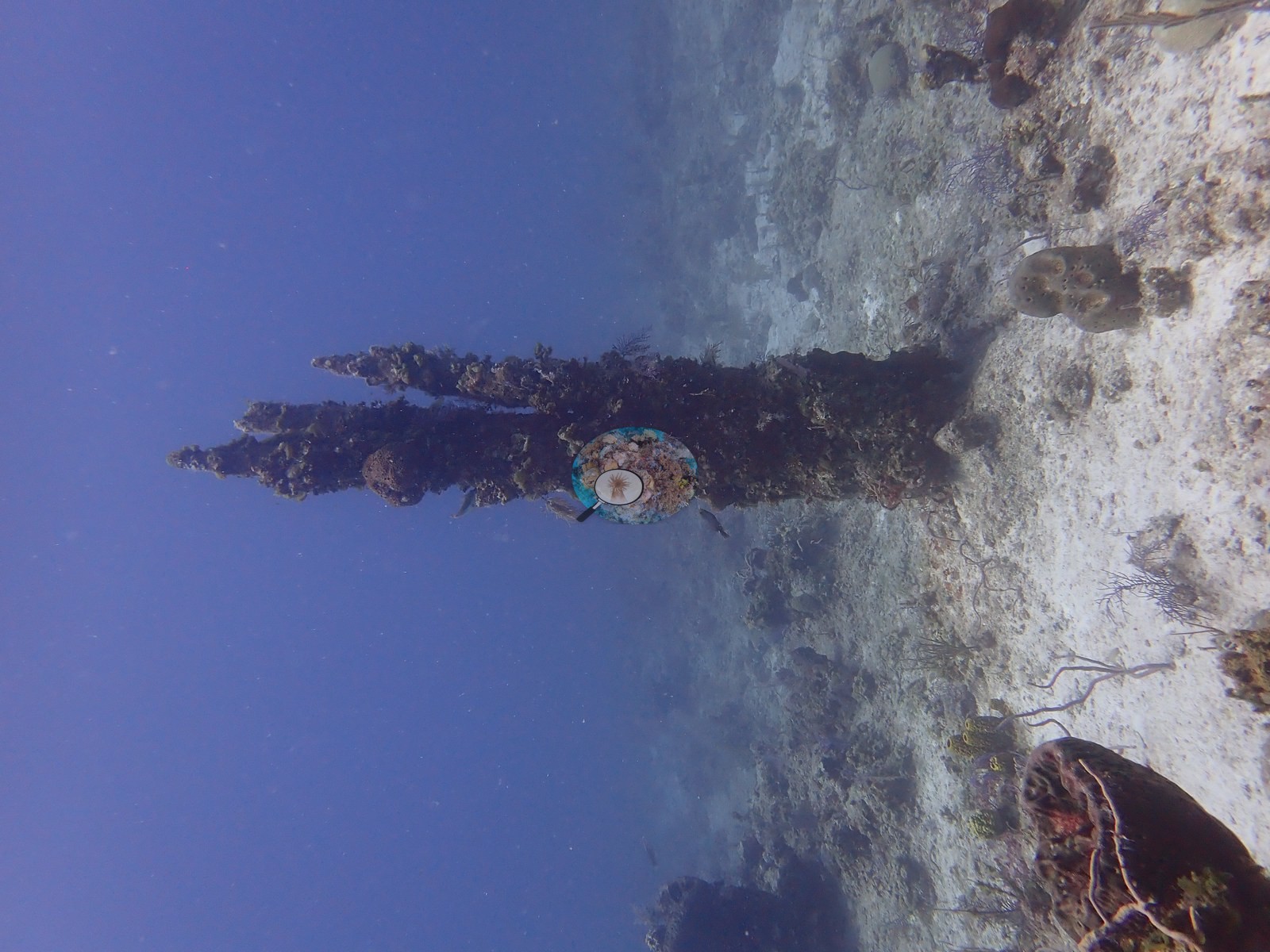
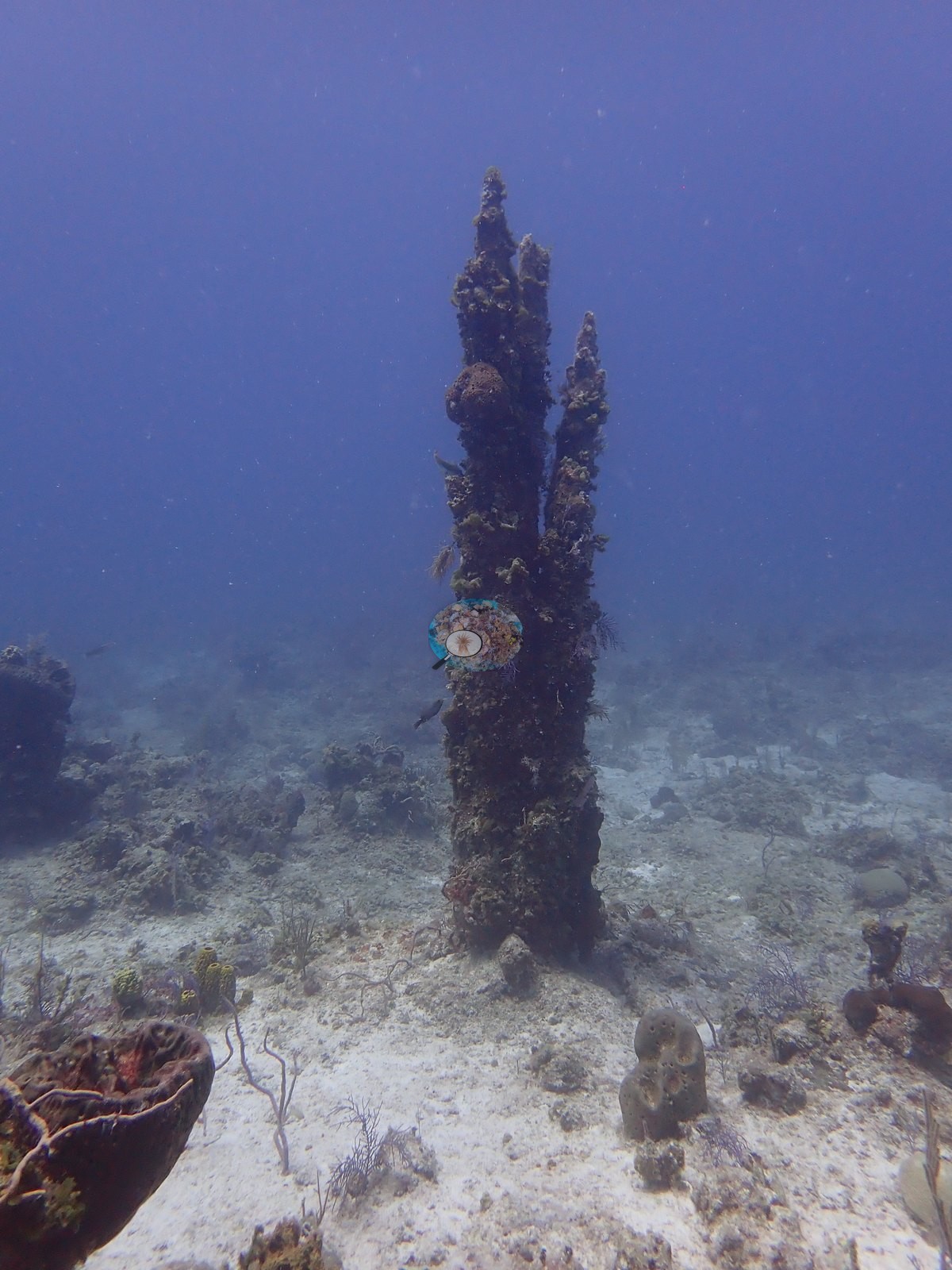
Below the wall a sand terrace runs out flat, punctuated by isolated limestone columns (e.g., Image 38, Image 45) and mushroom-shaped undercut remnants two meters high (e.g., Image 51, Image 54), brown barrel sponges (Xestospongia muta) (e.g., Image 17, Image 47), tube sponges, sea rods, and purple sea plumes (e.g., Image 15, Image 25, Image 33).
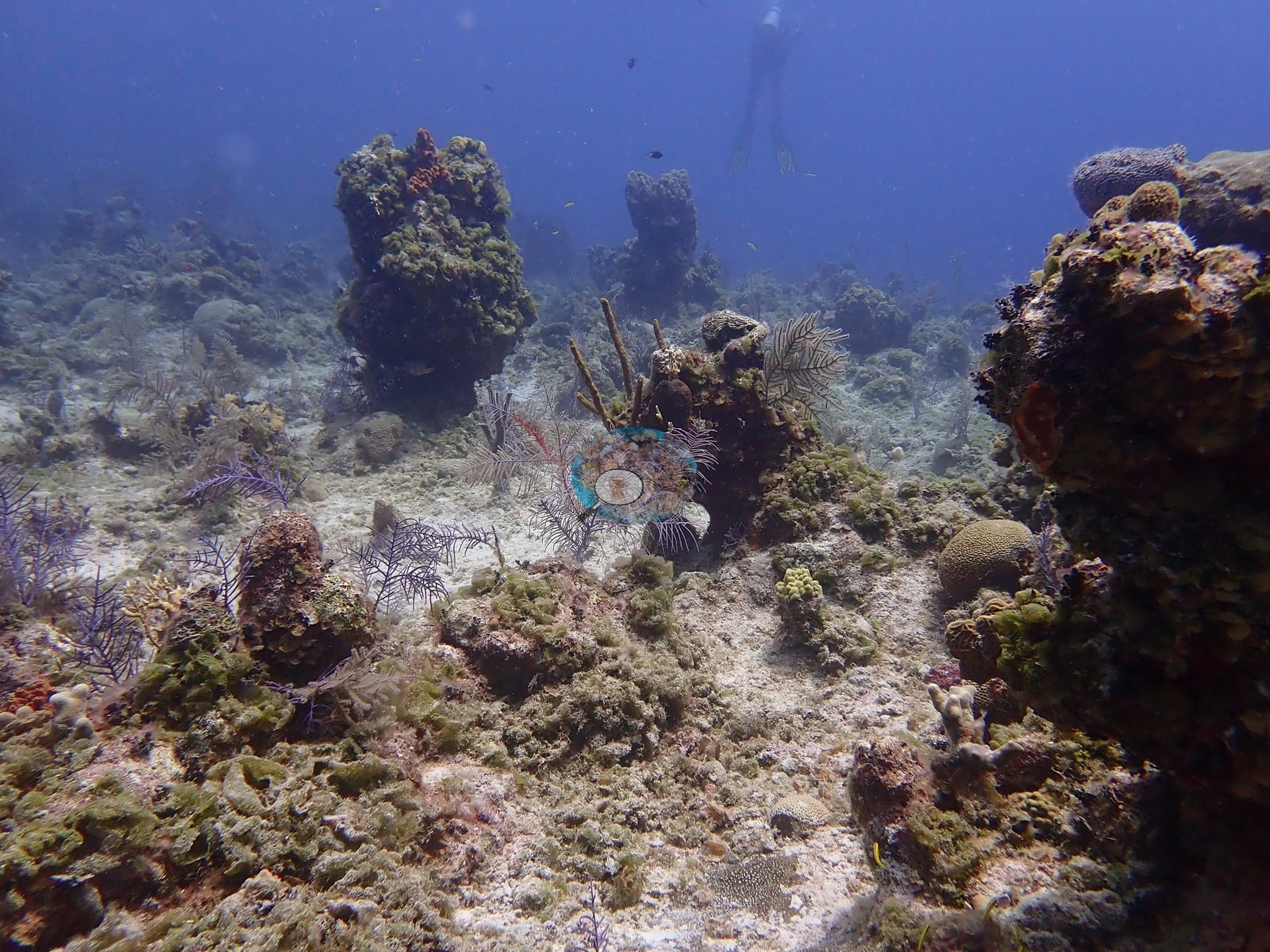
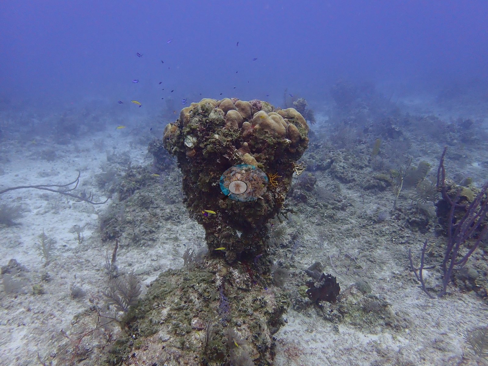
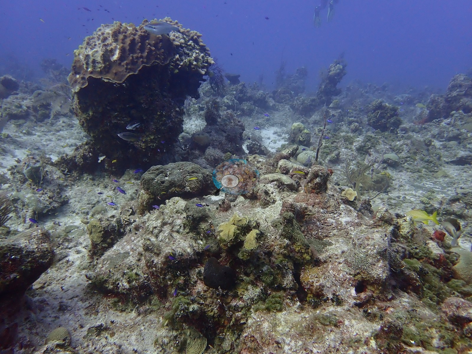
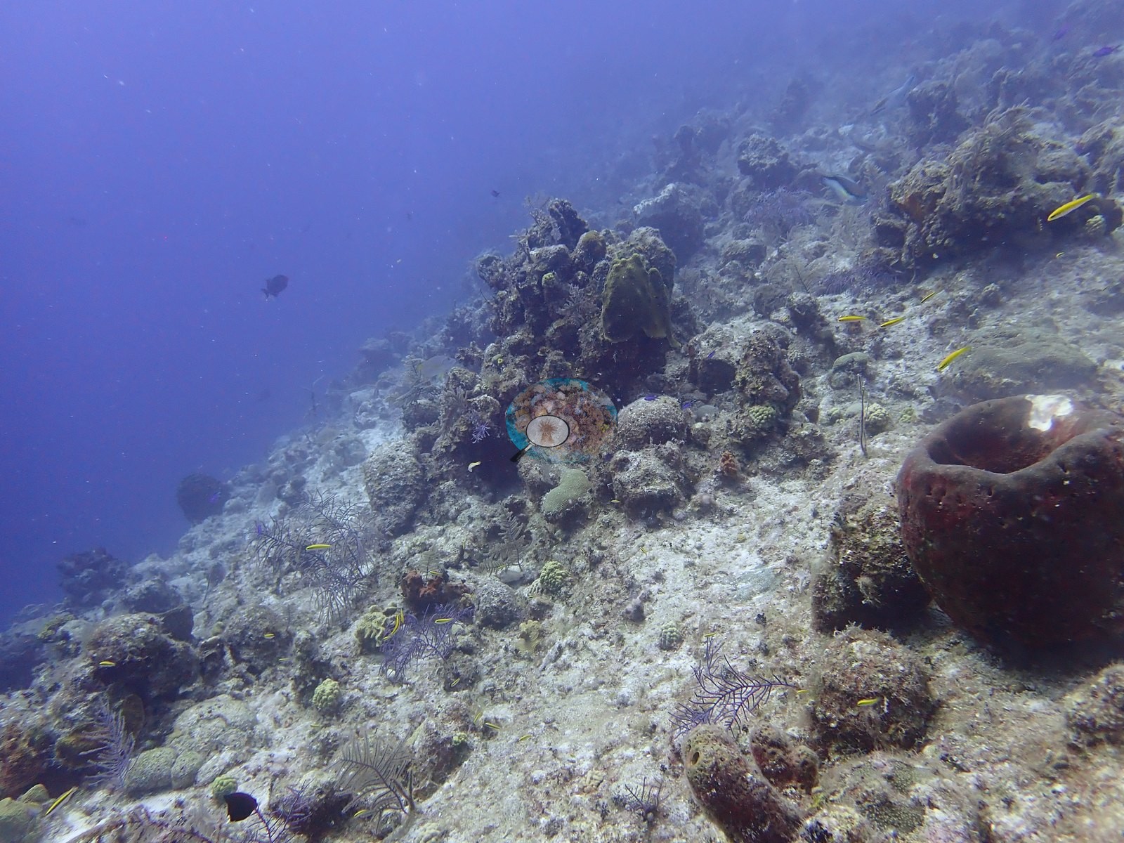
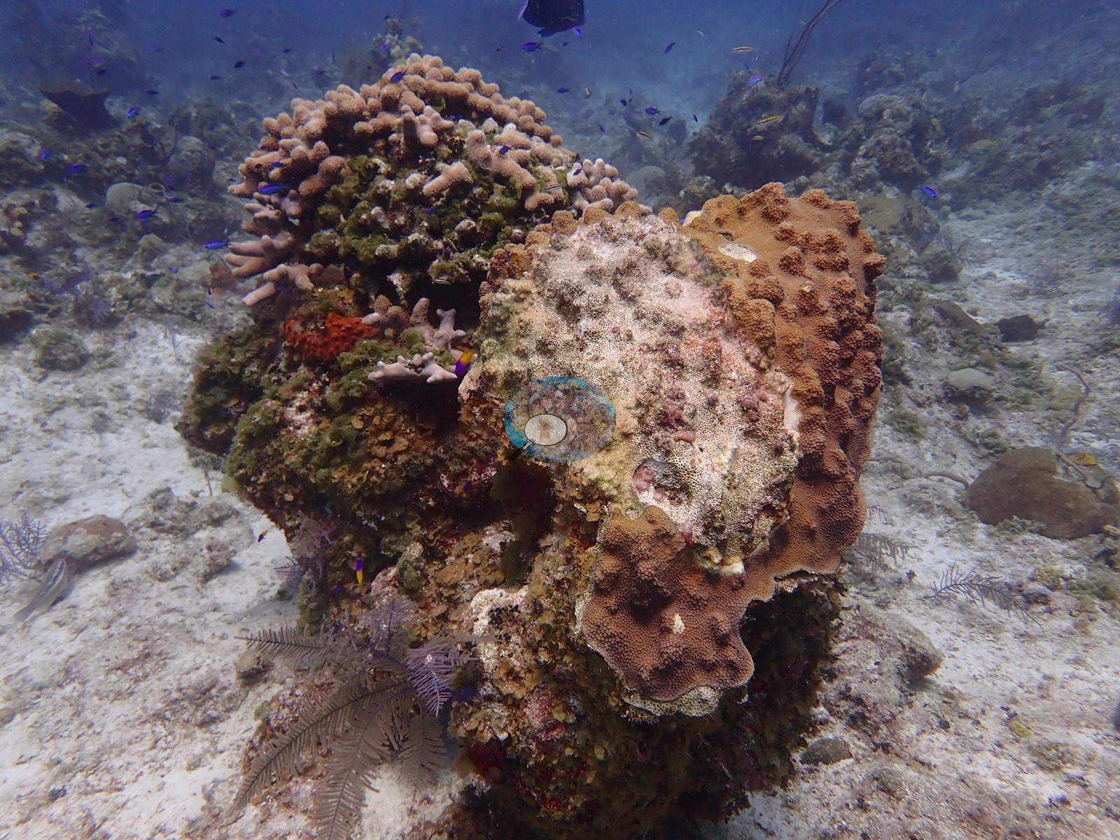
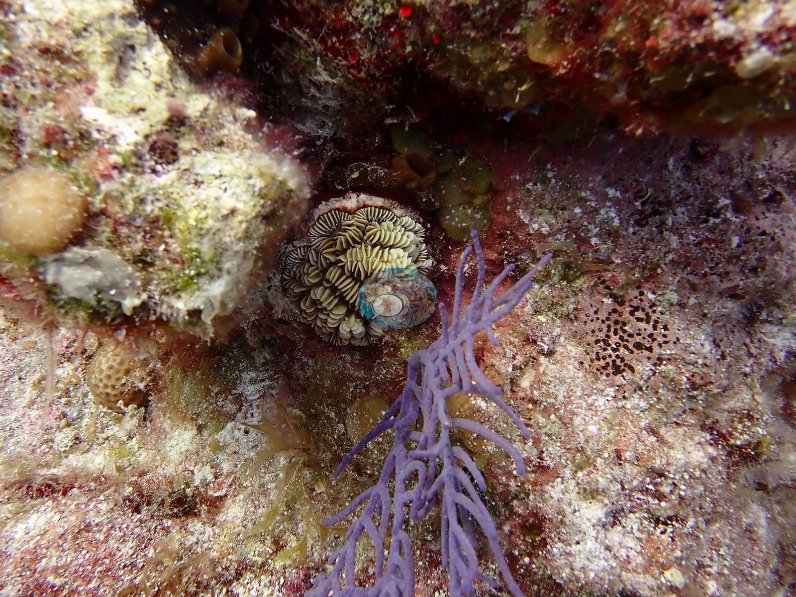
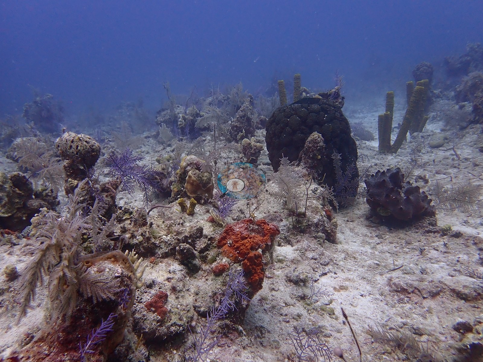
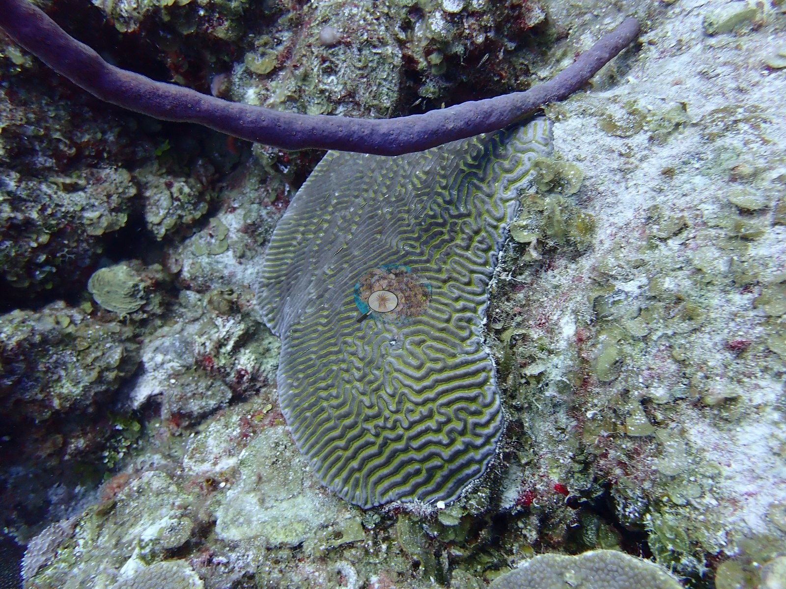
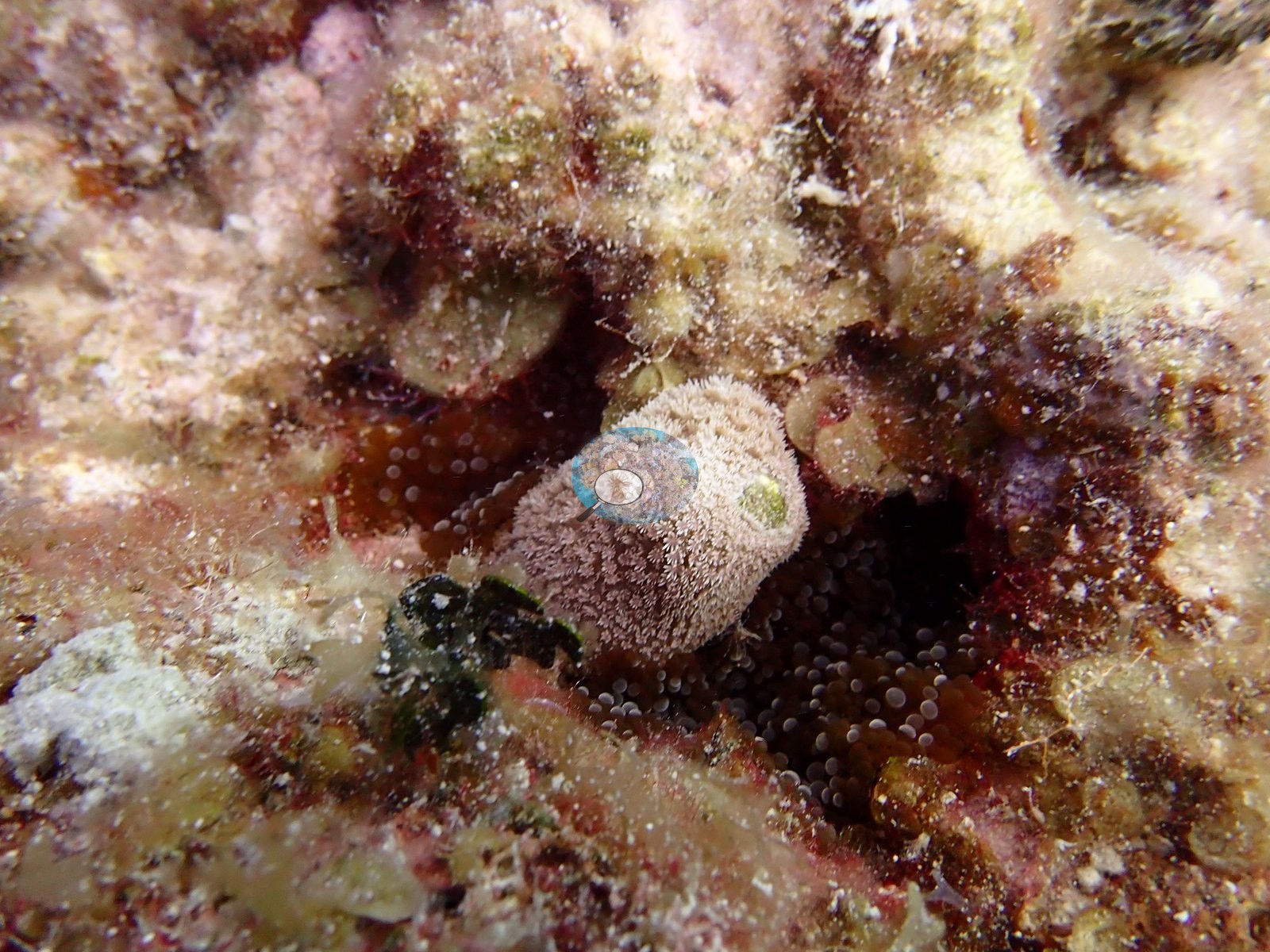
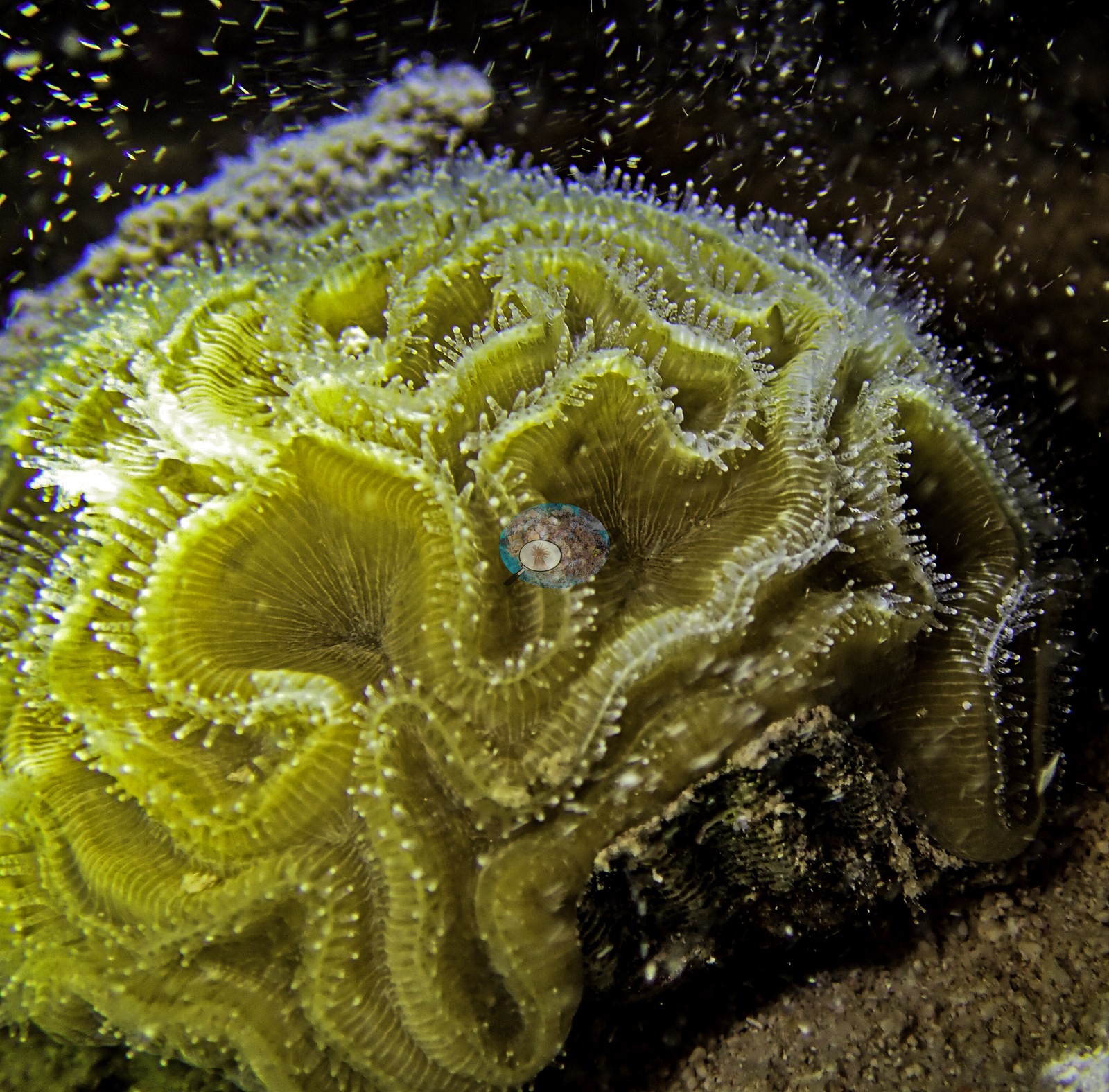
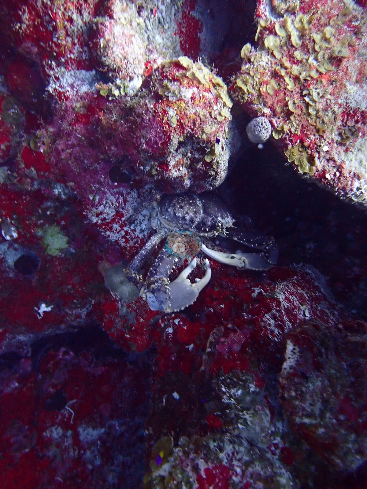
Corals identified (e.g., Image 1, Image 34, Image 46): plating and knobby Orbicella, Montastraea cavernosa as domes, columns, and plates, Agaricia plates, Porites astreoides mounds (e.g., Image 16, Image 39, Image 50), thick-fingered Porites porites (e.g., Image 56), Siderastrea-type domes, a clean Pseudodiploria strigosa, meandroid brains of the Colpophyllia/Diploria labyrinthiformis group (e.g., Image 28, Image 35), and fleshy Mussa- or Isophyllia-type polyps.
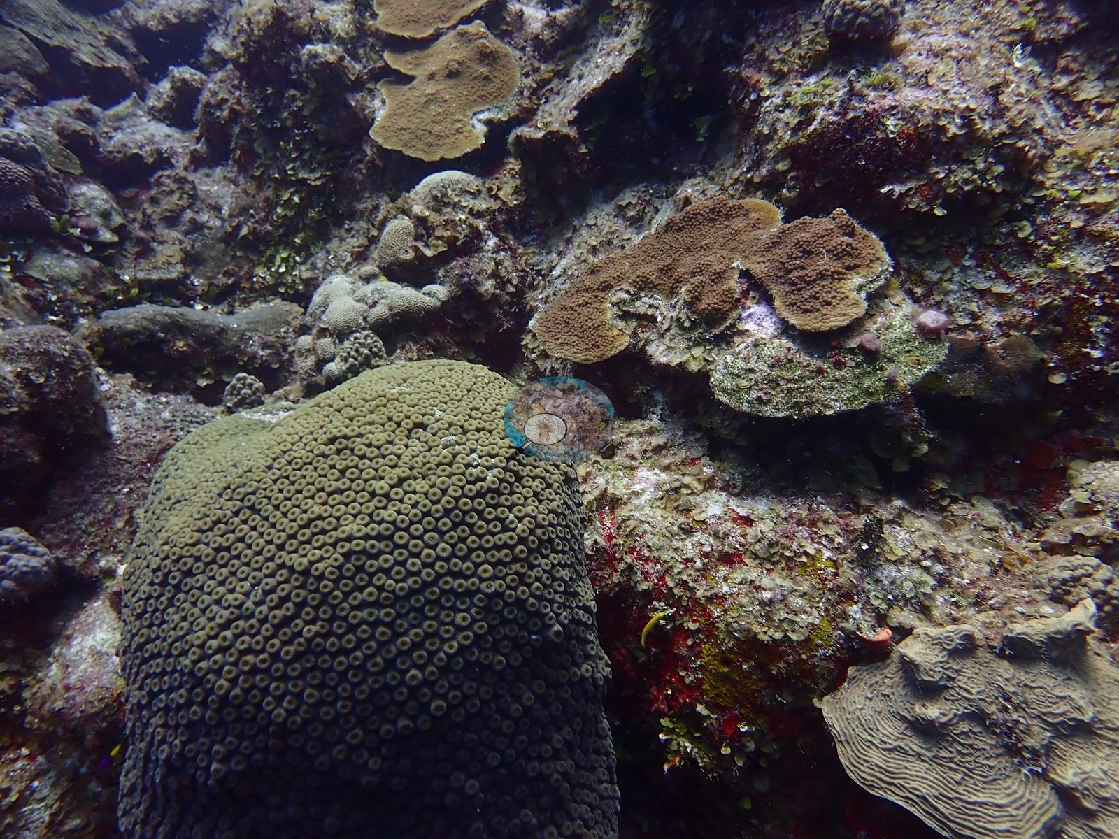
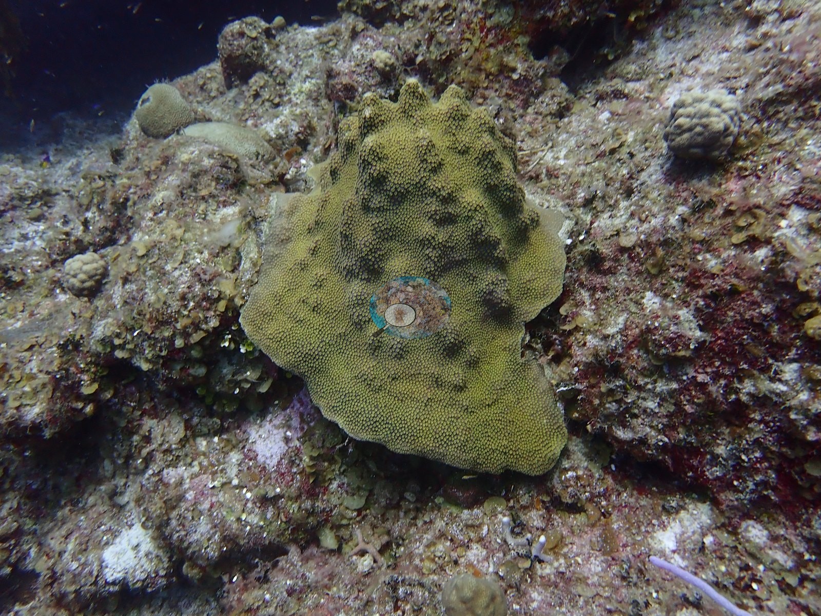
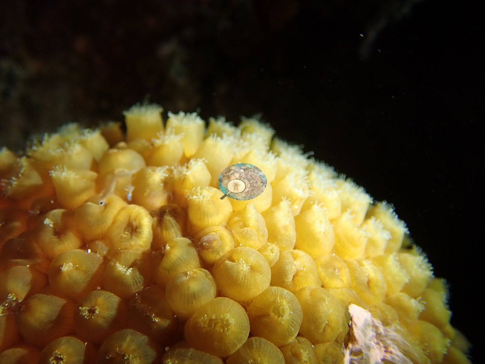
The night frames are all macro: M. cavernosa, Orbicella, Porites porites, and meandroid brains with polyps and tentacles fully extended (e.g., Image 6), plus channel clinging crabs on the sand (e.g., Image 7) and orange brittle stars threading the colonies.
Coral cover
- Reference: Claude CV point count (this site's photos)
- Coral cover: 16% — a blind Claude computer-vision point count of 44 of Anderson's edited photographs (the mean of the two dives' means; 20 points per photo; water, fish, and obstructions excluded). Other computer-vision methods are listed in the method table.
Stressors
Condition: roughly a dozen day frames show active tissue loss - bright bands and patches of freshly bared skeleton on Orbicella plates and on M. cavernosa, sometimes creamy white and unfouled (e.g., Image 18, Image 26), sometimes with turf already established behind the front (e.g., Image 40, Image 44).
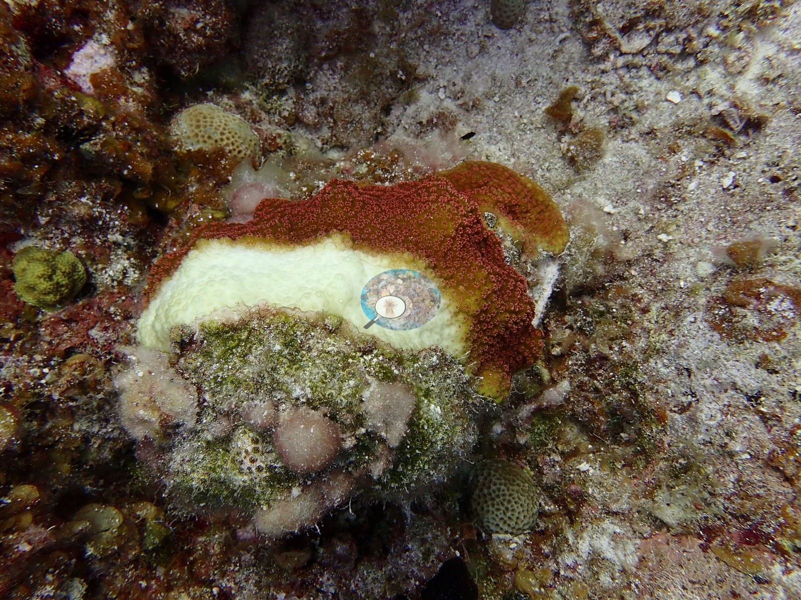
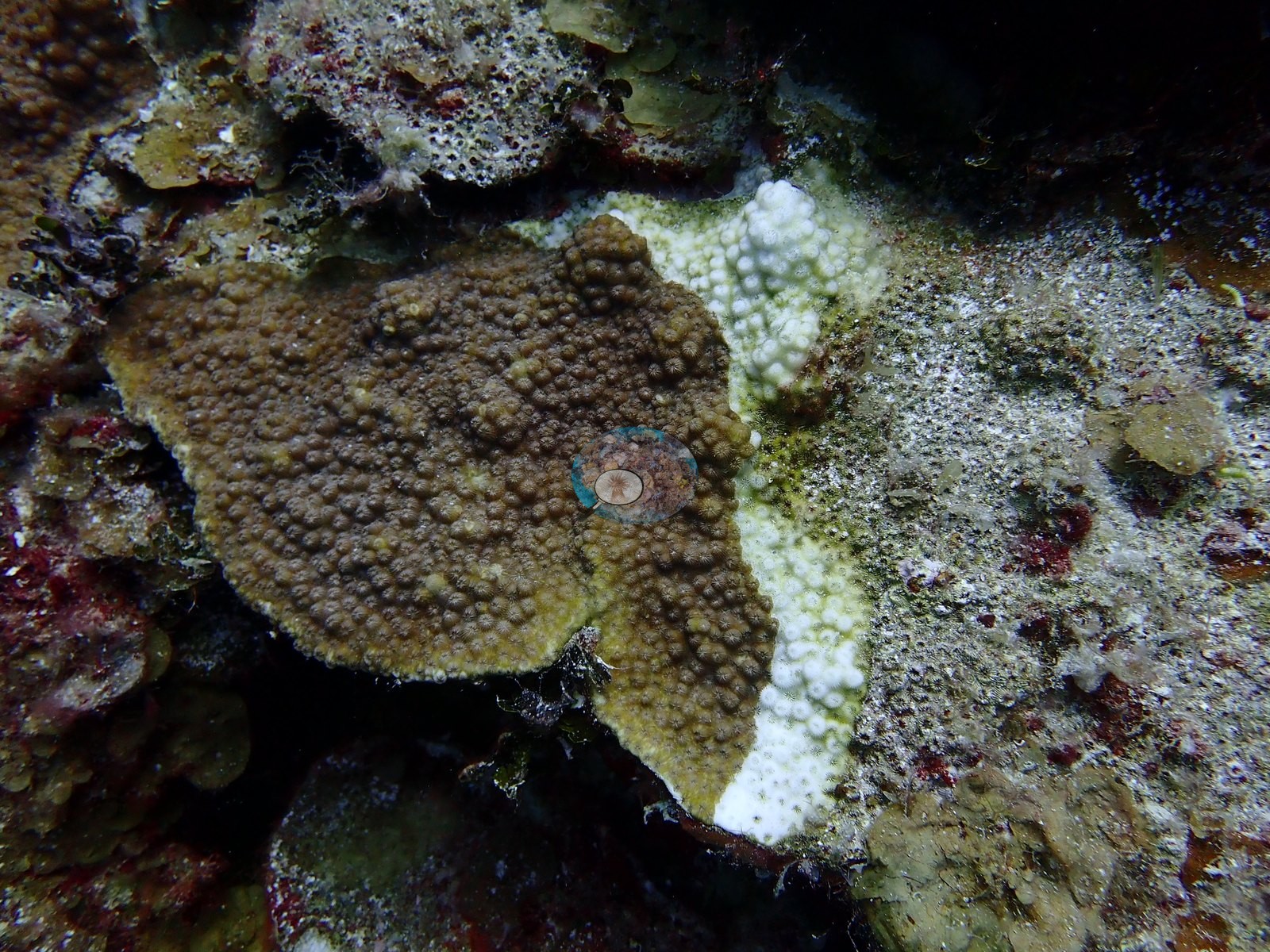
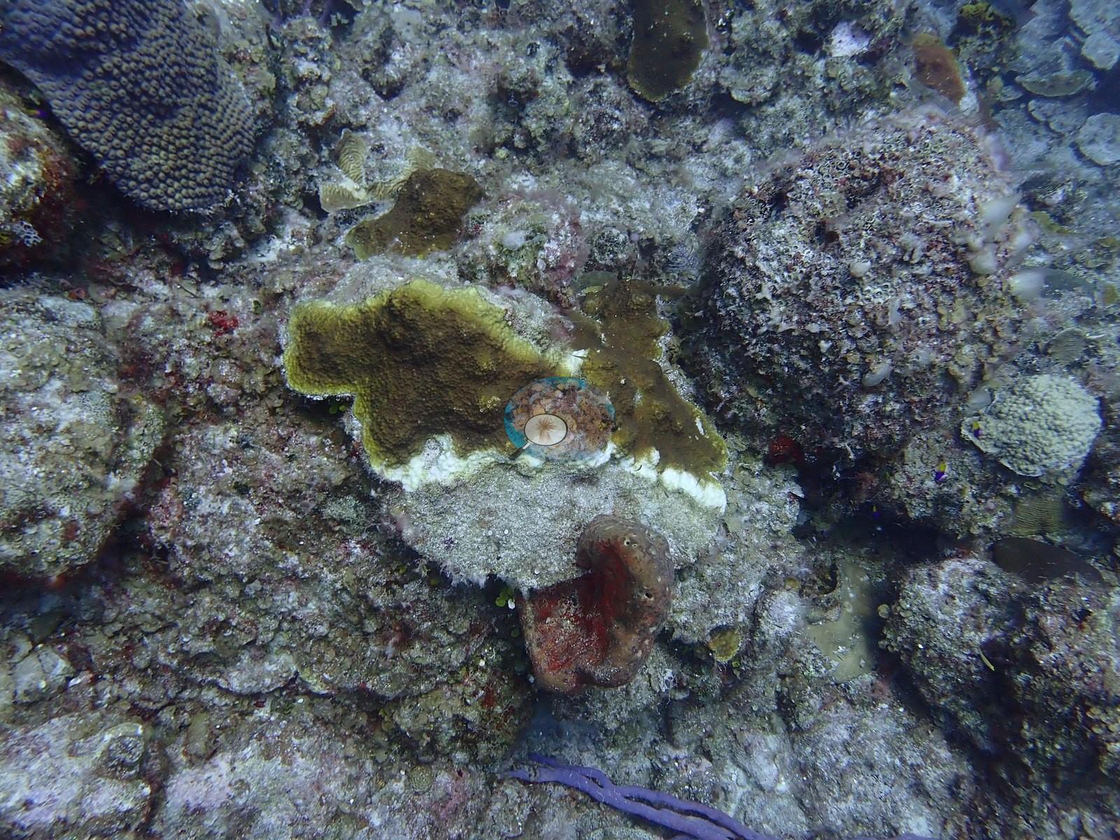
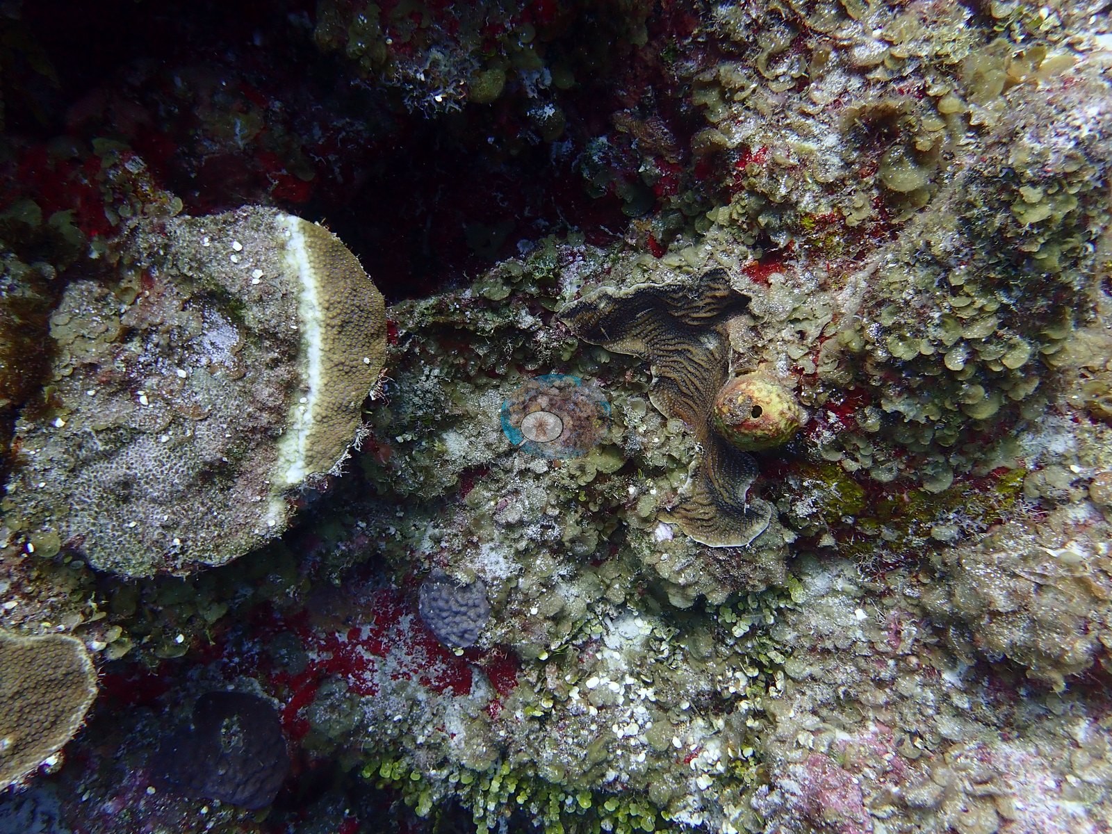
One down-shot catches ten or more small colonies each carrying its own white lesion. Standing dead skeleton is everywhere on the pavement (e.g., Image 21).
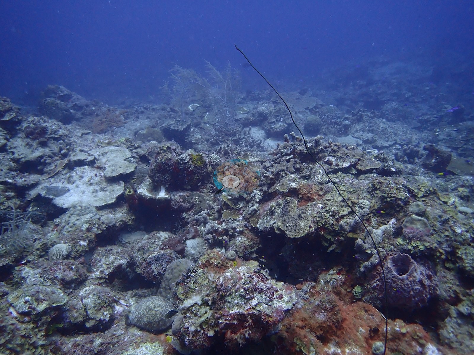
No corallivores evident.
Fish and other fauna
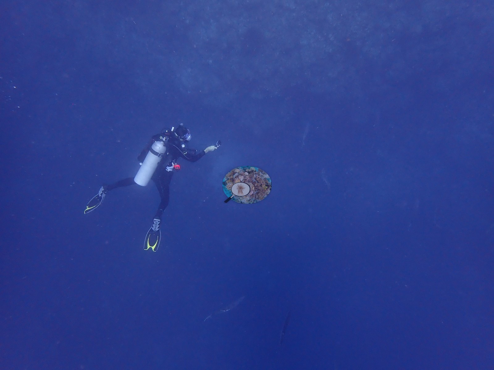
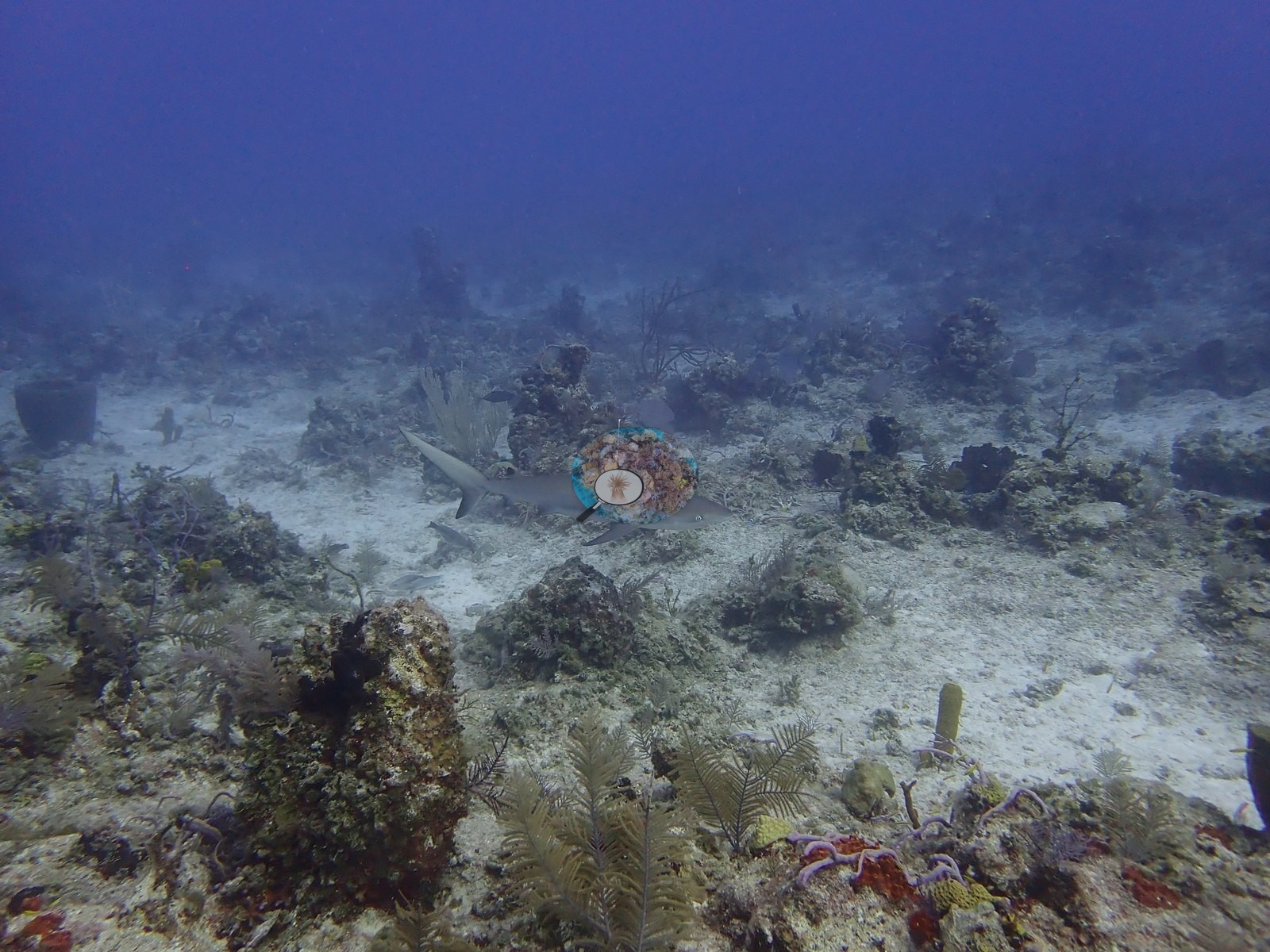
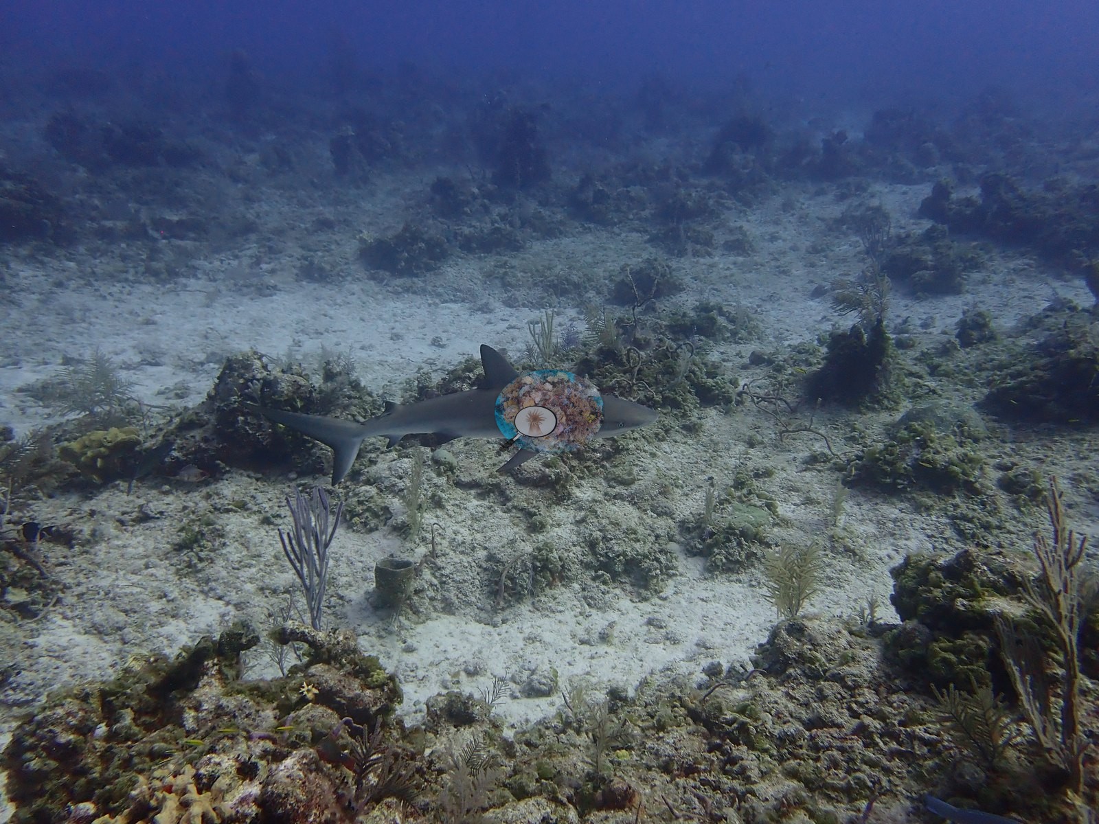
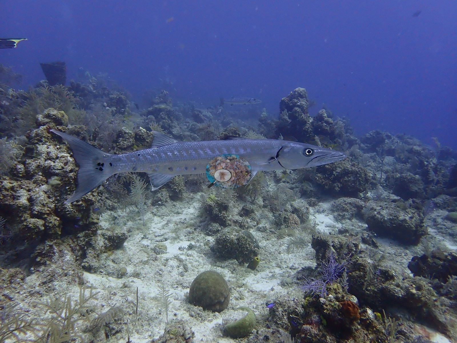
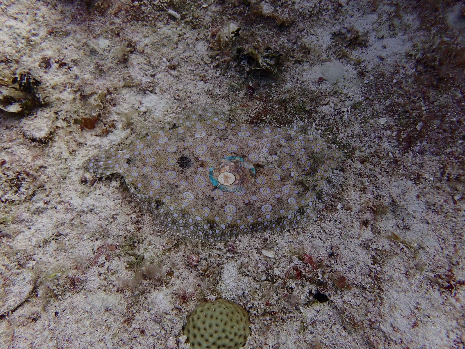
Fauna was the richest of this group: Caribbean reef sharks (Carcharhinus perezi) in five frames, four cruising low over the sand terrace (e.g., Image 8, Image 9); two southern stingrays; two great barracuda (e.g., Image 13); schoolmaster snapper and grunt schools; black durgon, blue chromis, creole wrasse, blue tang; a peacock flounder (e.g., Image 29); three spiny lobsters under ledges.
Comparisons with other data
Shark Hotel (Northwest Point / Columbus Landfall Marine Park, Providenciales) was dived from the Turks & Caicos Explorer II liveaboard on 31 July 2022. A wall dive starting around 40ft where a chimney swim-through drops to 80ft and exits back onto the wall face near 130ft.
Other notes
Corals identified: plating and knobby Orbicella, Montastraea cavernosa as domes, columns, and plates, Agaricia plates, Porites astreoides mounds, thick-fingered Porites, Siderastrea-type domes, a clean Pseudodiploria strigosa, meandroid brains of the Colpophyllia/Diploria labyrinthiformis group, and fleshy Mussa- or Isophyllia-type polyps (e.g., Image 1, Image 19, Image 28, Image 33, Image 39).
CV model comparison
More info
A few colony-focused photos raise the mean; the median is the typical photo. No computer-vision method yet estimates coral cover accurately on new images, so treat this as indicative rather than a measurement; it is revised as the methods improve.More info
Other rows are for comparison only and are never averaged. No diver transect exists for this trip, so agreement between rows is not accuracy.| Method | Hard-coral cover (%) |
|---|---|
| CoralSCOP edited | 19.0 |
| Claude (edited photos) | 15.5 |
| Reef Check 2007 (same site) | 10.0 |
See a more detailed, stand-alone version of this table.
Dive history
| Date | Time in | Max depth (m) | Bottom time (min) |
|---|---|---|---|
| 2022-07-31 | 14:02 | 25.3 | 62 |
| 2022-07-31 | 16:30 | 28.0 | 55 |
| 2022-07-31 | 20:38 | 17.1 | 51 |
Independent surveys nearby
| Agency / program | Year(s) | Stations / distance | Link |
|---|---|---|---|
| Reef Check | 2005–2007 | 4 stations, 0.00–1.11 km | Records ↓ |
| XL Catlin Seaview Survey | 2013 | 1 station, 0.13 km | Records ↓ |
 Coral Reef Diagnostics
Within 5 km, 5 independent survey stations recorded hard coral cover in 5 records between 2005 and 2013. The most recent nearby survey is from 2013, so these values predate recent heat stress. This site's own estimate (16%) sits within the range of these surveys (2.9–17%); the methods differ, so the comparison is context, not a like-for-like test.
Collected by other programs using different methods (Reef Check uses point-intercept transects; the XL Catlin Seaview Survey uses photo-transects with automated image annotation), so these are context for the figures above, not a like-for-like comparison. Where a station was surveyed more than once the range is shown. Station names are not dive-site names.
| Agency | Site / location | Date | Hard coral cover (%) | Distance | Link |
|---|---|---|---|---|---|
| Reef Check | Black Coral Forest | 2005 | 5.6% | 0.26 km | — |
| Reef Check | Chimney | 2005 | 6.2% | 0.69 km | — |
| Reef Check | Shark Hotel | 2007 | 10% | 0.00 km | — |
| Reef Check | Eel Garden | 2007 | 17% | 1.11 km | — |
| XL Catlin Seaview Survey | transect 24044 | 2013 | 2.9% | 0.13 km | doi:10.14264/uql.2019.930 |
Data & links
- HeatCRD database ↗
- van Woesik and Kratochwill (2024) ↗
- Photo portfolio ↗
- Coral Reef Diagnostics page for this area ↗
All images from Coral Reef Diagnostics. Data are from Coral Reef Diagnostics and the organizations named in the data sources above.
© Coral Reef Diagnostics.