
Triton Bay - Last Frontier
Part of the regional summary: Indonesia - Triton Bay
More info
West Papua (NOAA CRW virtual station)Last Frontier is a karst islet reef where soft corals like Sinularia and Sarcophyton dominate at depth, giving way to increasing Acropora diversity and cover toward the shallows, including one 3-4 m table. Fish biomass was very high, with bait balls, giant trevally, barracuda, and the trip's first turtle. No bleaching or disease. A blind Claude computer-vision point count puts coral cover around 7.8%, though the methods are known to fail in both directions here.
Penyelaman 4: “Last Frontier”. Diselami pada terumbu tepi yang mengelilingi pulau batu kecil. Jarak pandang turun hingga 5 m di sebagian tempat, meningkat menjadi 10 m di tempat lain. Arus lembut (masih mungkin dilawan dengan berenang).
Terjemahan ringkasan ini oleh AI; versi bahasa Inggris yang berlaku. Translated from the site’s shorter map summary.
Four other CRD dive sites within 5 km shown in orange — click a marker to open its page.
4 of these 24 photographs are shown beside the text that discusses them under "Site description" below.
 3
3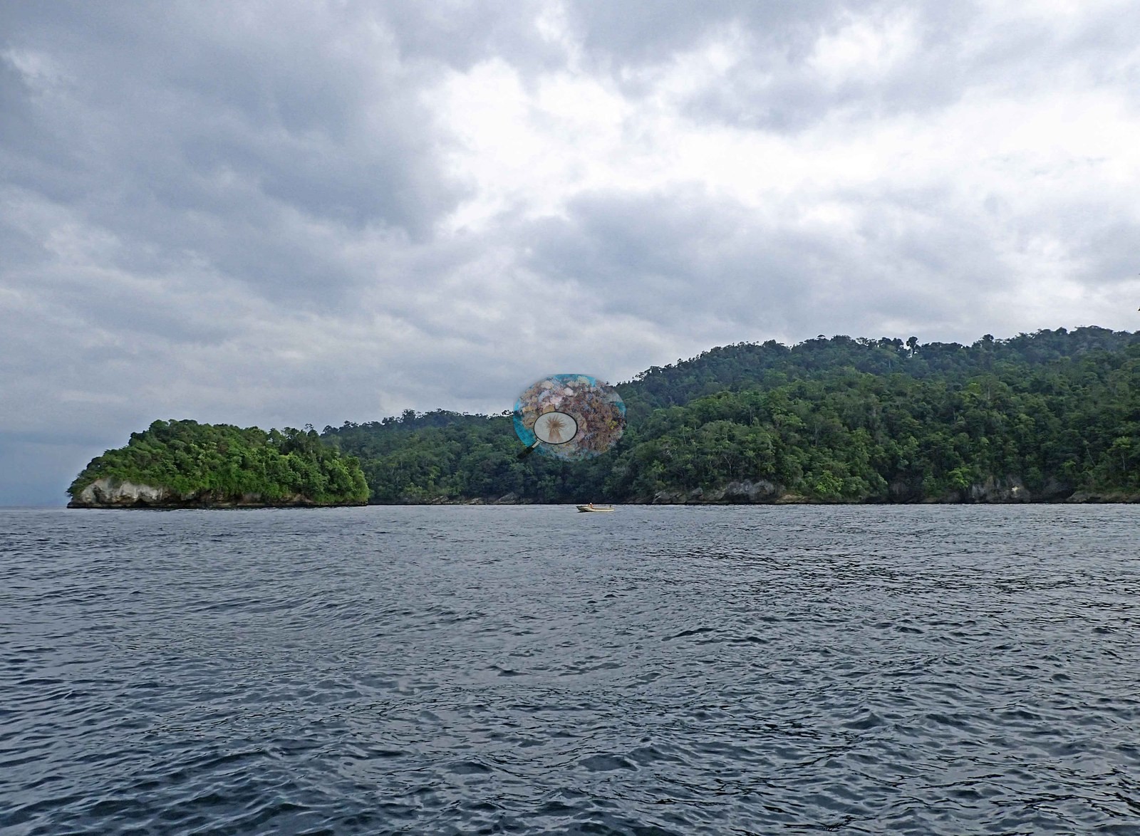 4
4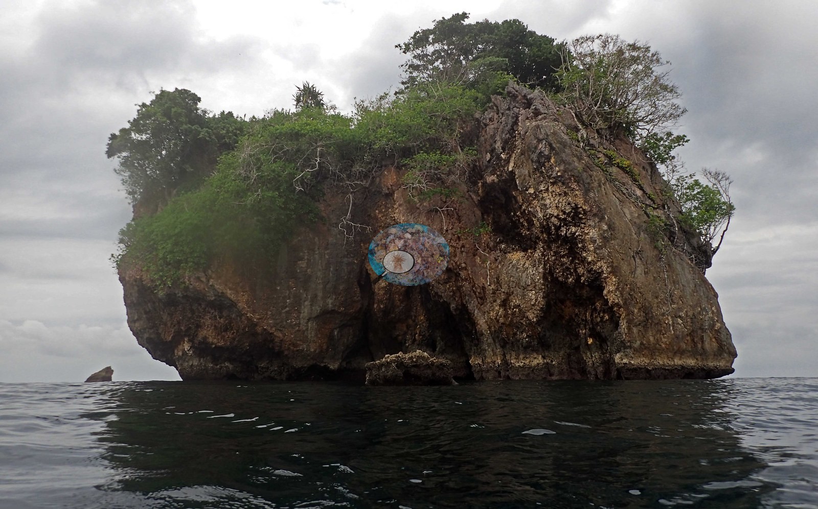 5
5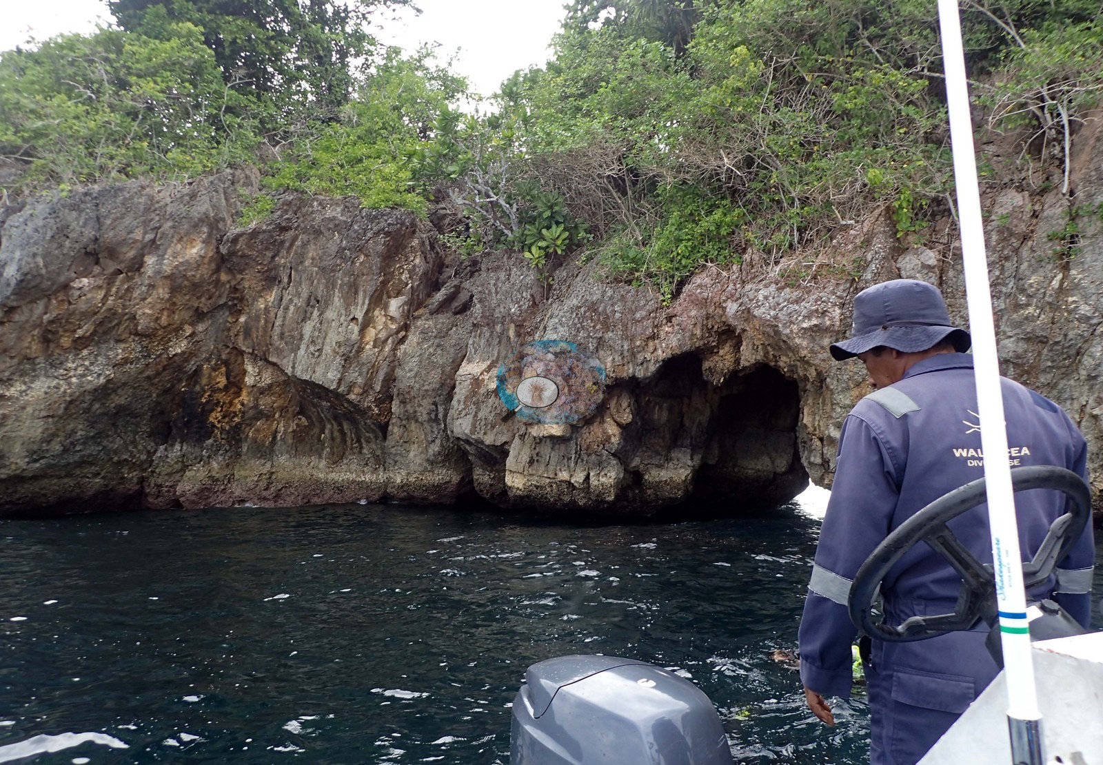 11
11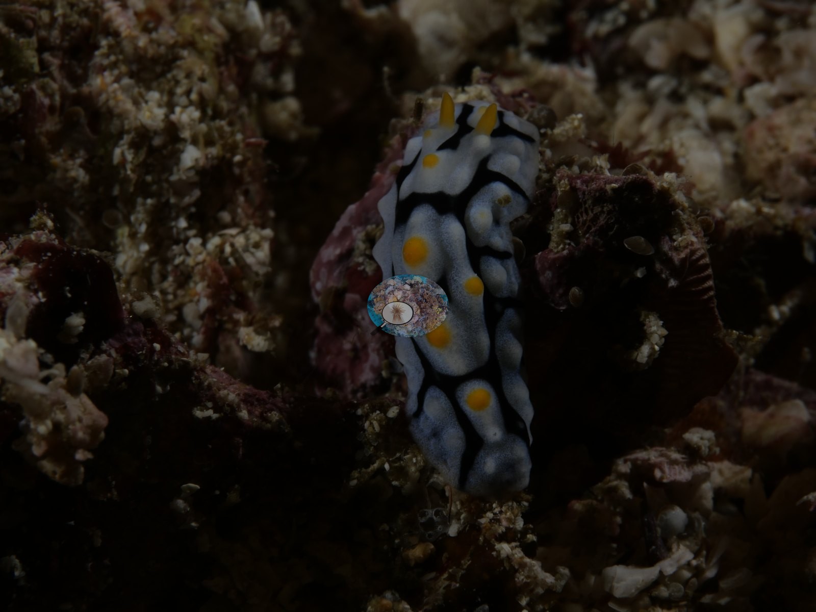 Photo: Pierre12
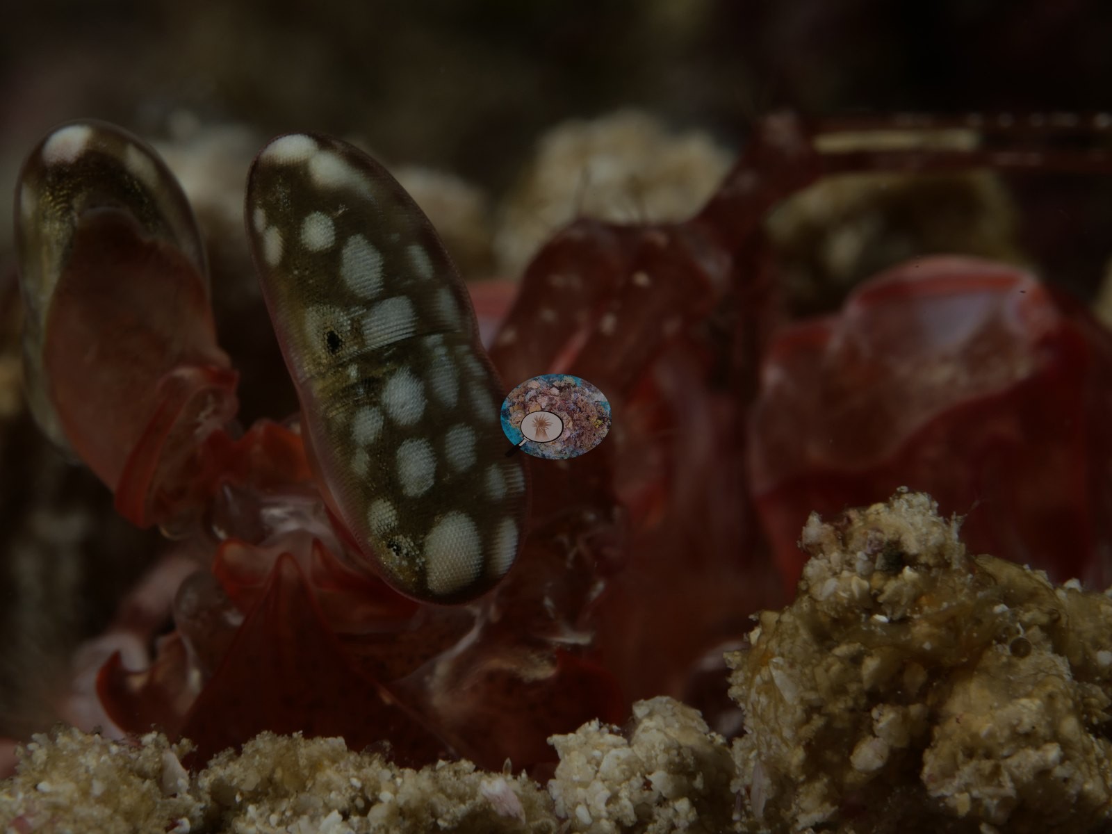 Photo: Pierre13
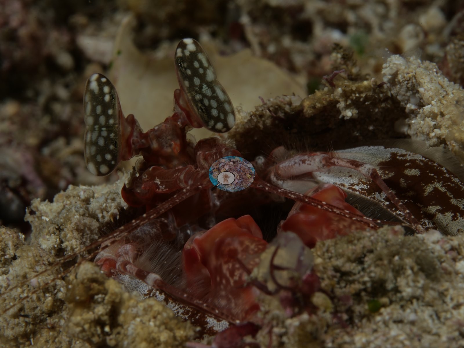 Photo: Pierre14
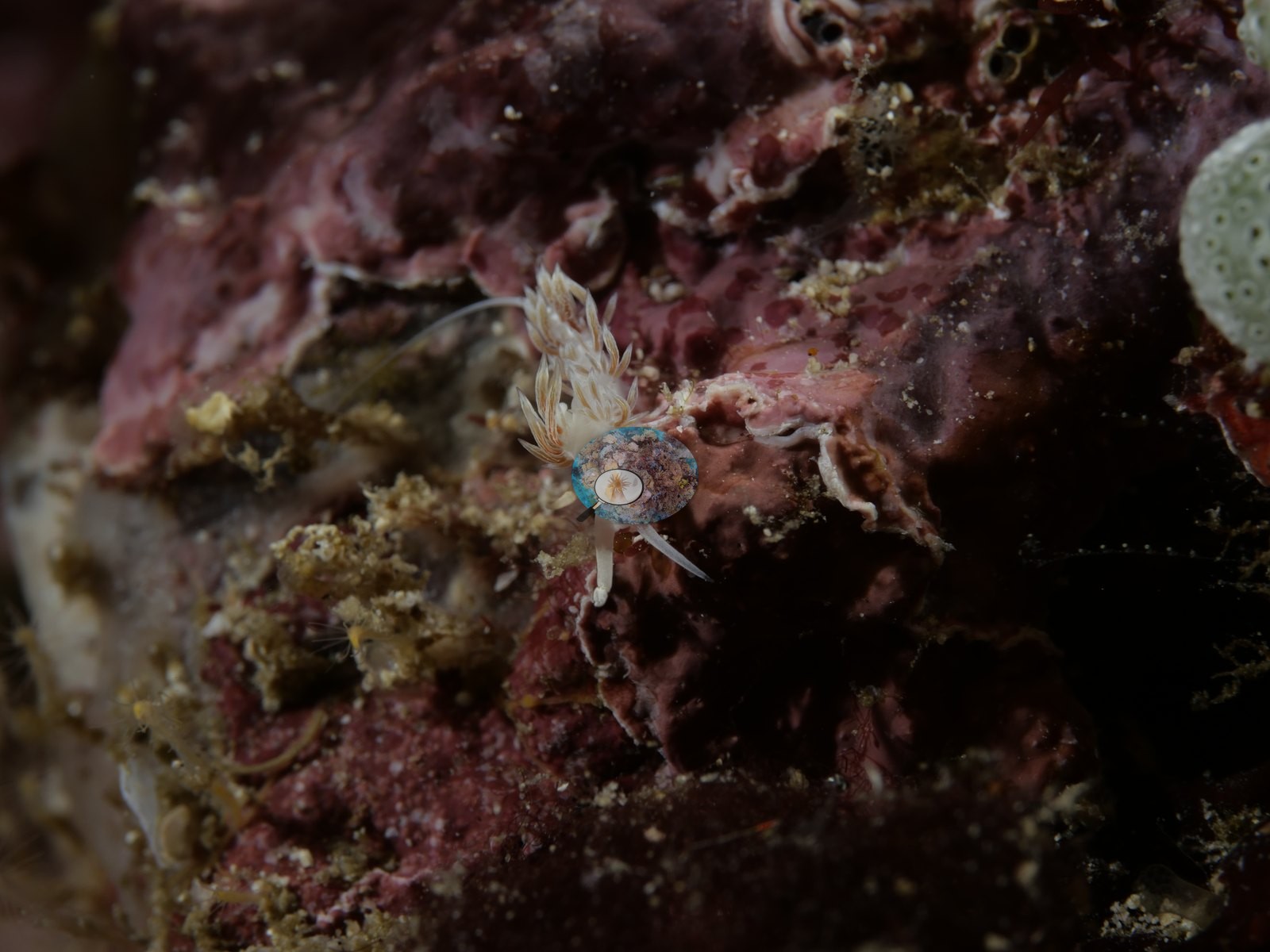 Photo: Pierre15
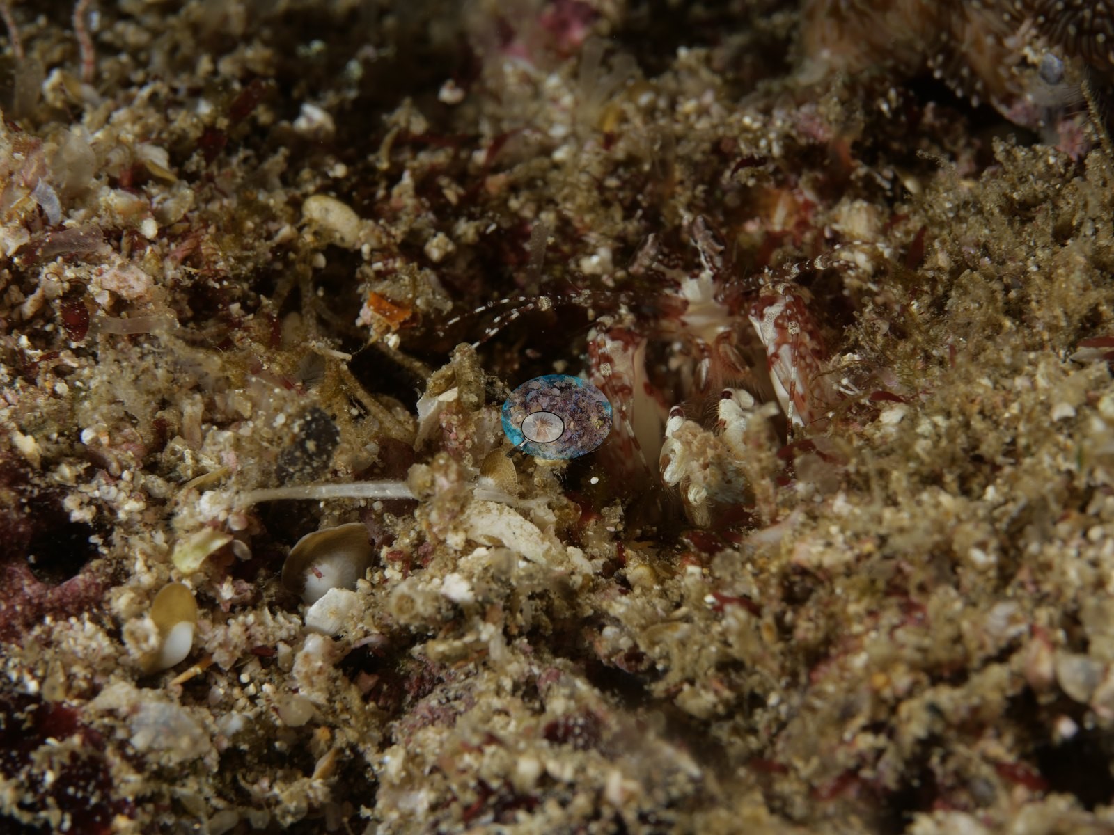 Photo: Pierre16
 Photo: Pierre17
 Photo: Pierre18
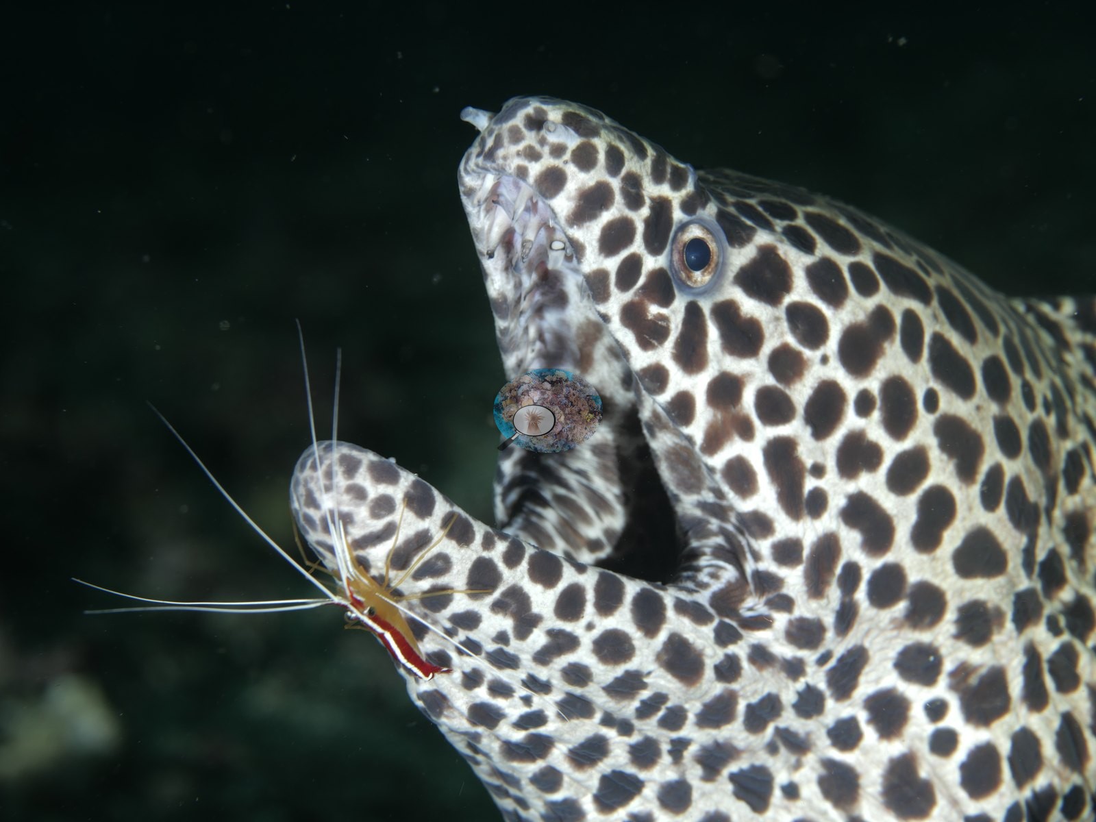 Photo: Pierre19
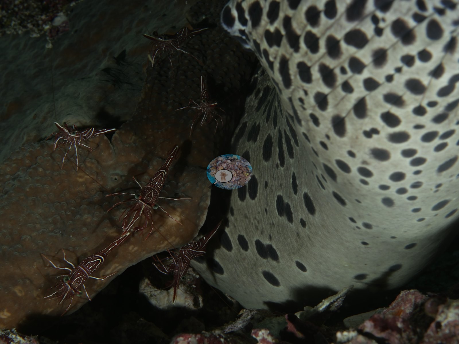 Photo: Pierre20
 Photo: Pierre21
 Photo: Pierre22
 Photo: Pierre23
 Photo: Pierre24
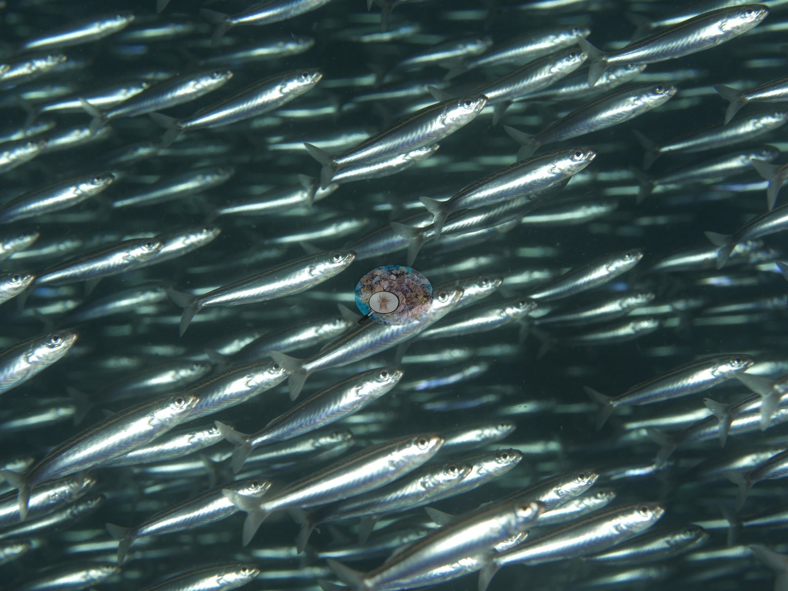 Photo: Pierre25
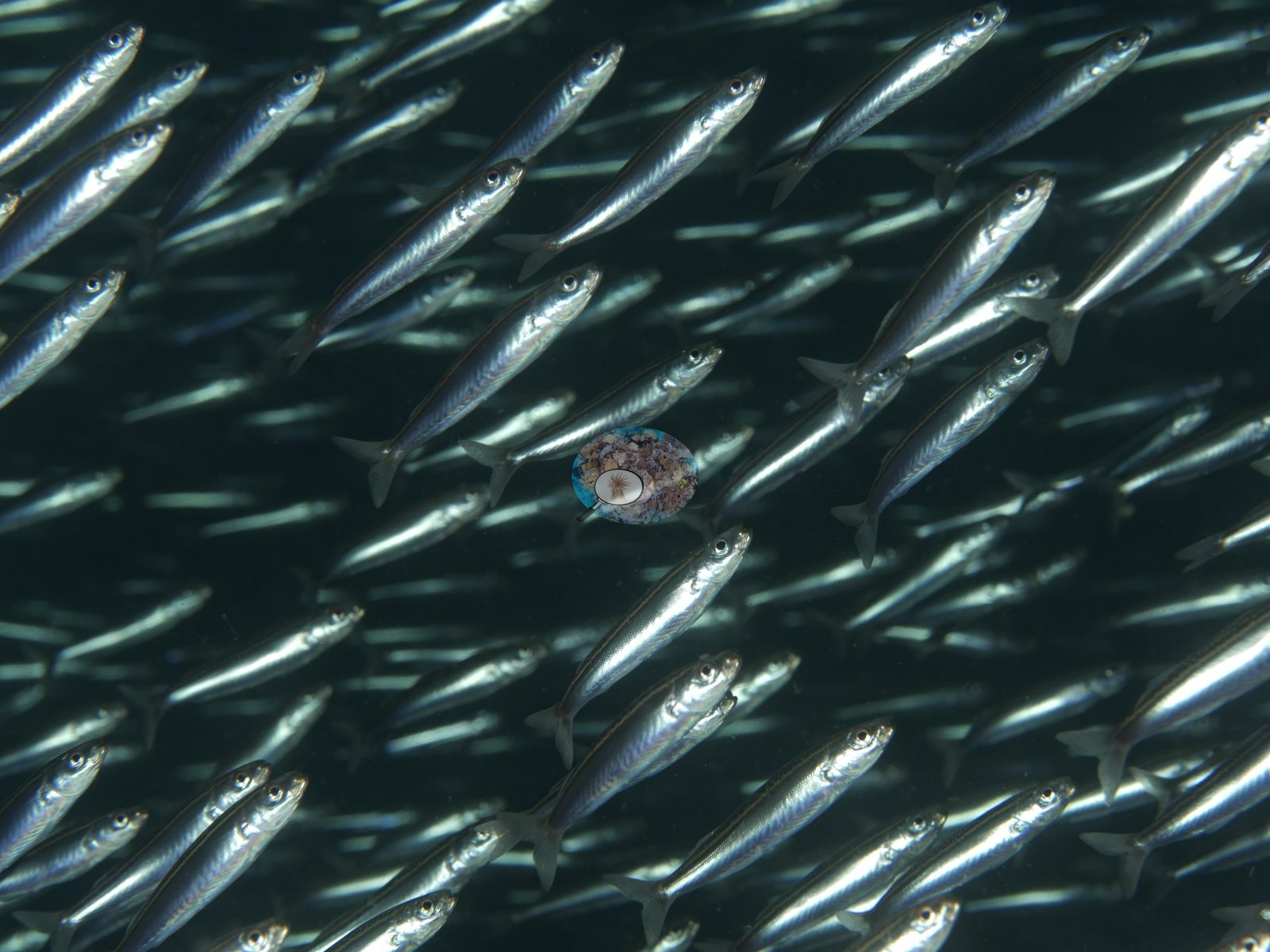 Photo: Pierre26
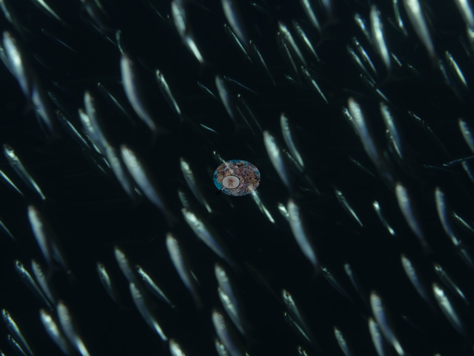 Photo: Pierre
Site description
Setting
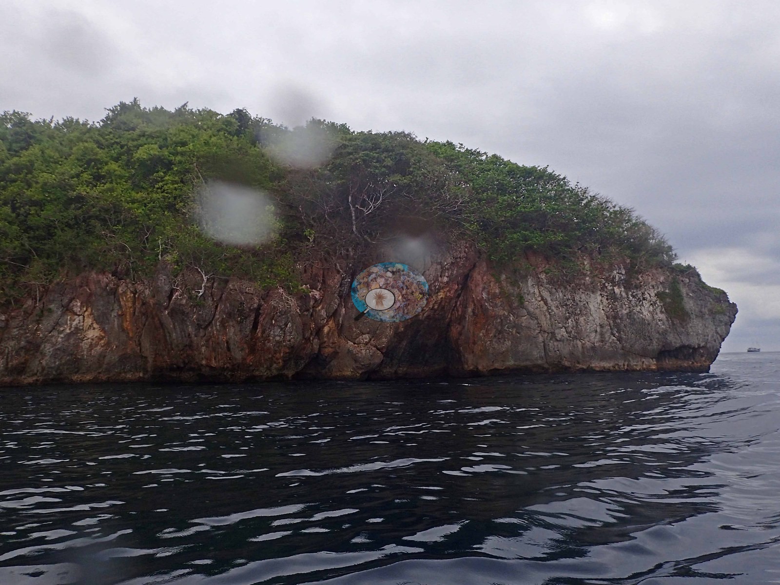
Last Frontier is a fringing reef surrounding a tiny rock island in Triton Bay, Papua: a steep limestone karst islet with a deeply undercut, notched base and sea caves at the waterline, off a forested mainland shore. Visibility was down to 5 m in places, increasing to 10 m in others, and a gentle current ran that was still possible to swim against. Light was very low, both because the water was murky and because the dive started so early; the notes say before 6:30, while the dive computer logged entry at 06:52. A thermocline sat at 12-13 m, with a 0.5°C change across it. Some areas had more complex structure and high rugosity, and an impressive swim-through ran through the middle of the rock island.
General benthic features
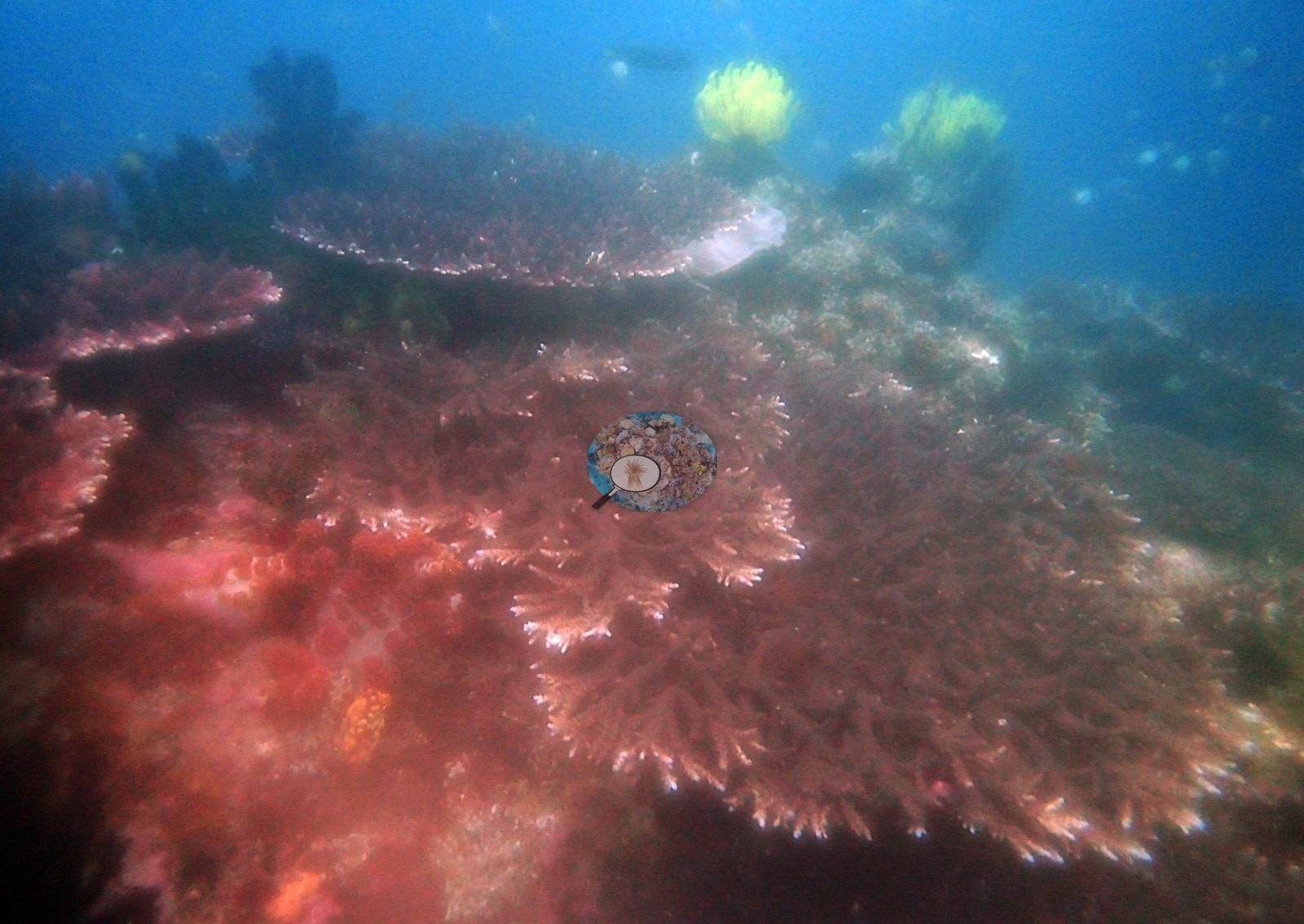
Soft corals dominated: Sinularia carpets ran throughout the site, Sarcophyton was very abundant, and soft-coral cover was very high everywhere, possibly because the water carries abundant nutrients and food. At 20 m there were few stony corals, only soft corals and sponges, with occasional plating coral. Orange and green Tubastrea grew throughout the site and was the dominant organism in some areas. Hard coral increased toward the shallows: acroporids grew on the bommies, larger Acropora tables stood at 9-10 m, and the dive eventually reached an area with high acroporid cover, with an impressive diversity of Acropora species at 5-8 m. In the shallows, one Acropora table measured 3-4 m across, with its polyps out. Pocillopora verrucosa occurred throughout the site, along with several Pocillopora meandrina colonies. One upper-slope frame was filled edge to edge with pink-purple corymbose branching colonies with pale growing tips, a large tabular plate behind them, red encrusting coralline algae and sponge between colonies, an orange-yellow encrusting sponge, and two bright yellow crinoids perched above the branches (e.g., Image 8).
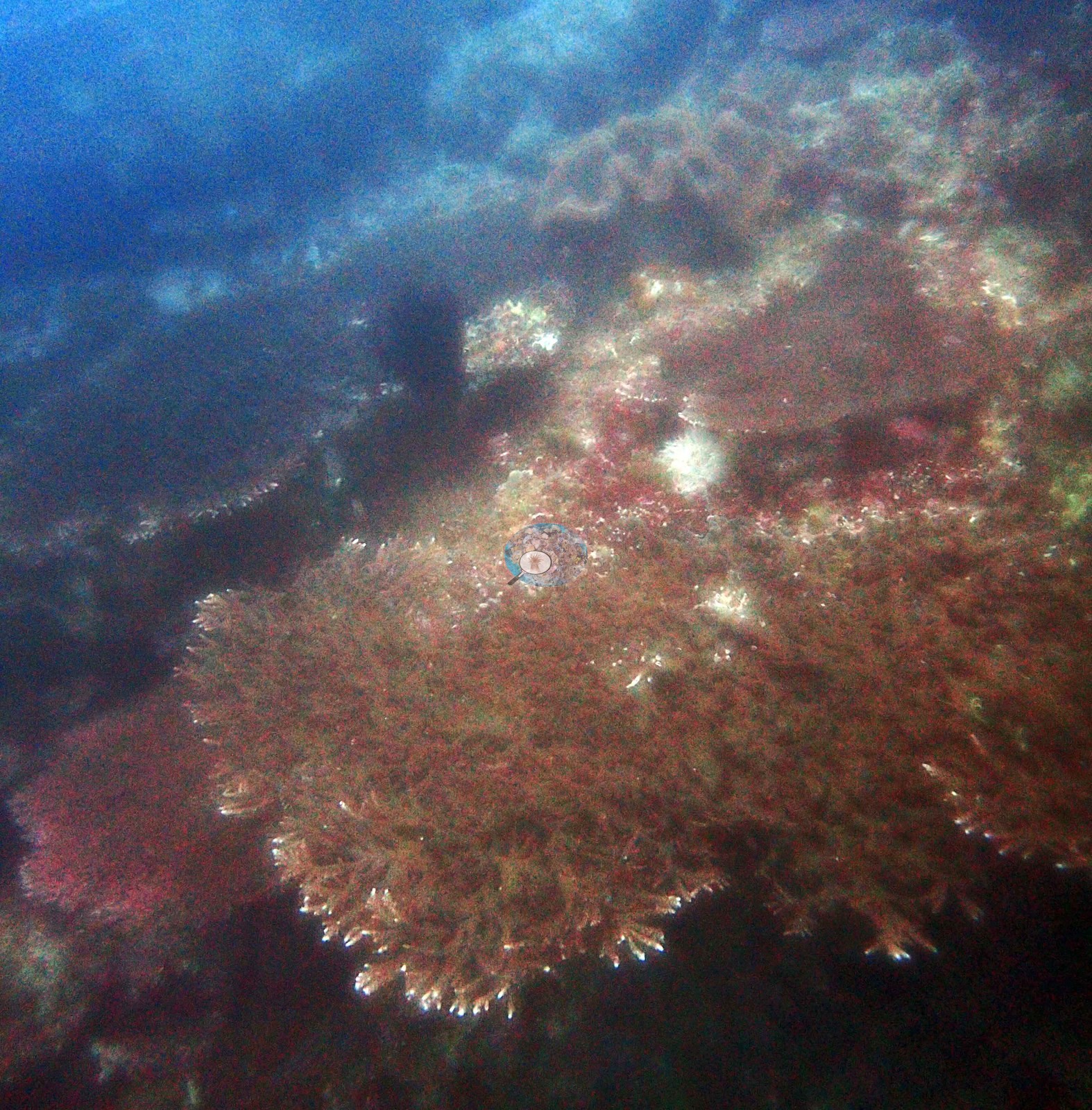
Another showed broad, fine-branched tabular plates stacked down the slope, their pale growing tips catching what little light there was, with red encrusting growth over the rock between them (e.g., Image 2).
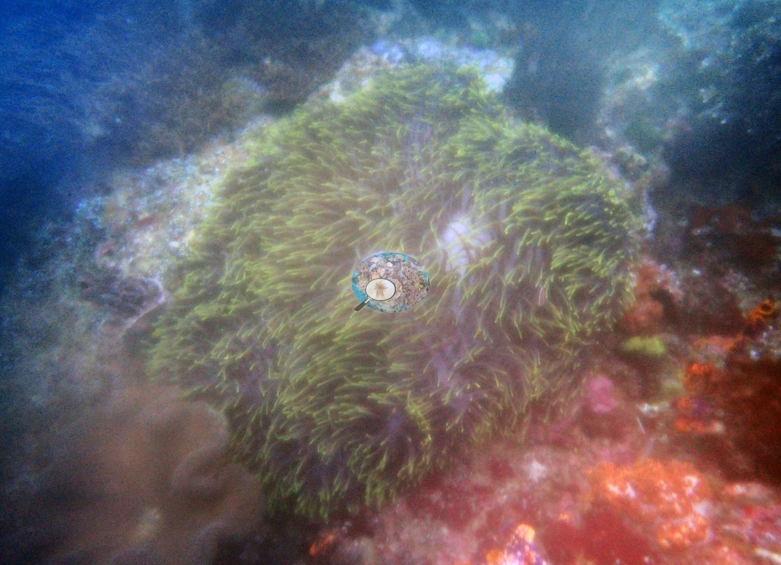
Patches of huge sea anemones gave the anemones high cover in places, and Naoko photographed an anemone species never seen before on these trips. One large yellow-green sea anemone sat on rock crusted with red and orange encrusting organisms (e.g., Image 10).
Photographs from the dive that are not shown here also recorded a single tabular colony two to three meters across seen dimly down the slope, and a second habitat of low-relief, sediment-veneered, rubble-strewn pavement carrying only small, scattered dark massive and encrusting colonies, a giant clam with a vivid blue mantle, a scatter of pale cream nodules, and coralline-crusted rock and turf with pale soft-coral clumps.
Coral cover
Coral cover: 7.8% — a blind Claude computer-vision point count of 14 of Anderson's edited photographs (20 points per photo; water, fish, and obstructions excluded). Other computer-vision methods are listed in the method table. This is based on a rudimentary computer vision analysis of diver photos. A more robust AI will corroborate this result in the coming months. The estimate inverts here and should not be trusted as cover: its highest frame, 47.2%, was the sea anemone, and its lowest, 2.2%, was a frame most of which is living branching coral. That is the over-read and the under-read failure happening in the same 15 photographs.
Stressors
No bleaching, disease, crown-of-thorns, or anchor and blast damage was seen in any frame. White subjects were checked at full resolution: the white branch tips are growing tips with pigmented tissue behind them, and the small pale tufts among the branches are fuzzy hydroid or coralline growths, not bleached tissue. Typical recent thermal stress for the Papua ecoregion is a five-year mean annual maximum of 10.1°C-weeks (Degree Heating Weeks, NOAA CRW).
Fish and other fauna
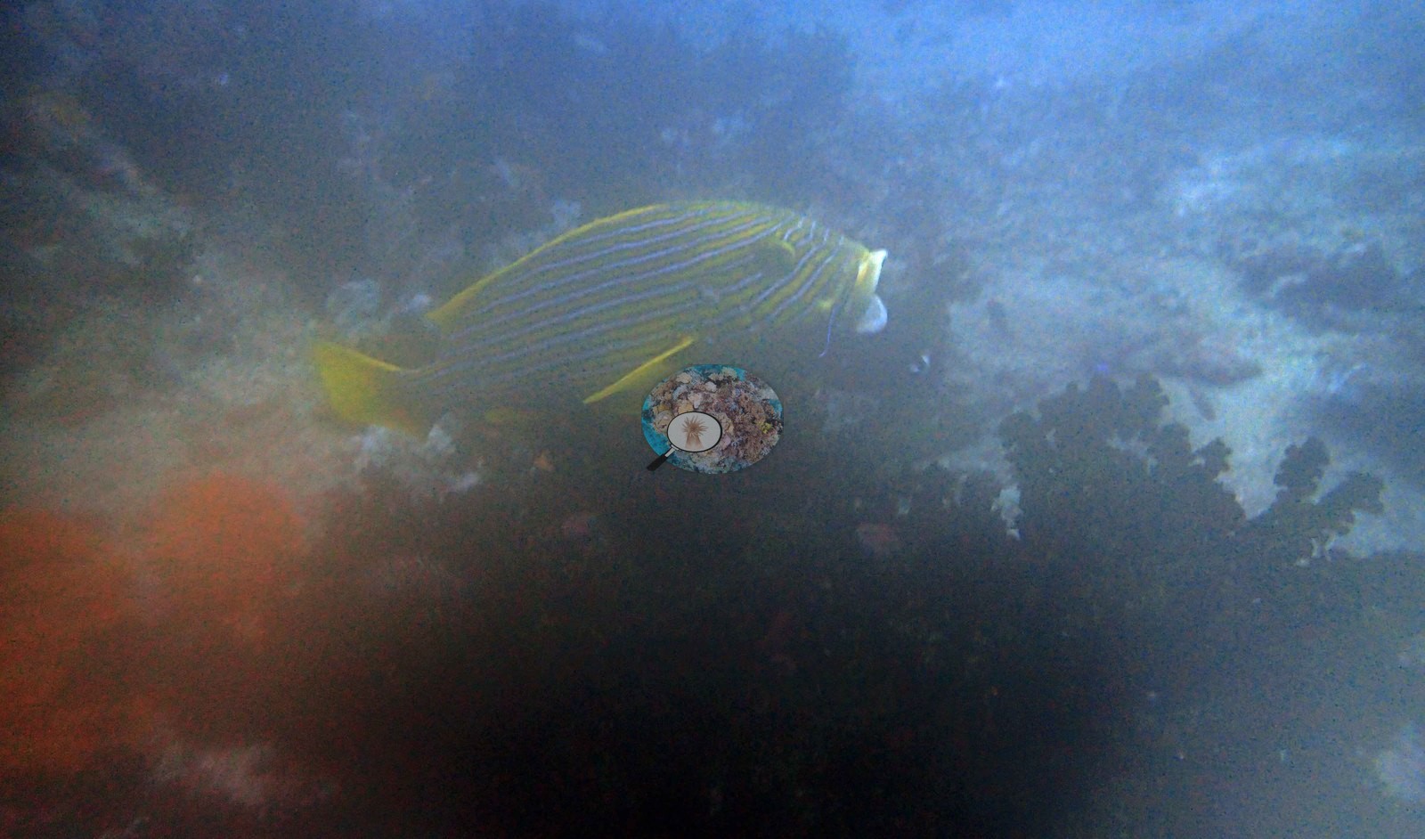
Fish biomass was very high, and many fish were still in sleep mode at that early hour. The descent dropped straight into a bait ball being stalked by giant trevally and large schools of barracuda, and another bait ball filled the shallows. Big fish also stalked bait-ball schools through the swim-through, with giant trevally and barracuda the dominant predators. Large bumphead parrotfish ranged throughout the site, alongside several Napoleon wrasse and the trip's first turtle. A sweetlips with a yellow ground and fine stripes cruised over the reef in the murk (e.g., Image 7).
Other photographs from the dive record a large trevally hunting through a dense bait ball in open blue water, a honeycomb moray, a tasselled wobbegong resting camouflaged on the reef, blue-and-yellow fusiliers, and damselfishes. A second wobbegong was seen but could not be photographed well because the camera housing fogged. A large mantis shrimp and a barramundi were also seen, and Naoko got good pictures of the mantis shrimp.
Comparisons with other data
No prior survey data from this site. If you would like to share yours, please email us.
Dive record
Dive 4, Last Frontier (also recorded as Cristi Island), was the first dive of the day on 15 Oct 2018, logged to 20.0 m for 66 minutes at 25°C under a flat gray overcast. It was logged as lifetime dive 45.
Other notes
The camera housing fogged on this dive, so the photographic record is sparse: fifteen frames survive, all fifteen were reviewed, and they are a small photo set, not a survey. They show what the reef looked like in a few places but cannot support site-wide claims about coral cover or condition; the field notes carry the broader picture.
CV model comparison
More info
DINOv3 and CoralSCOP were run on the same photos and are listed for comparison. Coralscapes v2 and v1 were also run but are not shown: Anderson does not consider Coralscapes reliable for these photos. No diver transect exists for this trip, so agreement between the rows is not accuracy, and as of September 2026 no computer-vision method estimates coral cover accurately on new images; treat every row as indicative.| Method | Hard-coral cover (%) |
|---|---|
| In-house DINOv3 | 10.2 |
| CoralSCOP edited | 16.0 |
| Claude (edited photos) | 7.8 |
Data & links
Images 11–26: Pierre; all other images from Coral Reef Diagnostics. Data are from Coral Reef Diagnostics.
© Coral Reef Diagnostics.