
Part of the regional summaries: Tonga - Vava'u and Tonga
More info
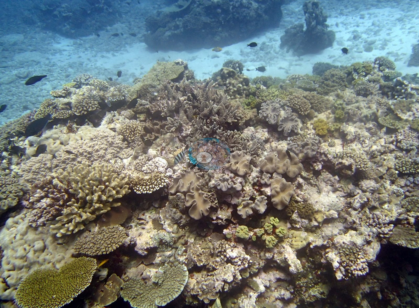
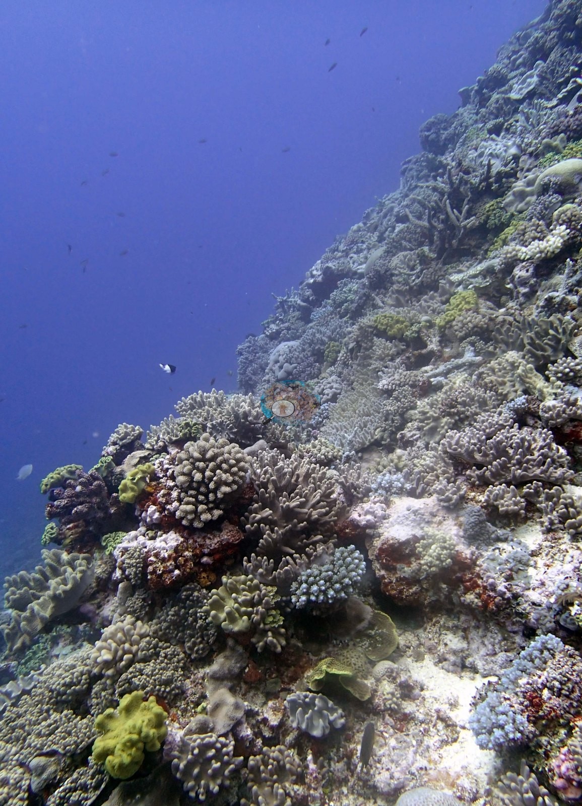
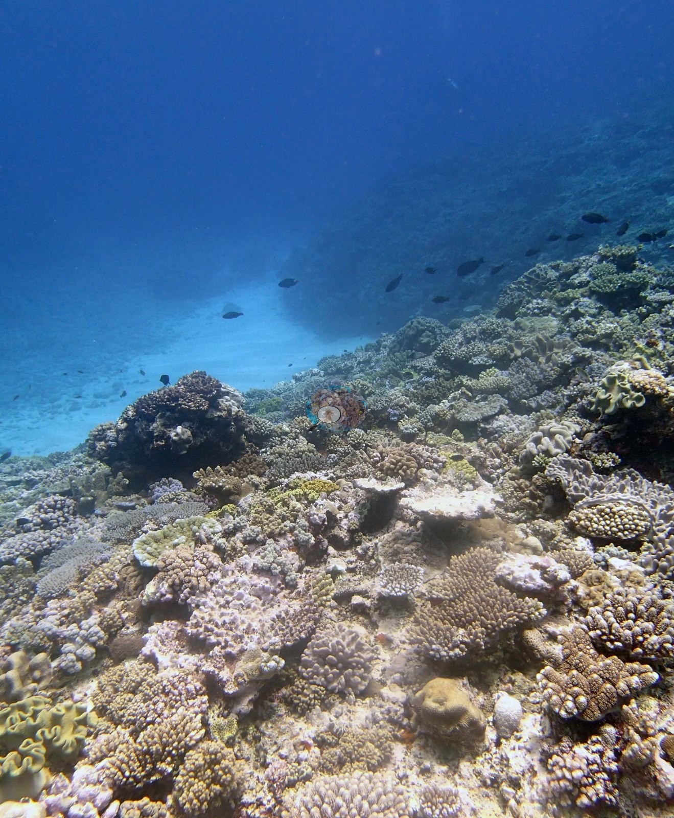
Southern Tonga (NOAA CRW virtual station)A mixed Vava'u reef of a steep face and a low terrace running out to a chute of white sand and rubble. The face is densely covered, corner to corner, with mustard and olive leathery soft corals packed among lavender and cream corymbose corals, with a long stream of banded damselfish along the reef edge. Surveyed on the Living Oceans Foundation's Global Reef Expedition.
Ko ha hakau fefiofiofi ʻi Vavaʻu ʻo ha mata tuʻu mo ha tuʻunga māʻulalo (low terrace) ʻoku aʻu atu ki ha hala ʻo e ʻoneʻone hinehina mo e maka momoa (rubble). Ko e mata ʻo e hakau ʻoku ʻufiʻufi fefuʻifuʻi, mei he tuliki ki he tuliki, ʻe he feo molū fakakili (leathery soft corals) lanu mastati mo ʻolive ʻoku fefuʻifuʻi mo e feo corymbose lanu laileki mo kulimi, mo ha laine loloa ʻo e ika damselfish (banded damselfish) ʻi he tafaʻaki ʻo e hakau.
AI translation of this summary; the English is authoritative. (low confidence)
CTD-cast midpoint of 26.0–26.1°C
Four other CRD dive sites with their own pages within 5 km shown in orange — click a marker to open its page.
🗺 Detailed map of the Global Reef Expedition dive sites, Vava'u, Tonga (Khaled bin Sultan Living Oceans Foundation; opens in a new window)
25 of the 32 photographs on this page are shown beside the text that discusses them under "Site description" below.
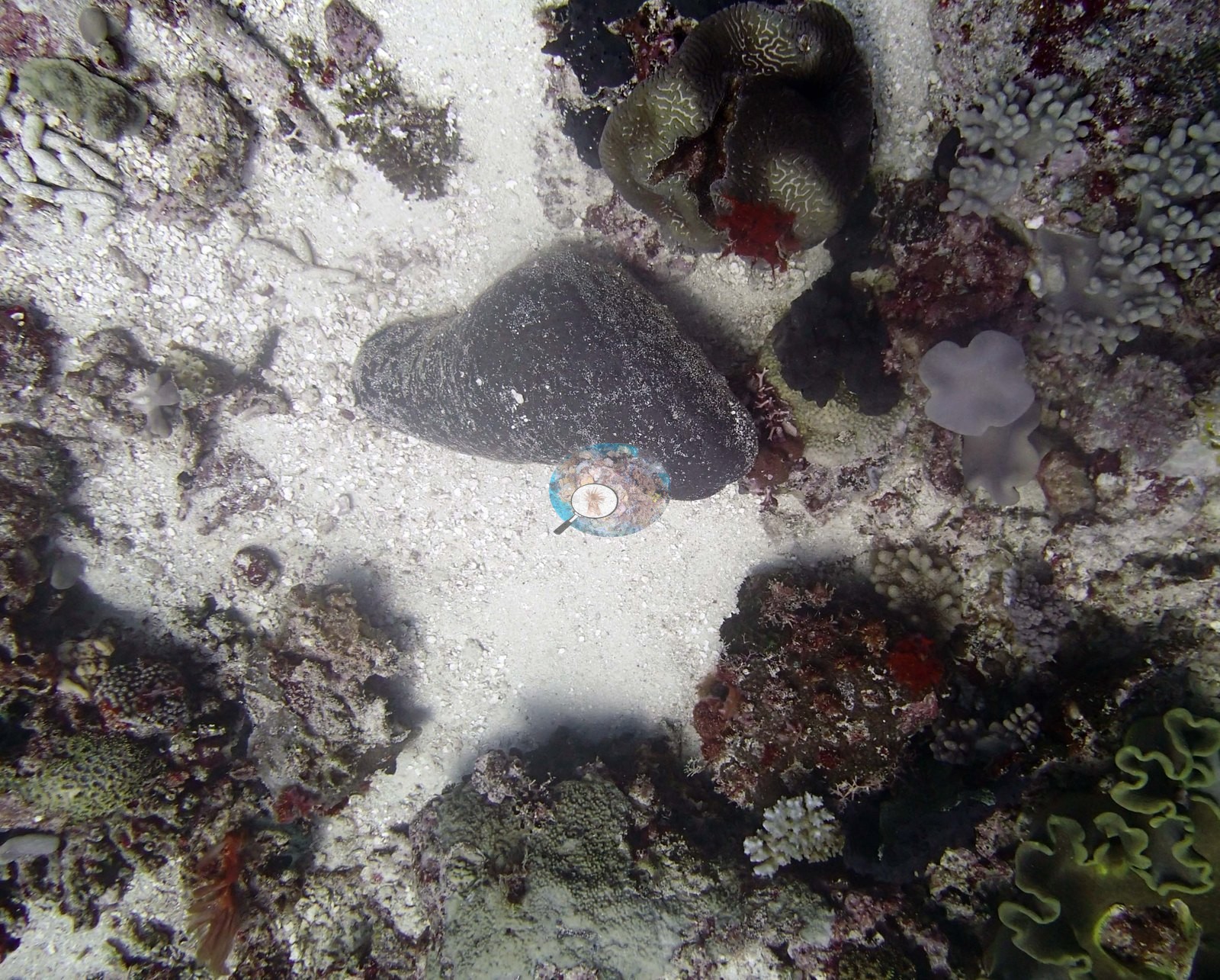
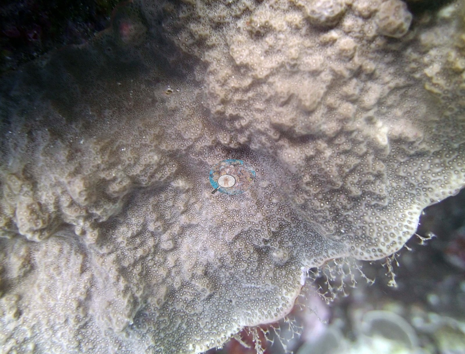
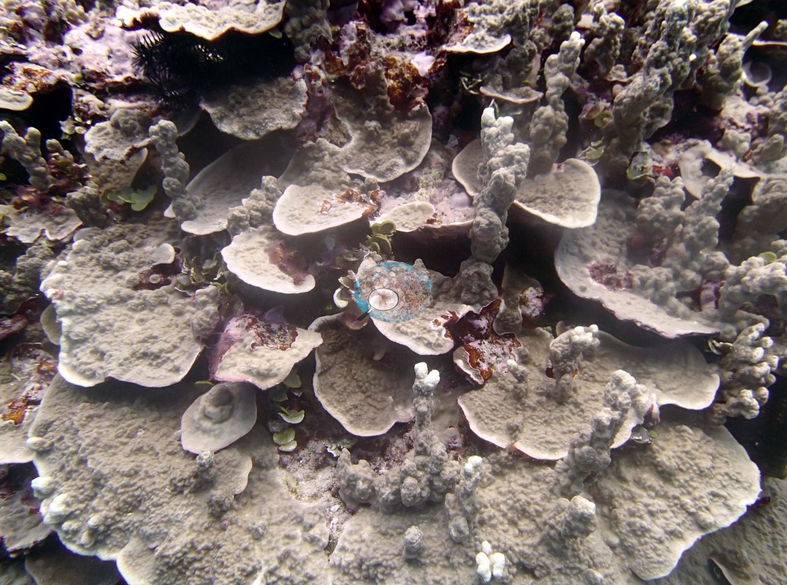
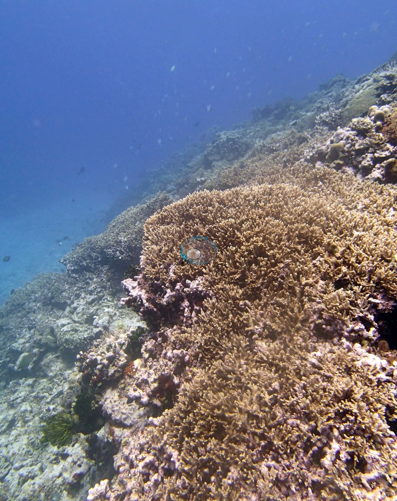
Site description
Setting
Roughly half the frames carry a milky green-white cast that washes the color out (e.g., Image 5, Image 14, Image 15, Image 16, Image 20, Image 21, Image 22), although Anderson's sheet logged calm conditions and 40 m visibility; the sun-lit shallow frames are flat and overexposed.
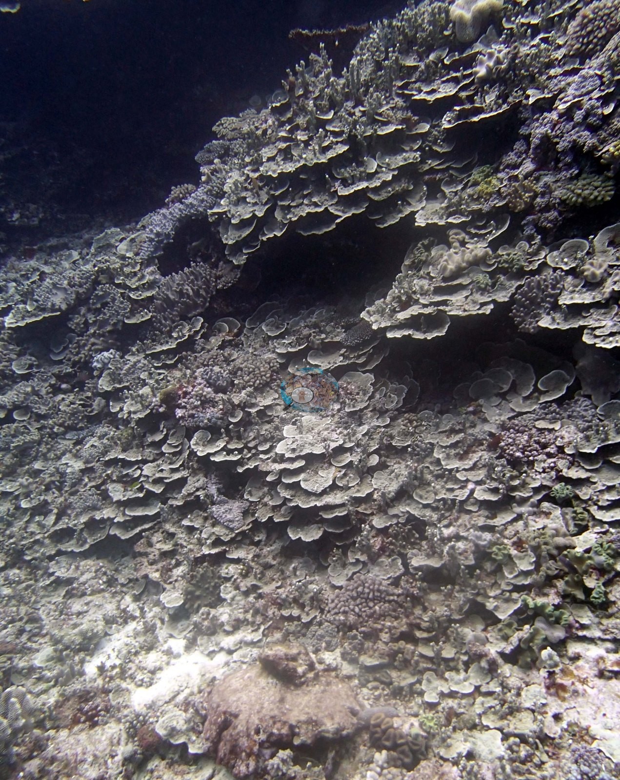
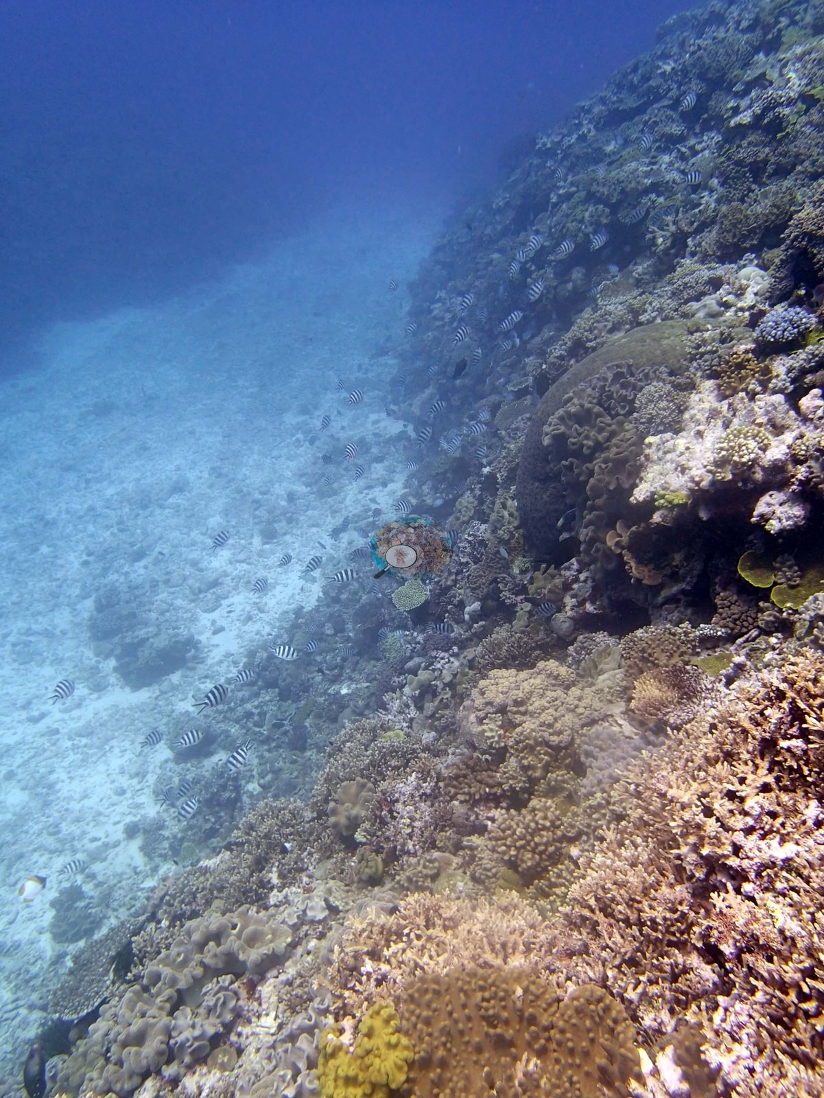
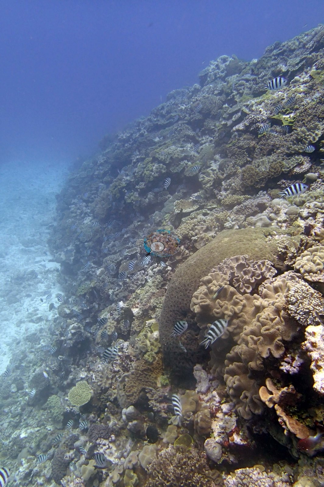
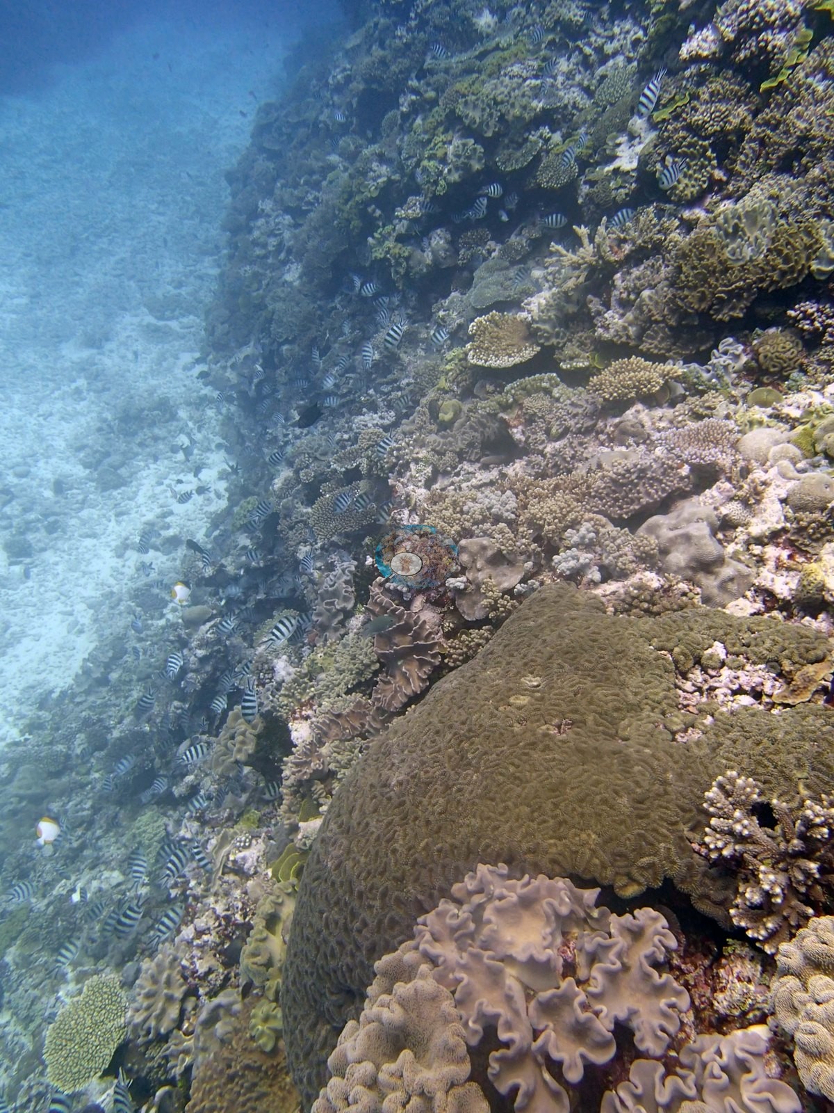
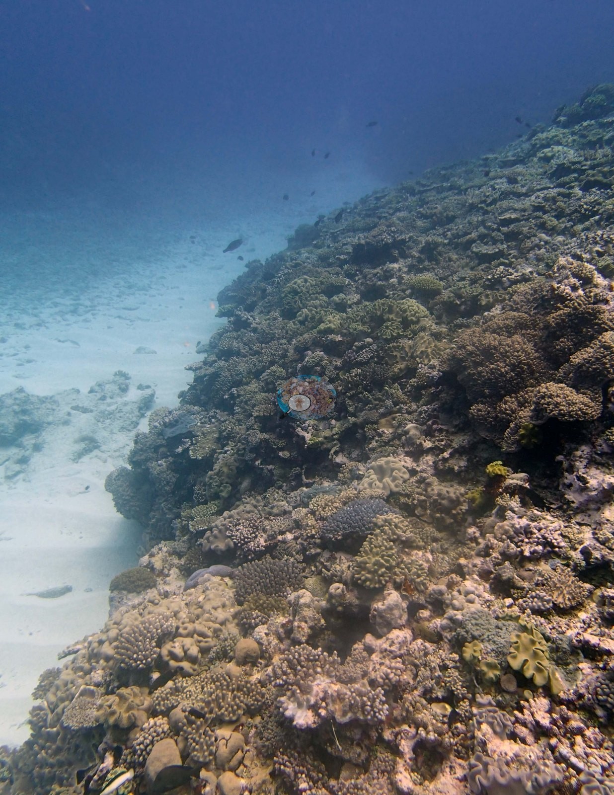
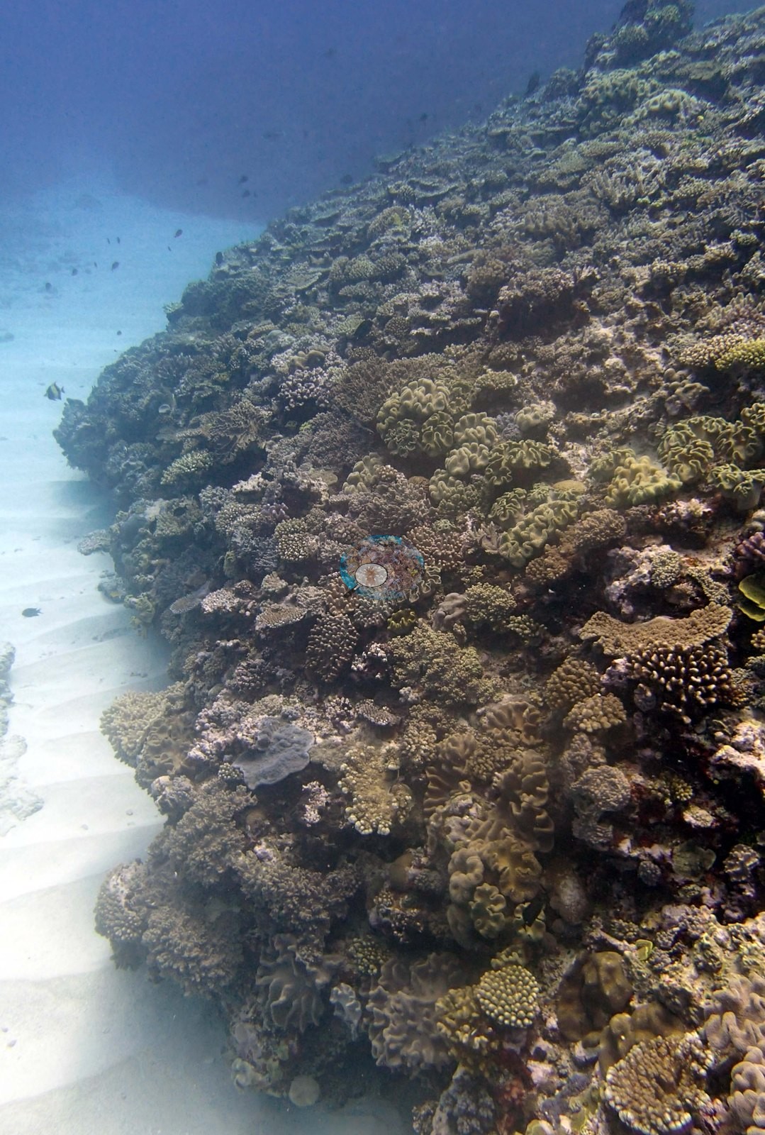
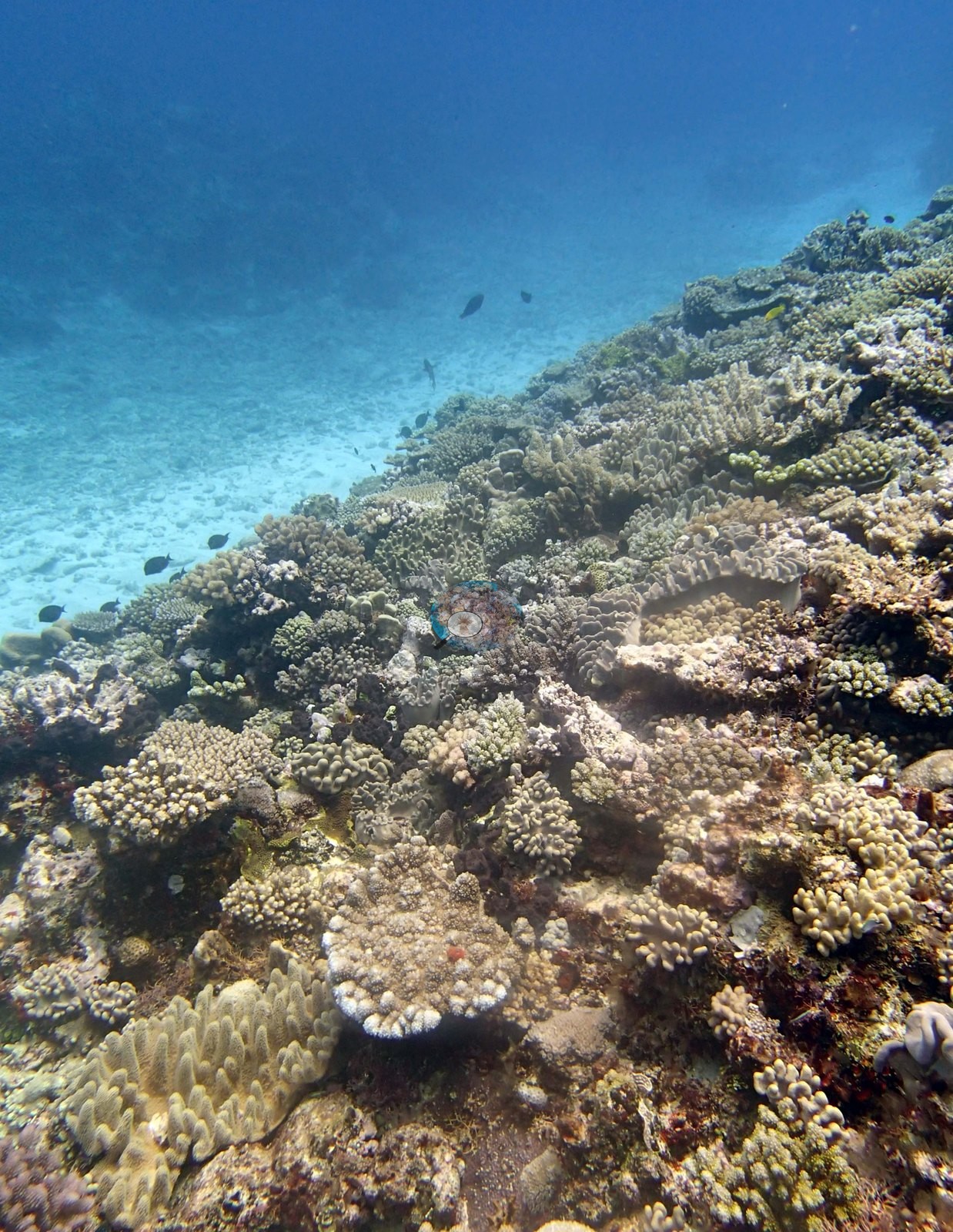
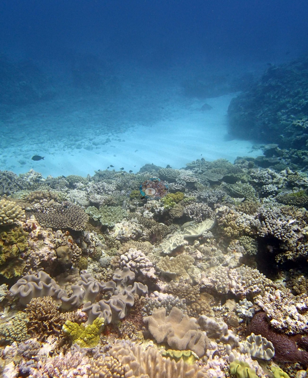
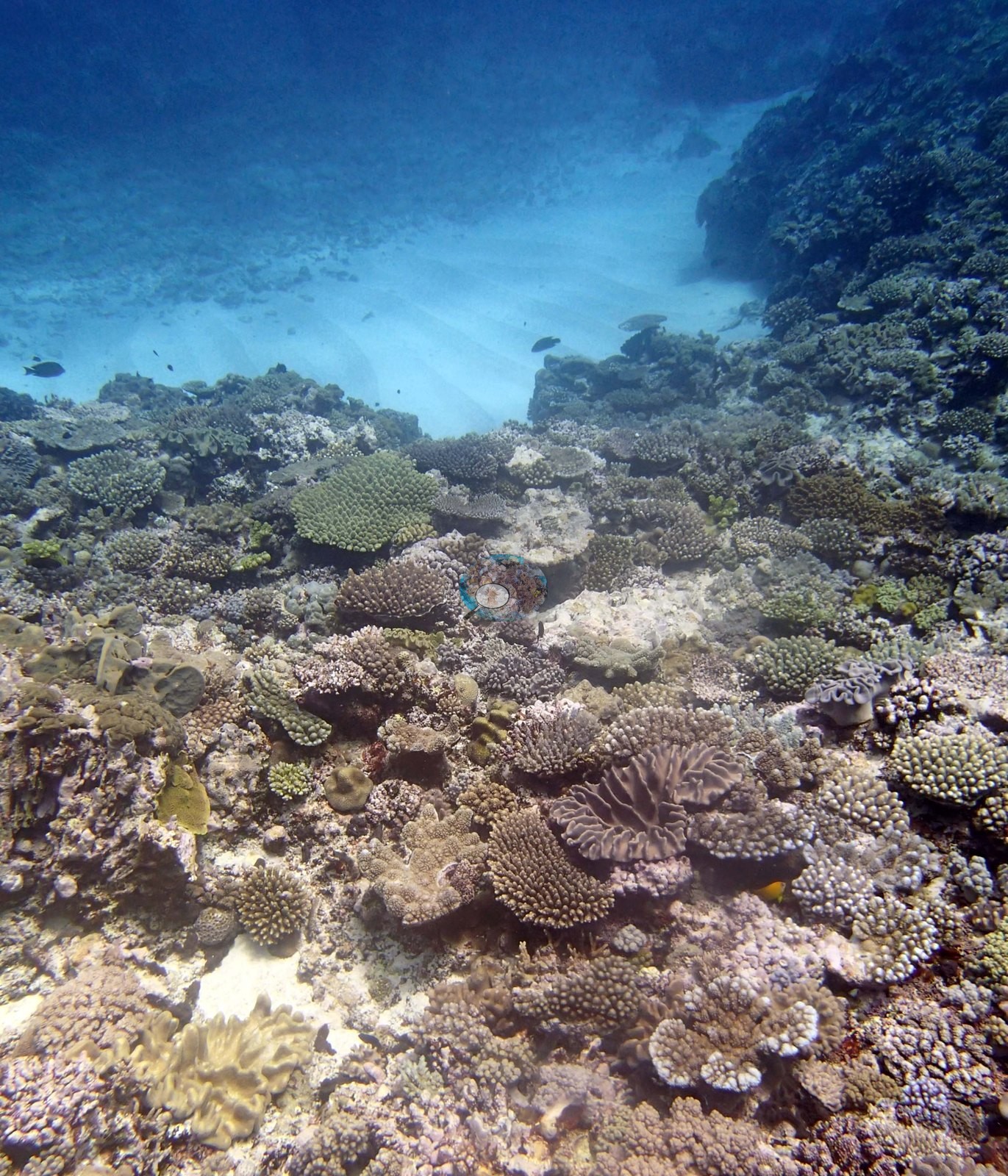
The shape of the site comes through clearly all the same. Several frames look along a steep face falling into blue (e.g., Image 5, Image 11, Image 12, Image 22), and several more look out across a low terrace to a broad chute or flat of clean white sand and pale rubble (e.g., Image 19, Image 20, Image 21, Image 26, Image 27).
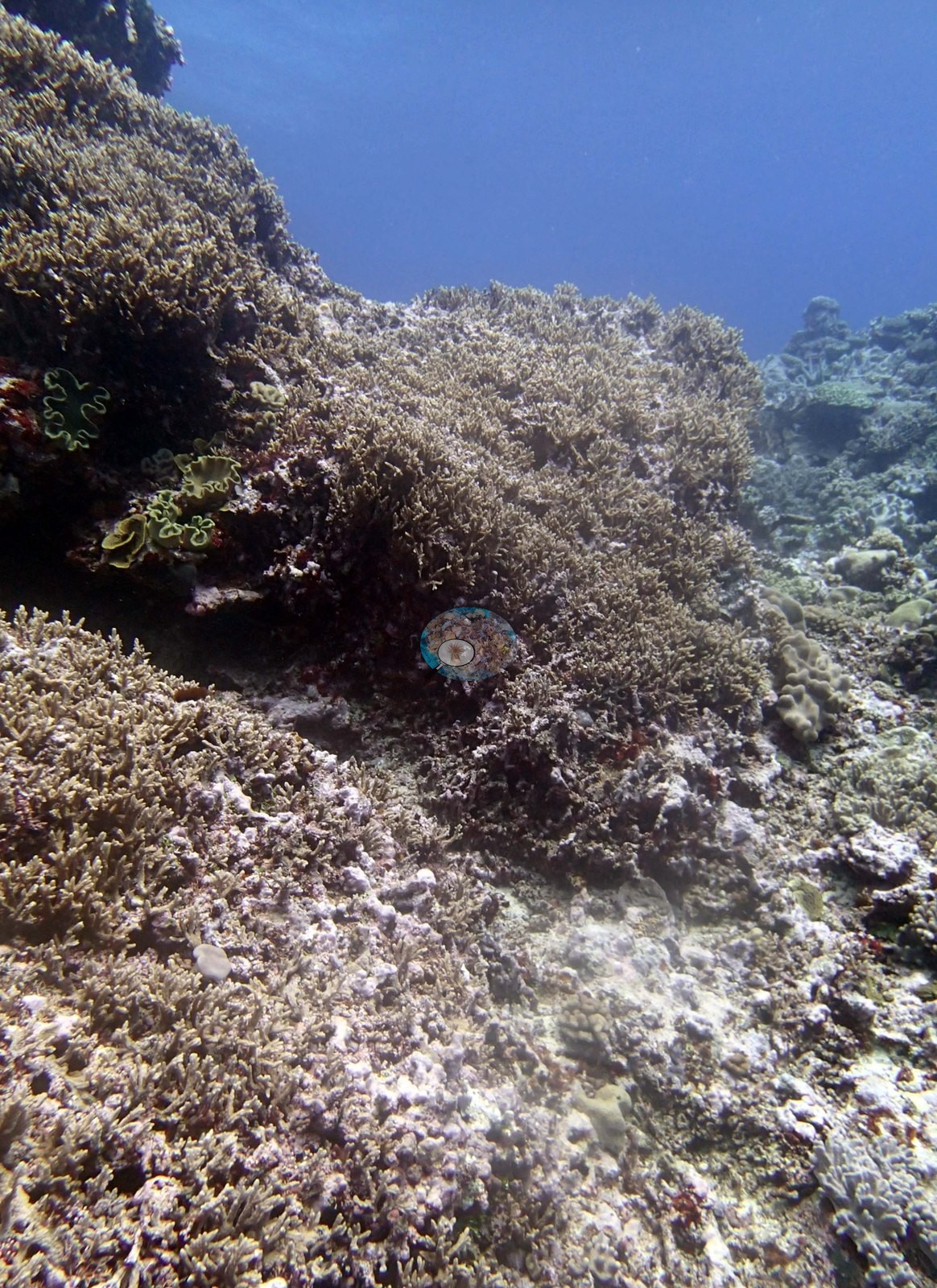
General benthic features
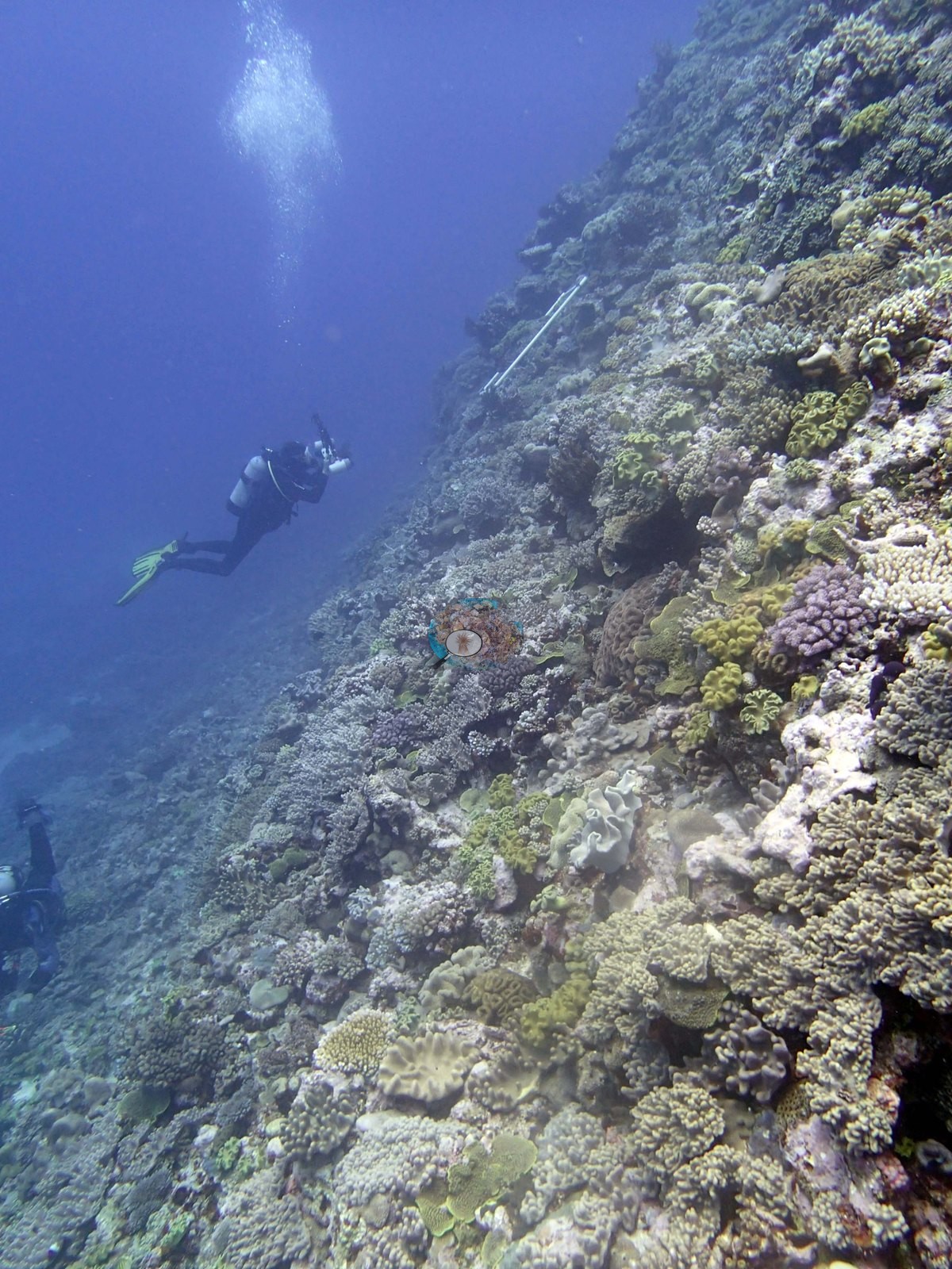
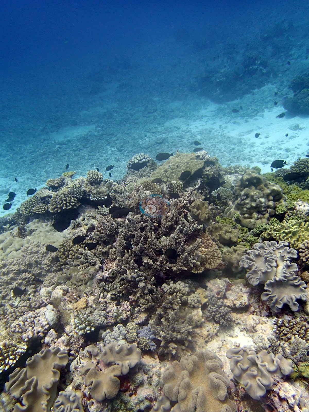
The face is densely covered: one frame taken over a survey diver's shoulder shows it covered corner to corner with mustard and olive leathery soft corals — mushroom-capped and lobed — packed among lavender and cream corymbose colonies, small digitate clumps, foliose plates, and encrusting sheets, with a thin white survey rod propped in the frame and a second diver working below (e.g., Image 11). Leathery soft corals are a genuine structural component here rather than an incidental: they recur at size in at least six frames (e.g., Image 15, Image 16, Image 17, Image 18, Image 19, Image 24, Image 25), in one case a frilled, mushroom-topped colony the size of a dinner plate sitting immediately below a large brain-textured massive with deep meandering valleys (e.g., Image 15).
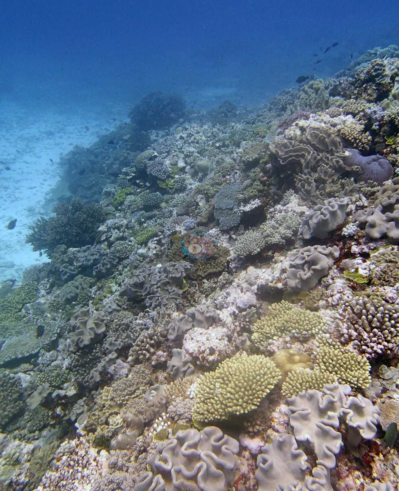
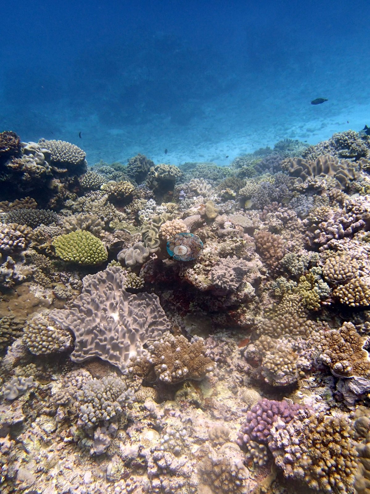
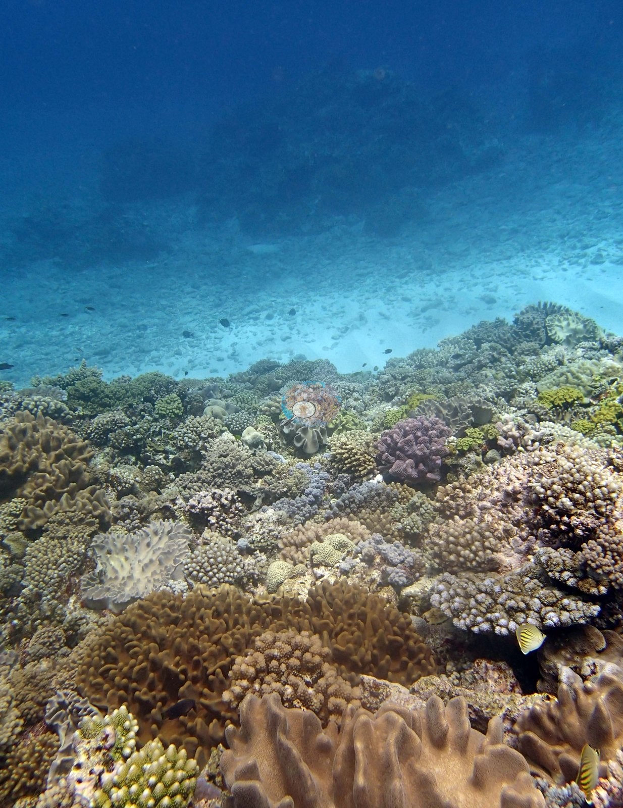
Elsewhere the terrace surface is a tightly packed low mosaic: corymbose and digitate colonies, knobby clumps, small tables, knobbly mounding massives, and encrusting sheets, with pink to mauve coralline crust between them (e.g., Image 8, Image 9, Image 17, Image 18, Image 23, Image 24).
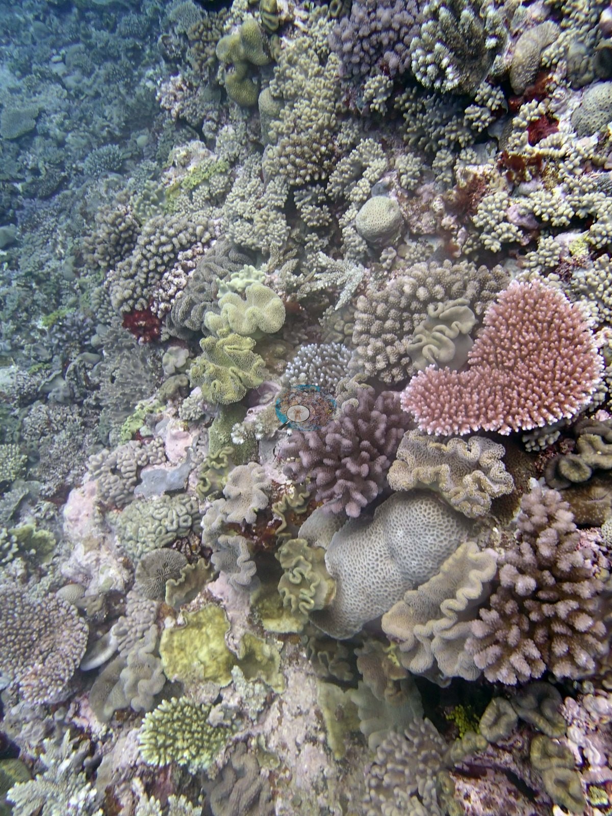
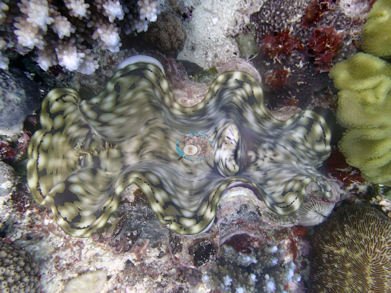
One frame is much the most colorful of the set, a close overhead view with a vivid pink corymbose plate at its center surrounded by mustard, lime, blue-gray, and pale cream colonies (e.g., Image 8). Large smooth mounding domes appear in two frames (e.g., Image 16, Image 25). Considerable turf- and algal-crusted non-living framework is visible between the colonies in the mid-depth frames, and the transition from reef to sand is abrupt rather than gradual. Three frames are close-ups: a giant clam with a boldly marbled olive, cream, and gray mantle wedged among corals and a mustard soft coral (e.g., Image 10); a pale plating-and-foliose colony with upright ruffled margins that Anderson tentatively identified as a montiporid; and a low, pale, nubbly encrusting colony left unidentified.
Coral cover
- Reference: Khaled bin Sultan Living Oceans Foundation, Global Reef Expedition (in situ transect)
- Transect mean 33 ± 6.3% SE (n = 5 transects; Living Oceans Foundation per-transect benthic data); the headline above is this all-transect mean.
Stressors
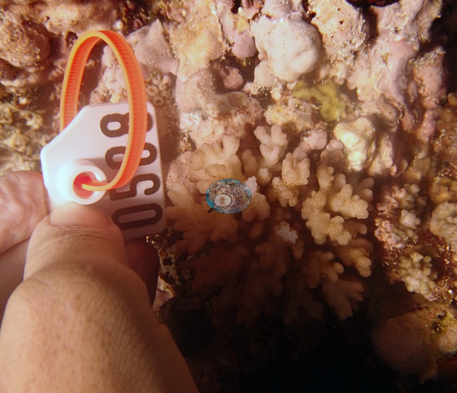
Anderson's sampling sheet records the Pocillopora colony sampled here (T55) as 80% bleached, and the photographs of it show pale cream-white branches with only scattered brown corallites (e.g., Image 28, Image 29, Image 30, Image 31, Image 32).
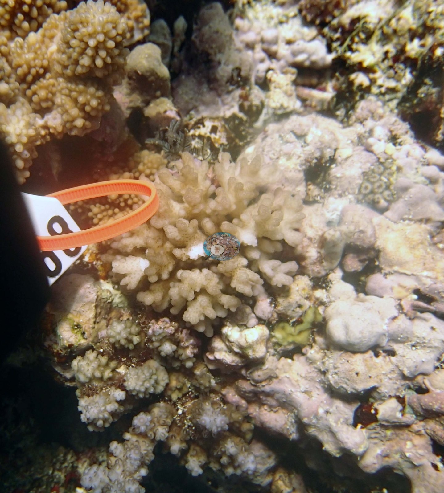
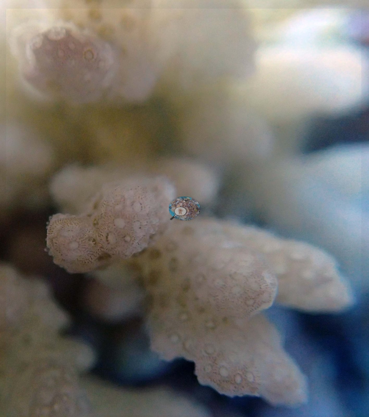
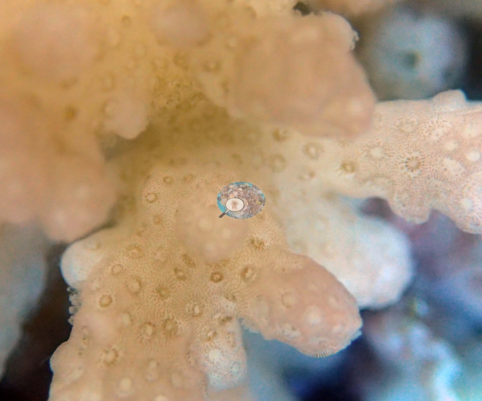
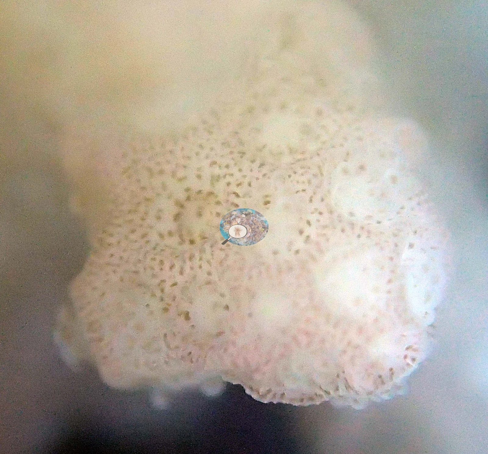
The habitat frames show no disease lesions, crown-of-thorns feeding scars, or fresh mortality, though the color cast in half the set means an absence of fine-scale lesions cannot be claimed with confidence. The Living Oceans Foundation transects here recorded 0.6% cyanobacteria and no recently dead coral.
Fish and other fauna
The same frame that shows the frilled soft coral below the brain-textured massive carries a long stream of black-and-white banded damselfishes running along the reef edge above a pale sand chute — the largest fish aggregation in the set (e.g., Image 15).
Comparisons with other data
Apart from the Living Oceans Foundation survey shown here, we found no other surveys at this site as of 2026. If you would like to share yours, please email us.
Other notes
The full set shows a mixed reef. Both of Anderson's calls above (the tentative montiporid and the unidentified colony) are left as recorded — the photographs cannot settle either one, and no identification is asserted here.
CV model comparison
More info
The photo row below is a methods comparison only: it is never averaged with the transect and never used as the headline. It compares site means only: the photos come from the same survey dive as the transect but not from the same points.| Method | Hard coral cover (%) | Photos (n) ± SE |
|---|---|---|
| Transect mean (all transects) | 33.4 | |
| CoralSCOP unedited | 36.3 | n = 21 · SE 3.2 |
| Claude (edited photos) | 32.5 | n = 21 · SE 2.7 |
The two computer-vision methods agree within 3.8 pp (32.5–36.3%) and read within 2.9 pp of the in-water transect (33.4%).
See a more detailed, stand-alone version of this table.
Data & links
- Download benthic survey data (CSV) ↗
- Download all LOF-GRE data (933 sites) ↗
- Photo portfolio ↗
- Coral Reef Diagnostics region page ↗
All images from Coral Reef Diagnostics. Data are from the Khaled bin Sultan Living Oceans Foundation Global Reef Expedition and Coral Reef Diagnostics.
 © Coral Reef Diagnostics.