
Part of the regional summaries: Fiji - Northern Lau (Vanua Balavu / Cicia / Nayau) and Fiji - Lau
More info
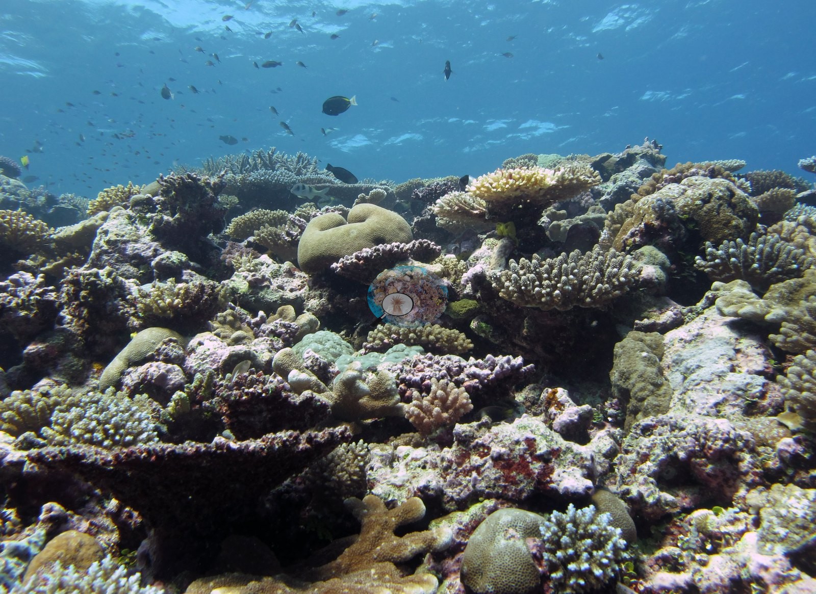
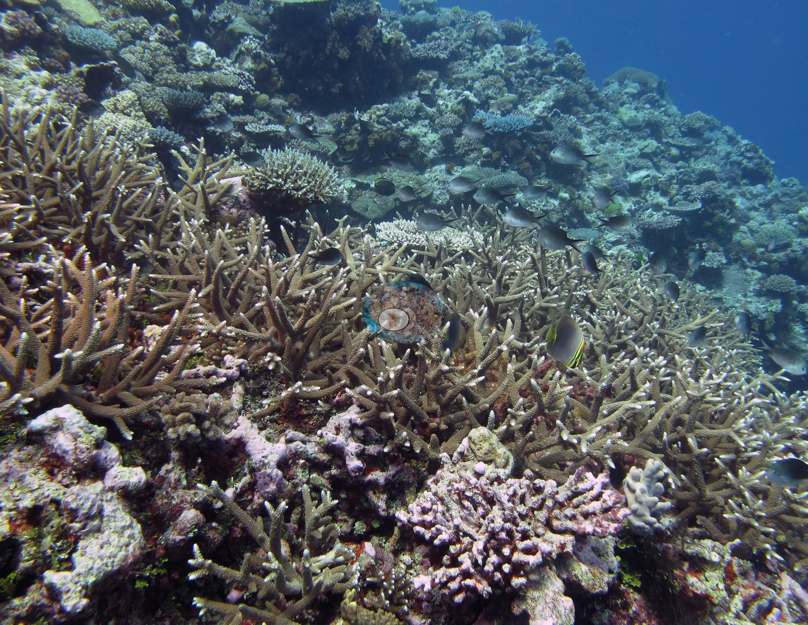
Fiji (NOAA CRW virtual station)A Tuvuca reef with a main terrace of near-continuous staghorn Acropora meadows under clouds of damselfish, and mixed stretches of violet, lilac, gold, and orange corals with meter-wide table corals. Surveyed on the Living Oceans Foundation's Global Reef Expedition.
E dua na cakau e Tuvuca e tiko kina e dua na vanua raraba (terrace) levu ni staghorn Acropora e voleka ni tomani tiko e ra ni ilawalawa levu ni ika lalai (damselfish), kei na vanua veiwaki ni lase e roka (violet, lilac, koula, oranse) kei na lase tepeli e dua na mita na kena raba.
AI translation of this summary; the English is authoritative. (low confidence)
CTD-cast midpoint of 26.8–27.1°C
One other CRD dive site with its own page within 5 km shown in orange — click a marker to open its page.
🗺 Detailed map of the Global Reef Expedition dive sites, Tuvuca, Fiji (Khaled bin Sultan Living Oceans Foundation; opens in a new window)
20 of the 24 photographs on this page are shown beside the text that discusses them under "Site description" below.
Site description
Setting
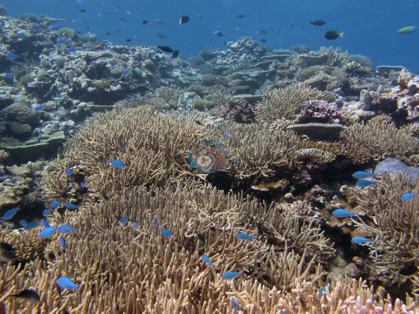
Anderson worked this reef from the shallows down the slope: the frame labels place three frames in the shallows at 3 to 5 m (e.g., Image 16, Image 23, Image 11) and three among the Acropora stands across the main terrace (e.g., Image 19, Image 12, Image 14).
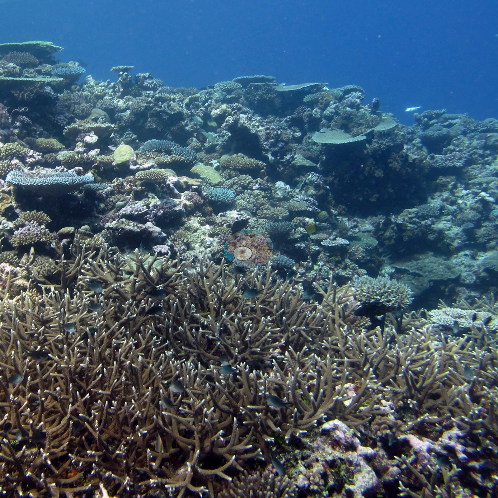
General benthic features
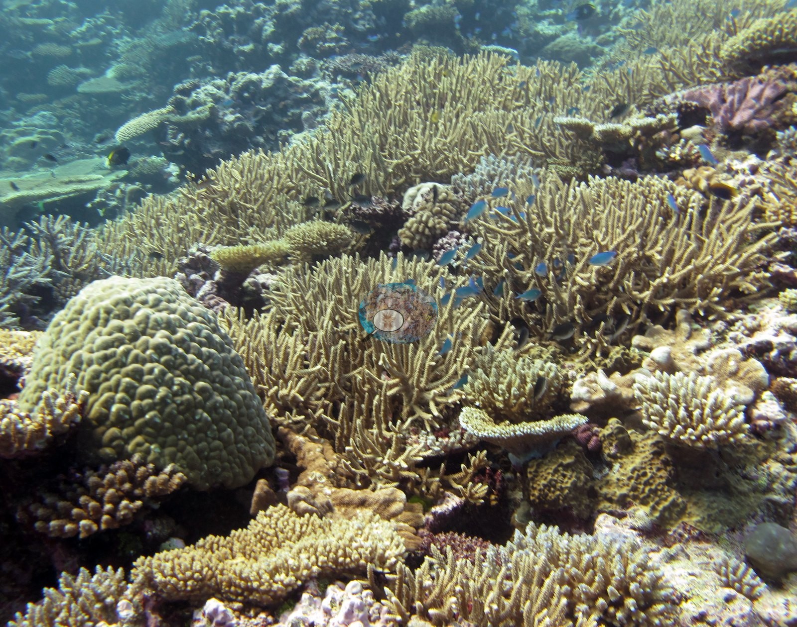
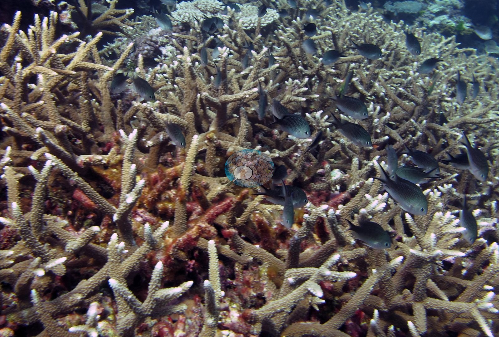
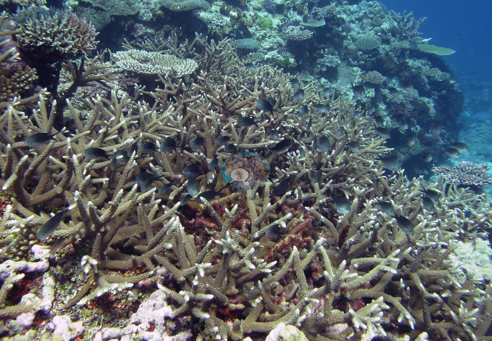
The terrace frames are the heart of the set: Image 15, Image 19, Image 12, and Image 14 show near-continuous stands of thin-branched staghorn Acropora, branches packed edge to edge and running out of frame, with clouds of small blue-green damselfish hanging over them and surgeonfish and butterflyfish working through; Image 14 is shot from inside the stand, looking along the branches with red encrusting algae and sponge deep in the interstices.
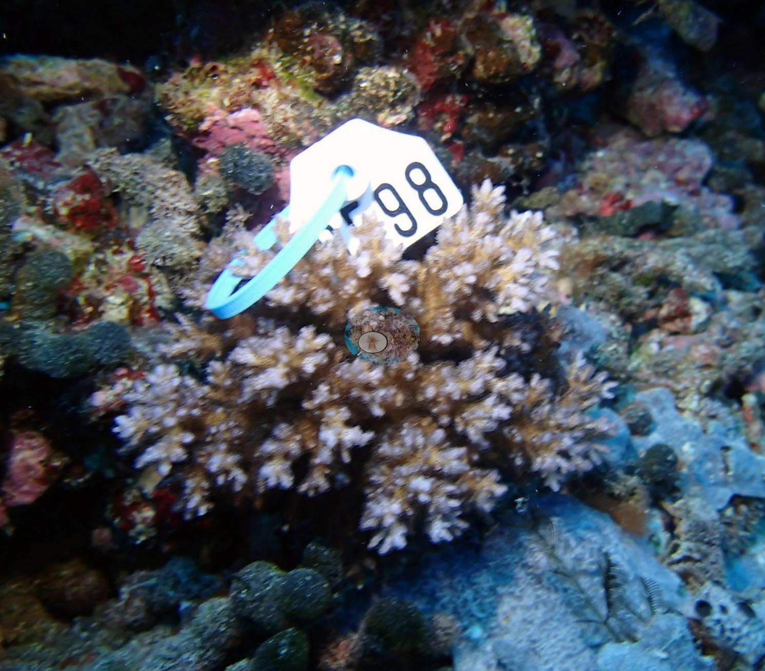
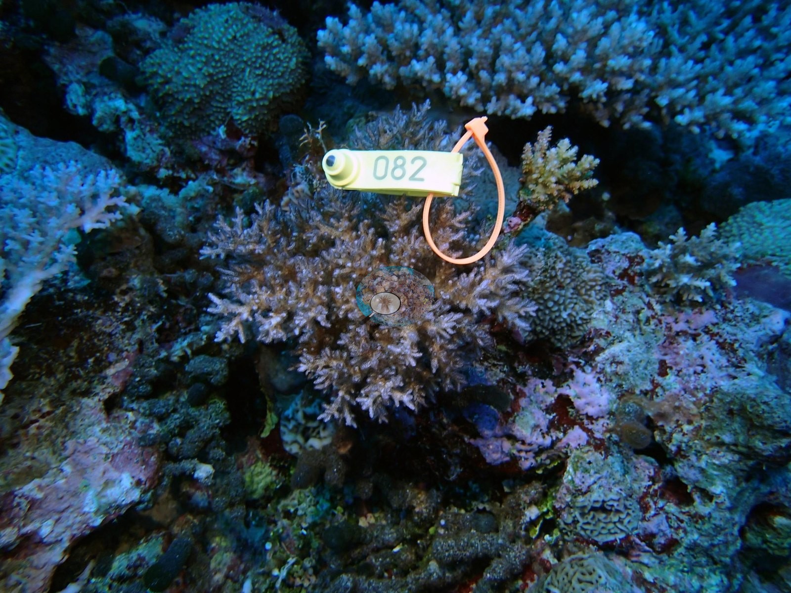
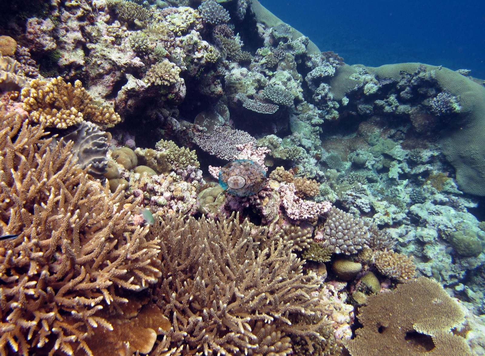
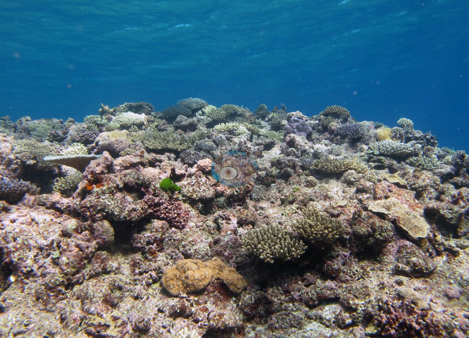
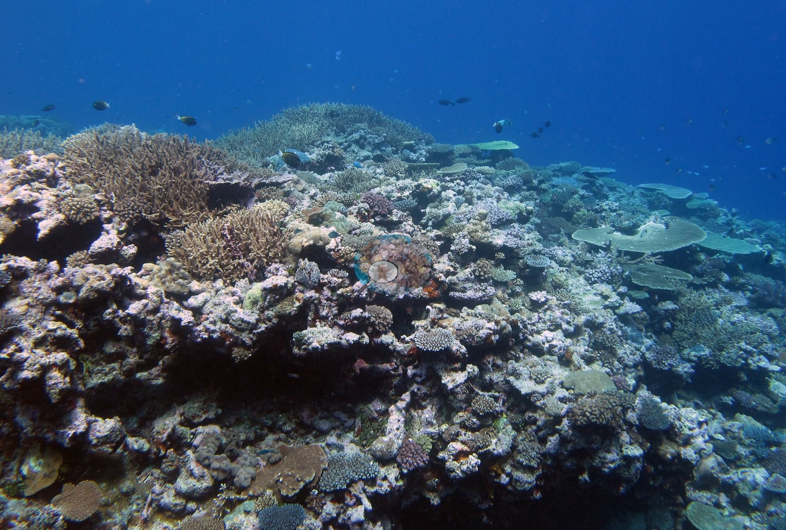
Where the reef is more mixed it is strikingly diverse and colorful — Image 10, Image 9, Image 8, and Image 17 hold tan and brown branching Acropora bushes, deep violet and lilac corymbose plates, golden-yellow and orange corymbose colonies, brown and tan tabulate tables, ruffled gray foliose colonies, and small rounded massives, all bedded in pink-lilac coralline crust.
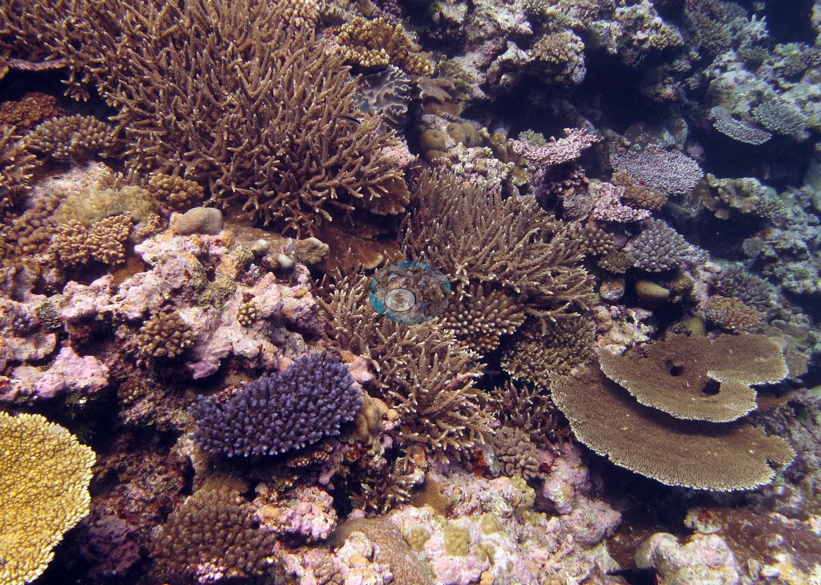
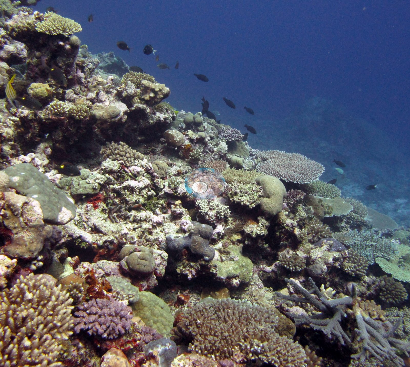
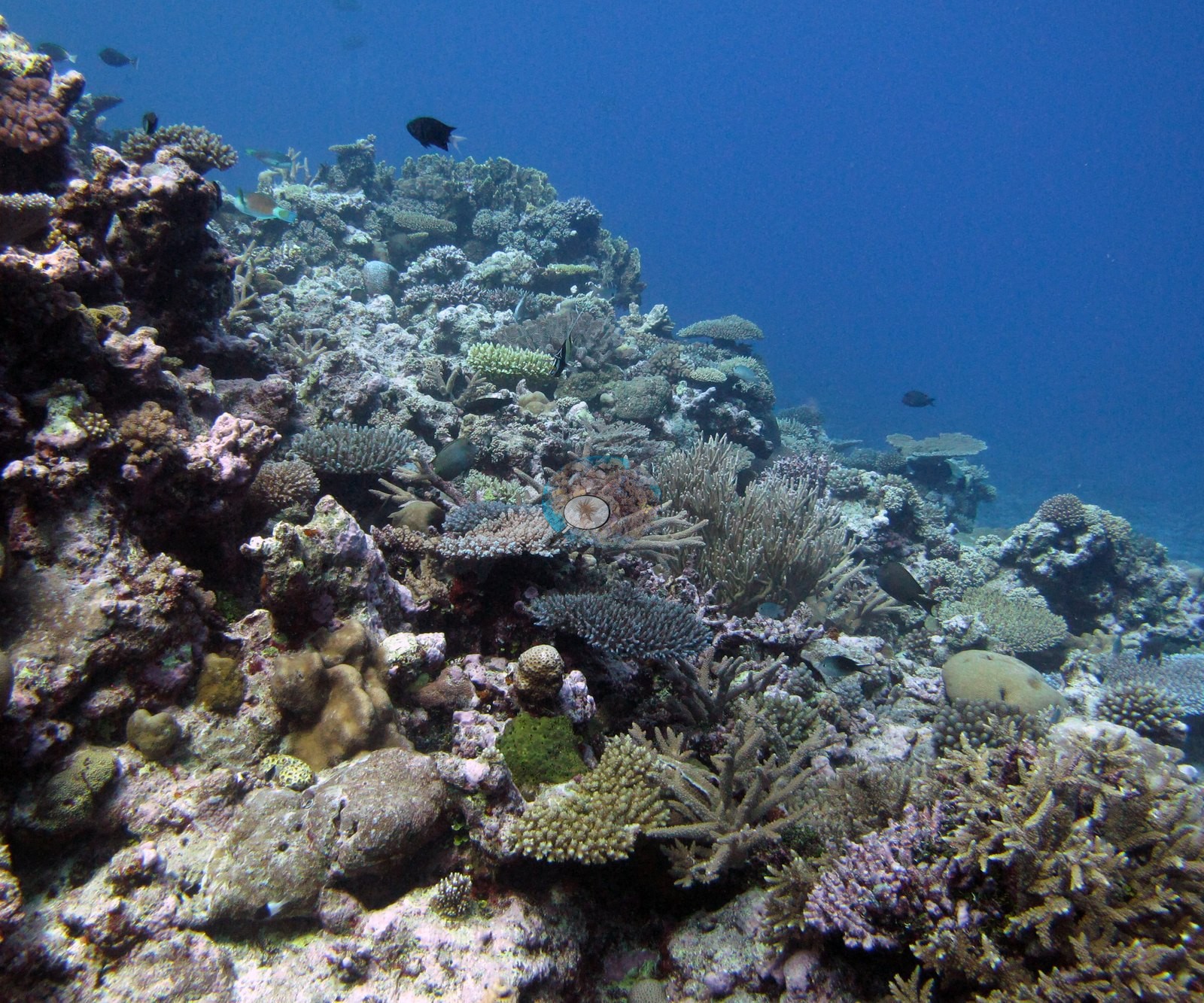
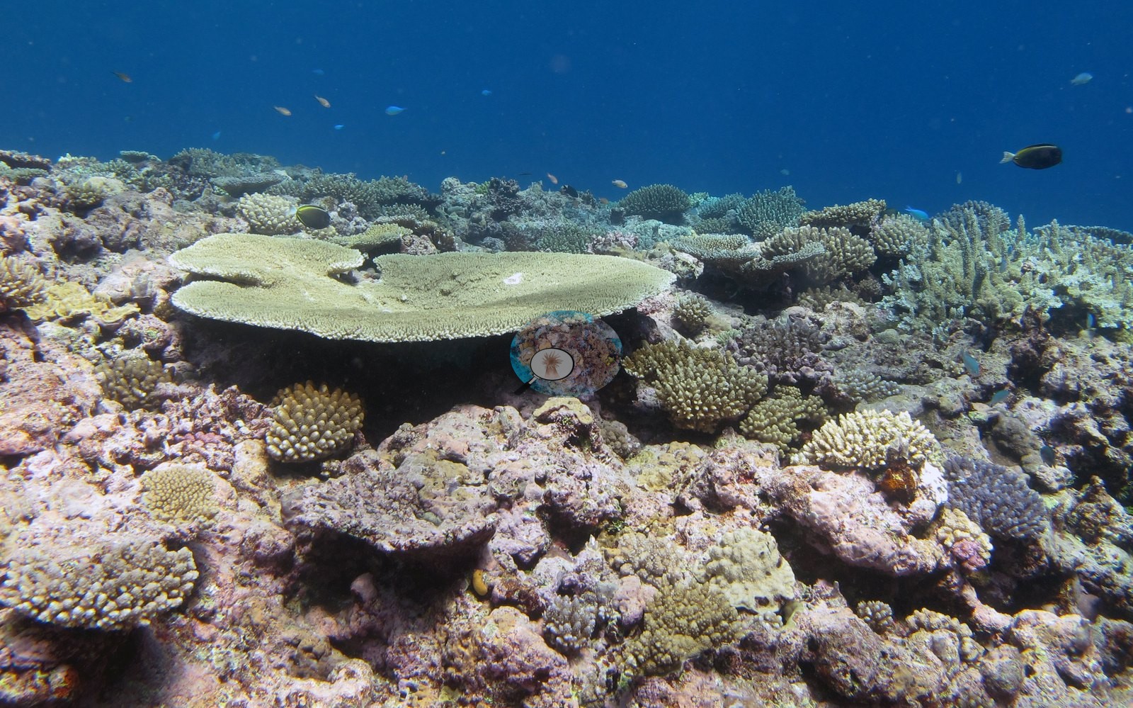
Large flat tables are a recurring landmark: a pale cream-green tabulate Acropora well over a meter across anchors both Image 21 and Image 23, and Image 18 shows another on the slope.
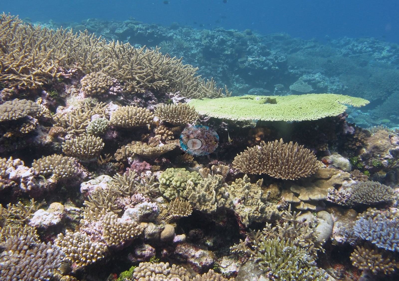
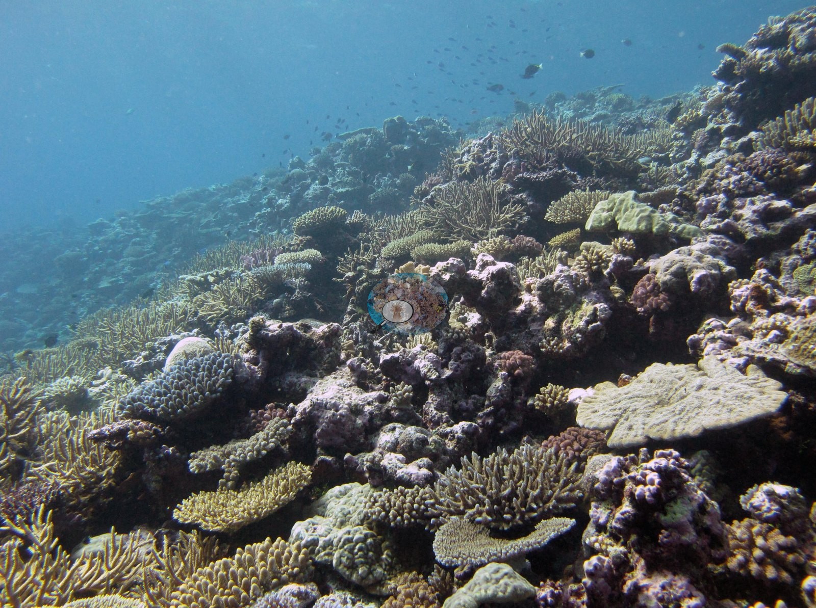
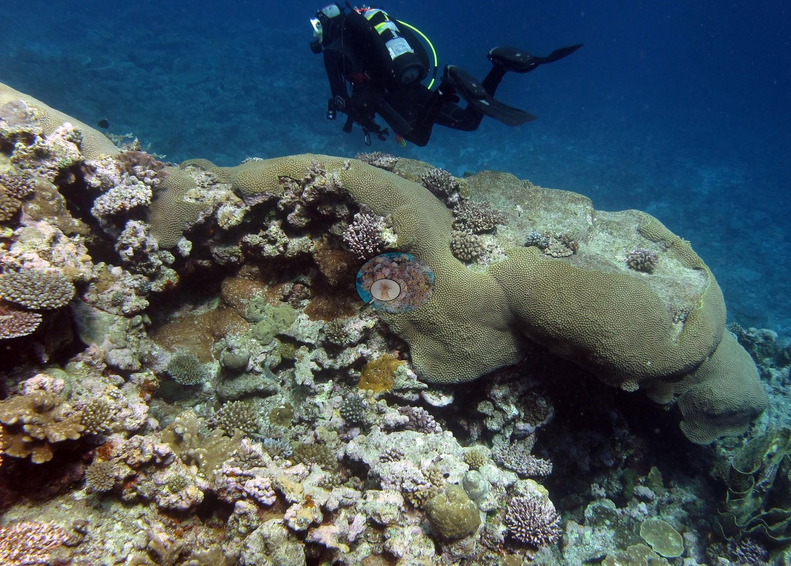
Image 11 is different again — a single large tan lobate-columnar massive with the smooth, evenly porous surface of Porites, big enough to fill the foreground, with a diver above it. The shallow frames (Image 16, Image 22, Image 24) are lower-relief: small digitate and corymbose colonies and scattered massives on rubbly, coralline-crusted pavement in ambient light.
Coral cover
- Reference: Khaled bin Sultan Living Oceans Foundation, Global Reef Expedition (in situ transect)
- Coral cover 37%, from the LOF-GRE in situ benthic transect — a diver transect, and the authoritative figure for this site. Transect mean 37 ± 5.5% SE (n = 5 transects; Living Oceans Foundation per-transect benthic data, which lists these five transects, labeled FJTV42, under TOVA42's station code).
Stressors
No bleaching front, disease lesion, or crown-of-thorns seastar appears in any of the 22 frames reviewed, and table and branch margins look clean and actively growing. Some gray-white branch tips within the densest staghorn (Image 15, Image 12) cannot be separated from exposed skeleton at this range, and the frames cannot settle how much old dead framework underlies the live canopy. The one coral colony from this site with an outlier score was not flagged as a physiological outlier (Mayfield et al. 2017, PLoS ONE). The Living Oceans Foundation transects here recorded 9.0% cyanobacteria and no recently dead coral.
Fish and other fauna
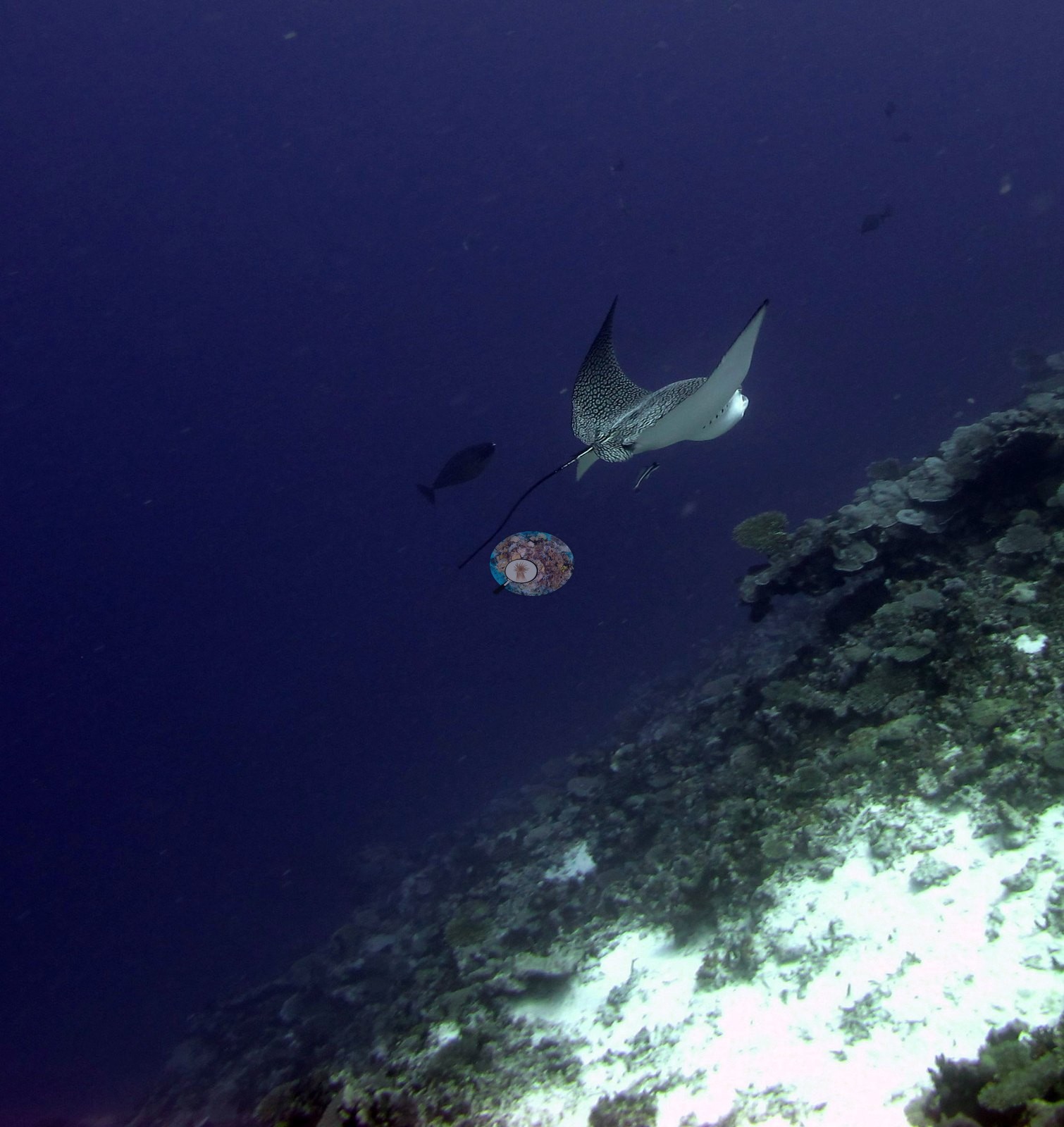
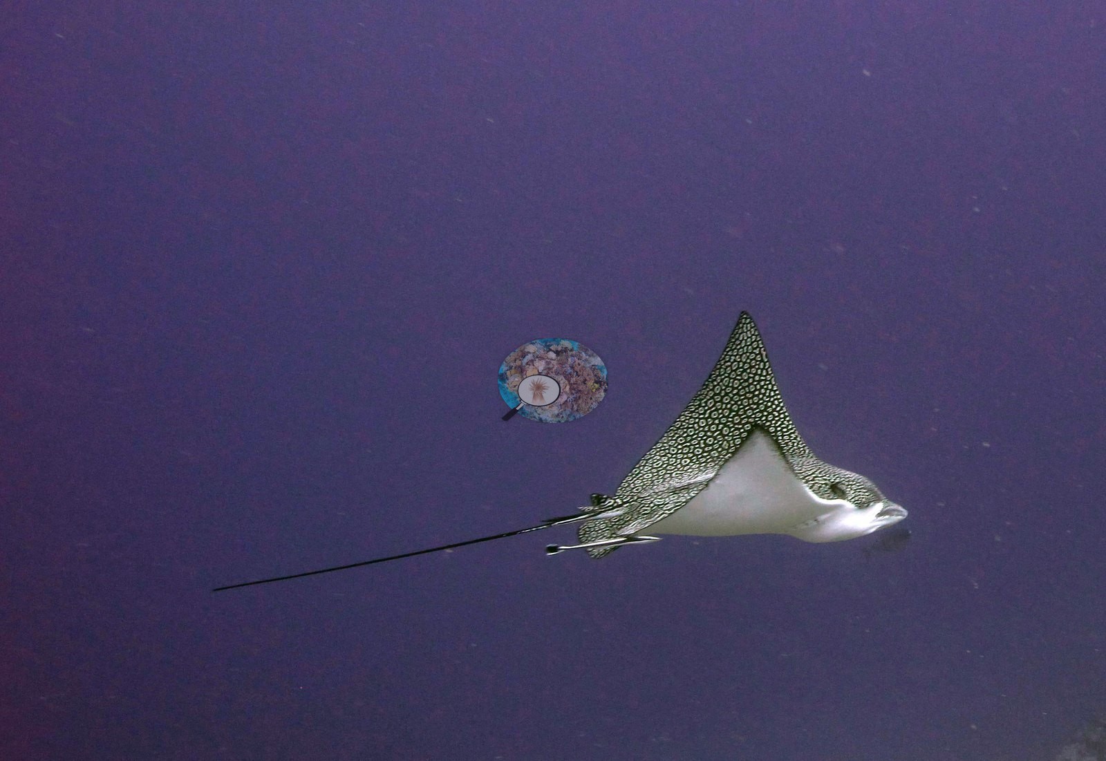
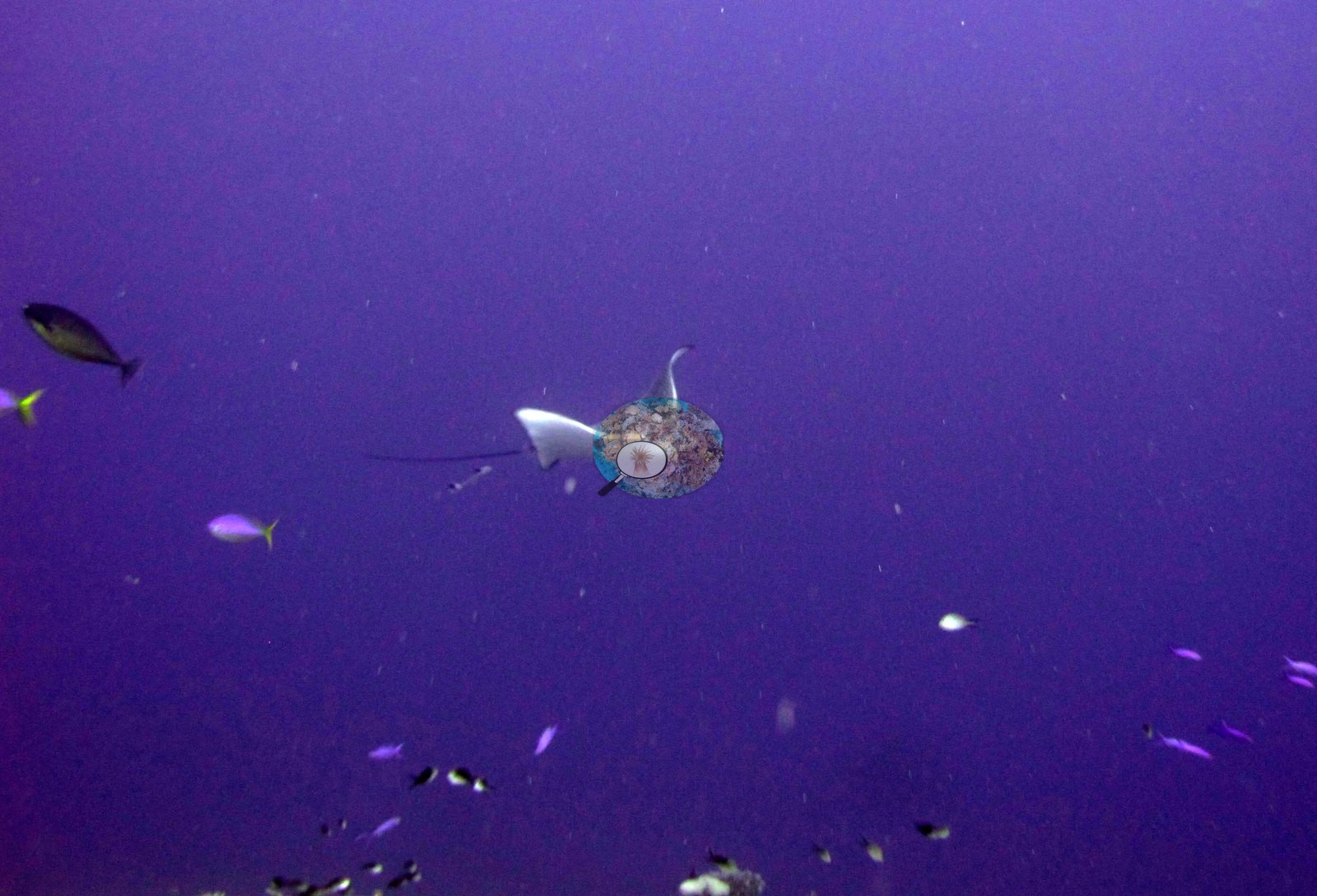
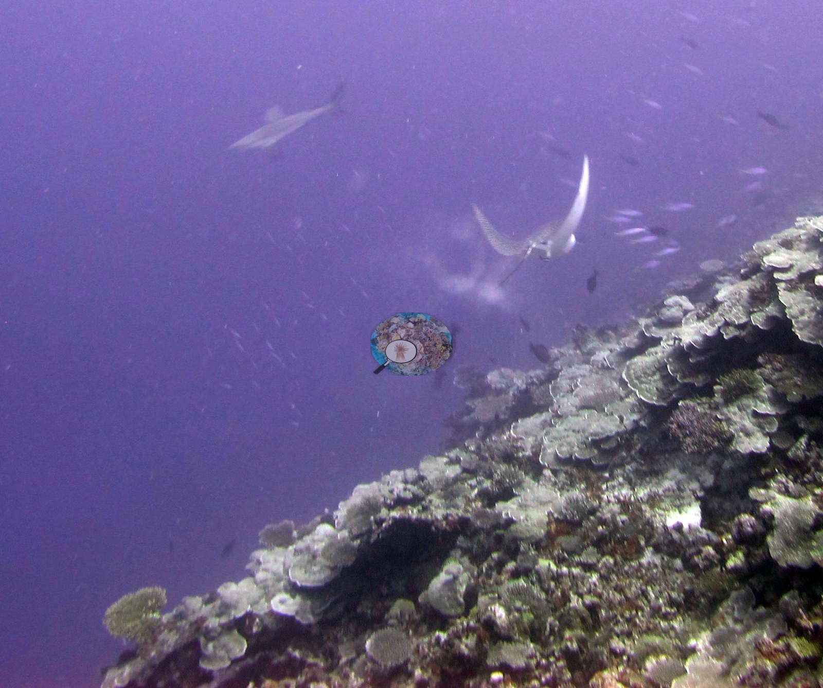
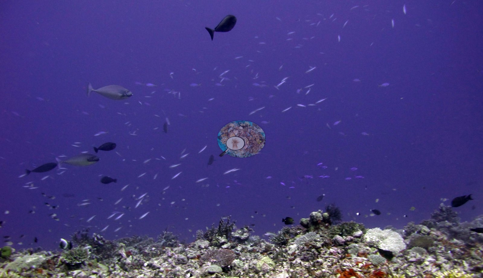
Four frames are pelagic rather than benthic — Image 5, Image 4, Image 3, and Image 6 are spotted eagle rays in open water (Image 6 also has a shark), one carrying attached remoras — and Image 7 is a school of small fish over the slope.
Comparisons with other data
Apart from the Living Oceans Foundation survey shown here, we found no other surveys at this site as of 2026. If you would like to share yours, please email us.
Other notes
Survey team's site notes: Isolated monospecific Isopora stands were found around 16 m. Again like other sites, shallow fore reef slope had coral assemblages of Pocillopora, Stylophora, and Acropora.
CV model comparison
More info
The photo row below is a methods comparison only: it is never averaged with the transect and never used as the headline. It compares site means only: the photos come from the same survey dive as the transect but not from the same points.| Method | Hard coral cover (%) | Photos (n) ± SE |
|---|---|---|
| Transect mean (all transects) | 36.8 | |
| CoralSCOP edited | 38.5 | n = 23 · SE 4.0 |
| Claude (edited photos) | 45.5 | n = 23 · SE 4.3 |
The two computer-vision methods agree within 7.0 pp (38.5–45.5%) and read 1.7–8.7 pp above the in-water transect (36.8%); the headline is the in-water transect because a diver transect is independent of the photographs.
See a more detailed, stand-alone version of this table.
Data & links
All images from Coral Reef Diagnostics. Data are from the Khaled bin Sultan Living Oceans Foundation Global Reef Expedition and Coral Reef Diagnostics.
 © Coral Reef Diagnostics.