
King Reef (Nassau north coast)

More info
Central Bahamas (NOAA CRW virtual station) · Bahamian ecoregion mean: 9.1 degree heating weeksAn elkhorn coral site where live Acropora palmata ranges from a "monster" colony and tall candelabra stands to a small recruit, side by side with standing dead elkhorn framework. Brain corals and gorgonians fill the low hardbottom between, and Anderson flagged one diseased Porites colony and some pale star corals.
Three independent survey stations shown with the program’s logo — click one to see how far it is from this site.
This page shows 66 of the 73 photographs from this site, including every one the text cites.
Site description
General benthic features
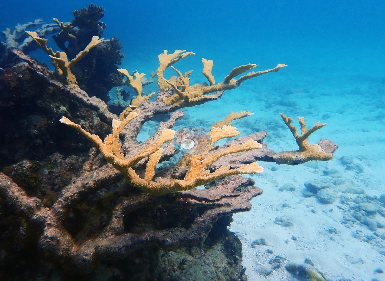
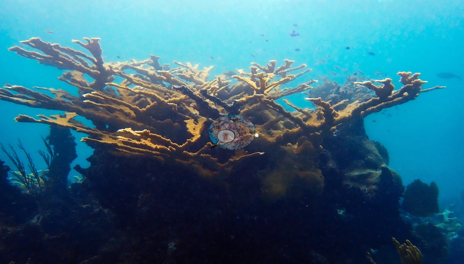
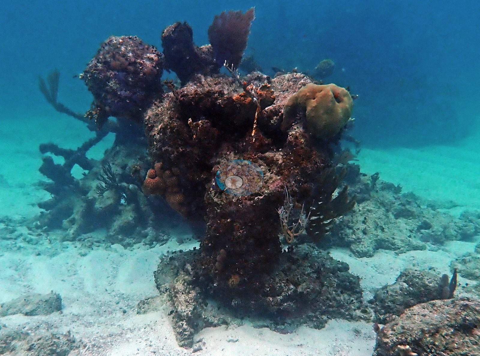
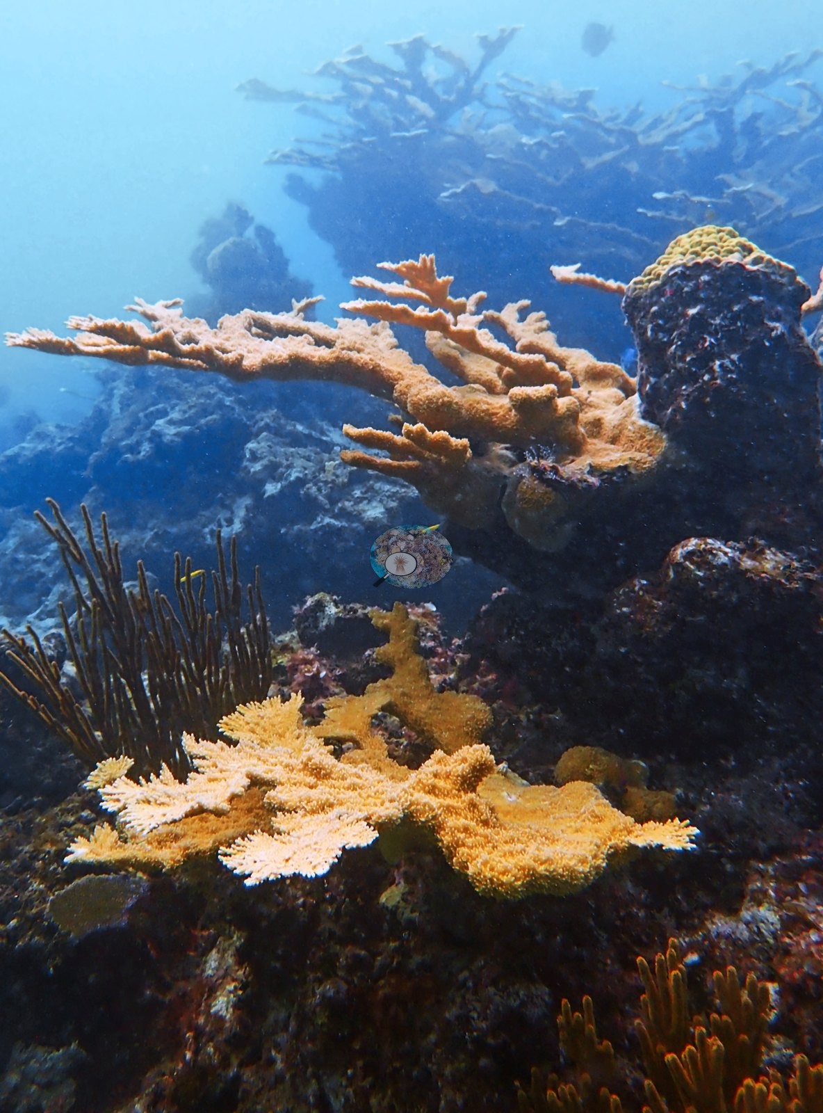
This is an elkhorn site, and the whole set turns on Acropora palmata (e.g., Image 48, Image 60, Image 71).
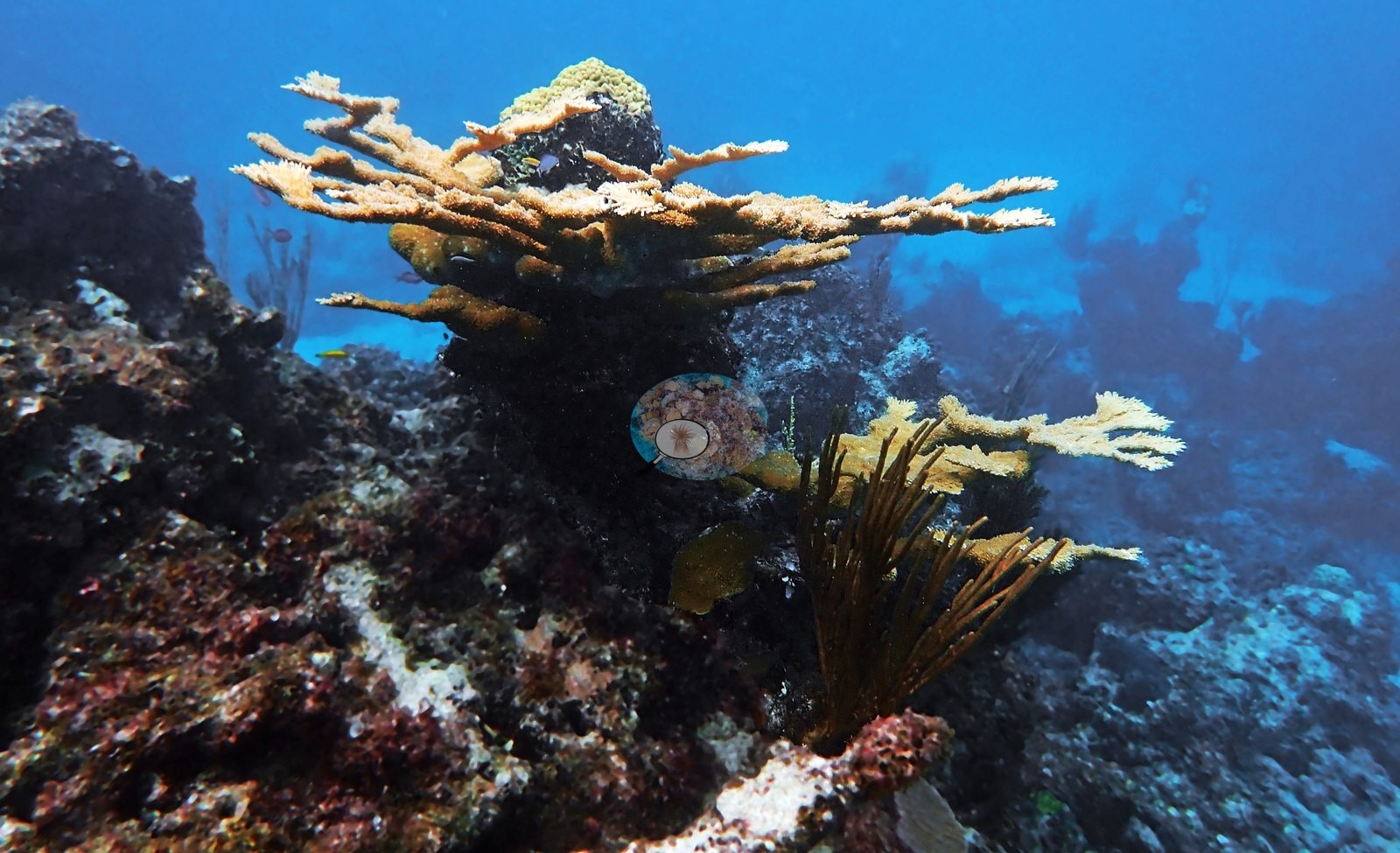
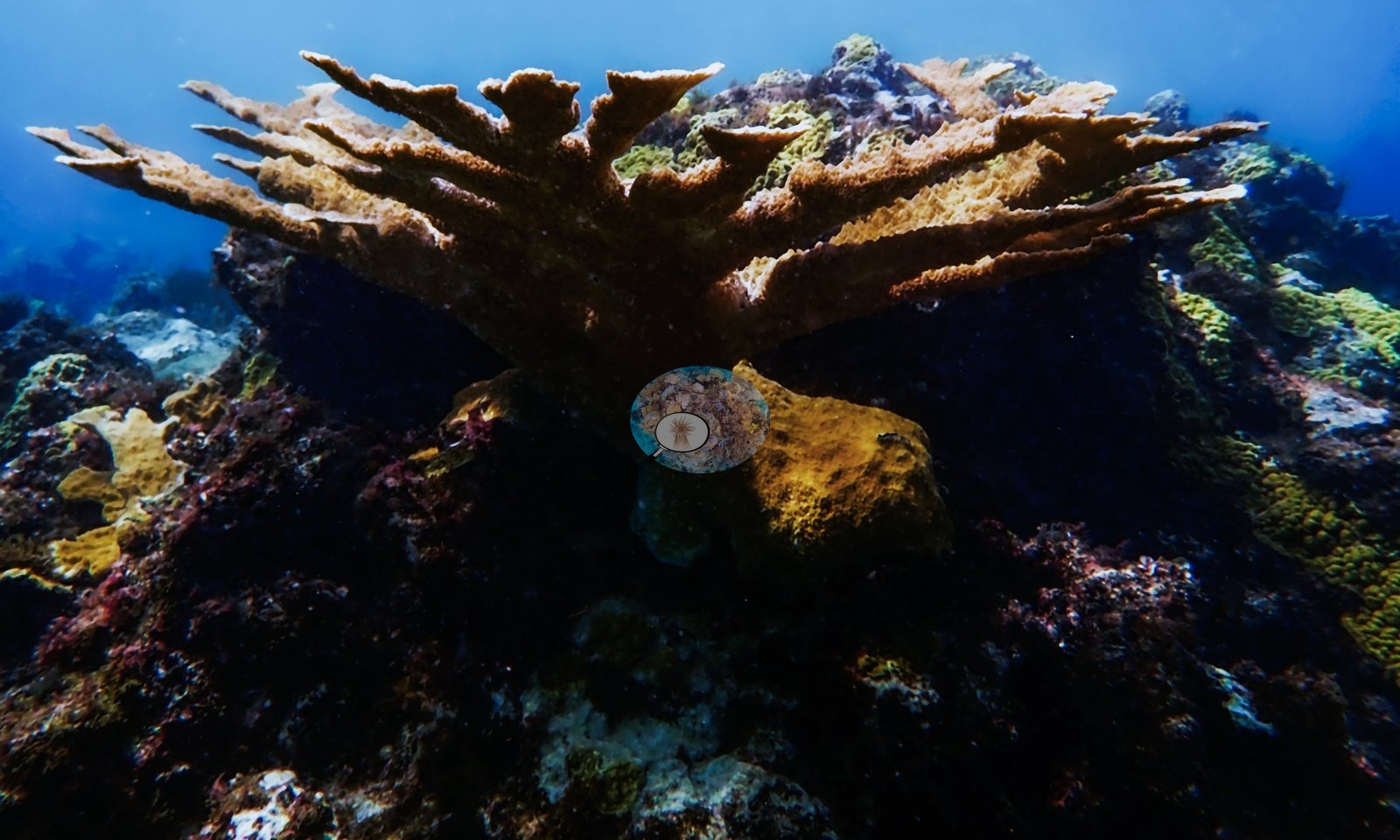
Live colonies are not incidental - they appear in something like a fifth of the frames. Broad golden-tan blades fan out over low hardbottom and white sand in Image 47, Image 48, Image 49, and Image 53; a spreading multi-branched colony stands clear of the bottom in Image 58, Image 61, Image 64, and Image 67; Image 60 and Image 71 show tall candelabra colonies with clean growing margins silhouetted against bright blue water; Image 65 catches a wide, densely branched colony from below.
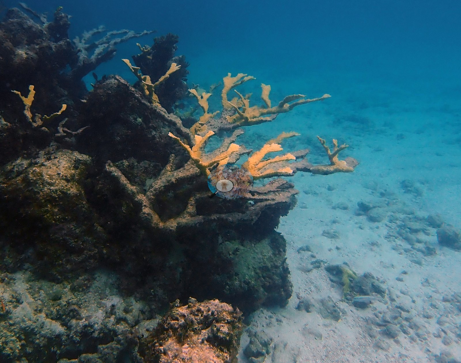
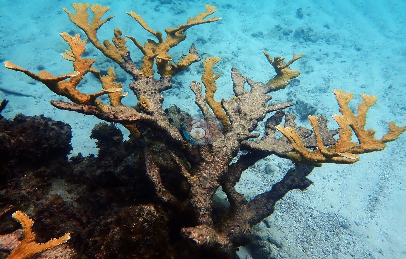
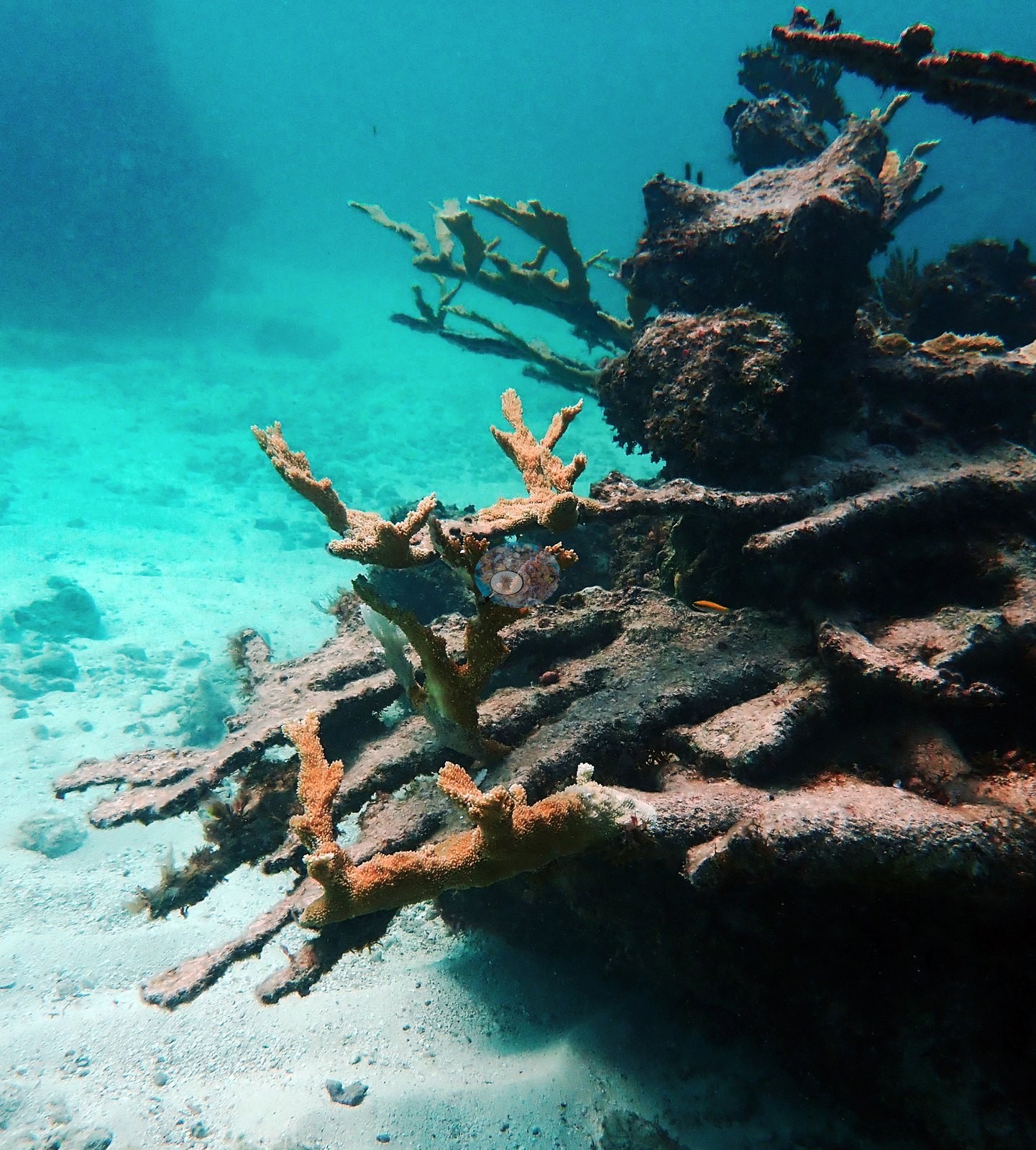
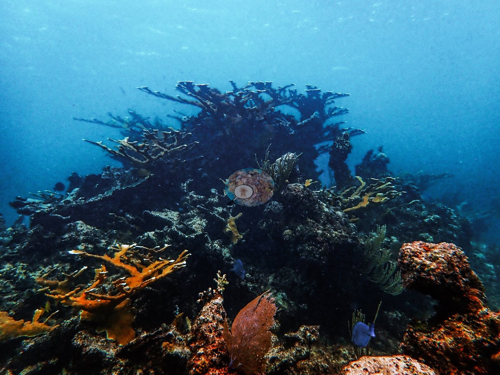
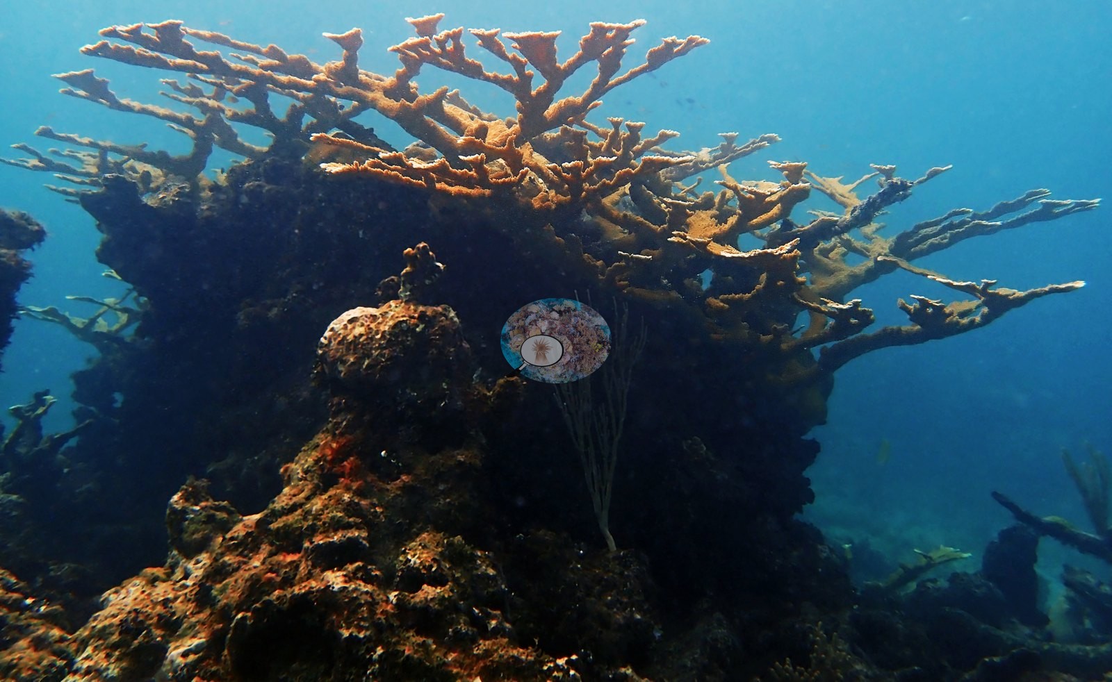
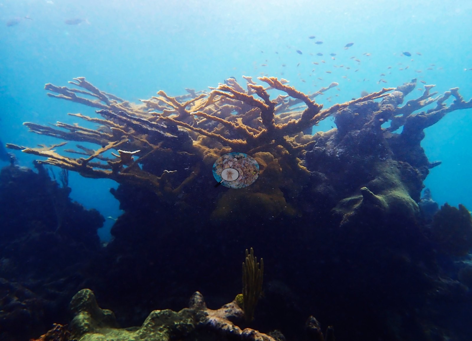
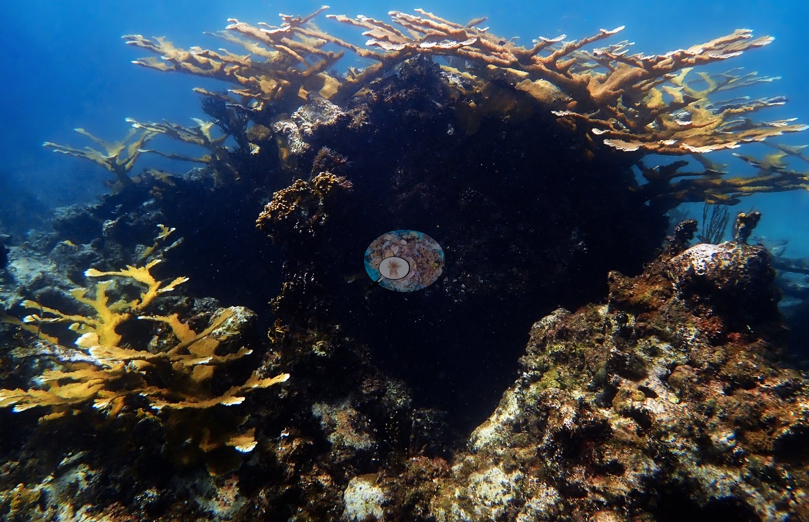
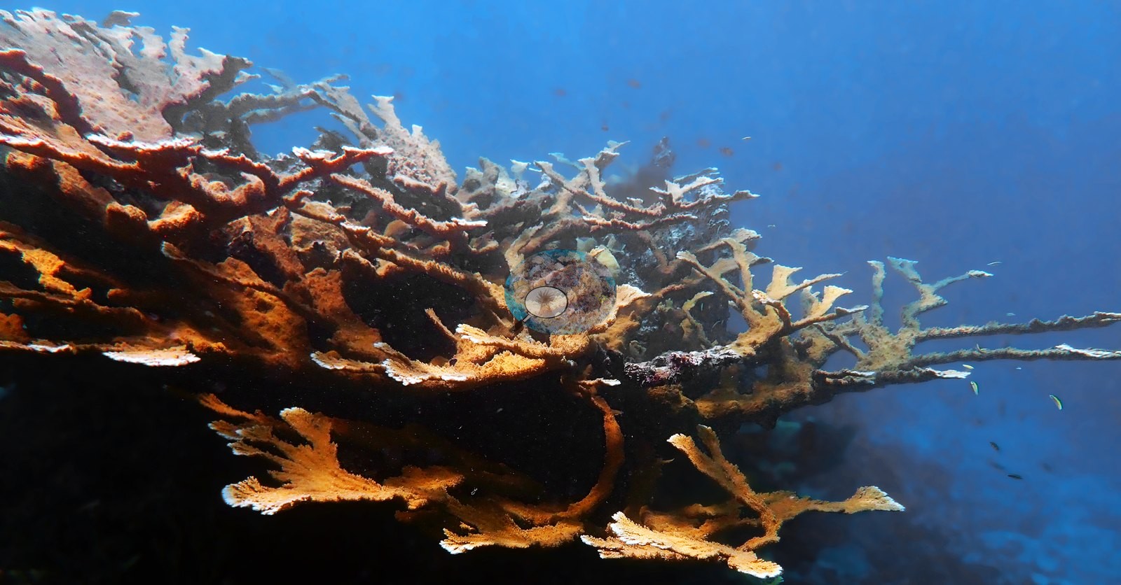
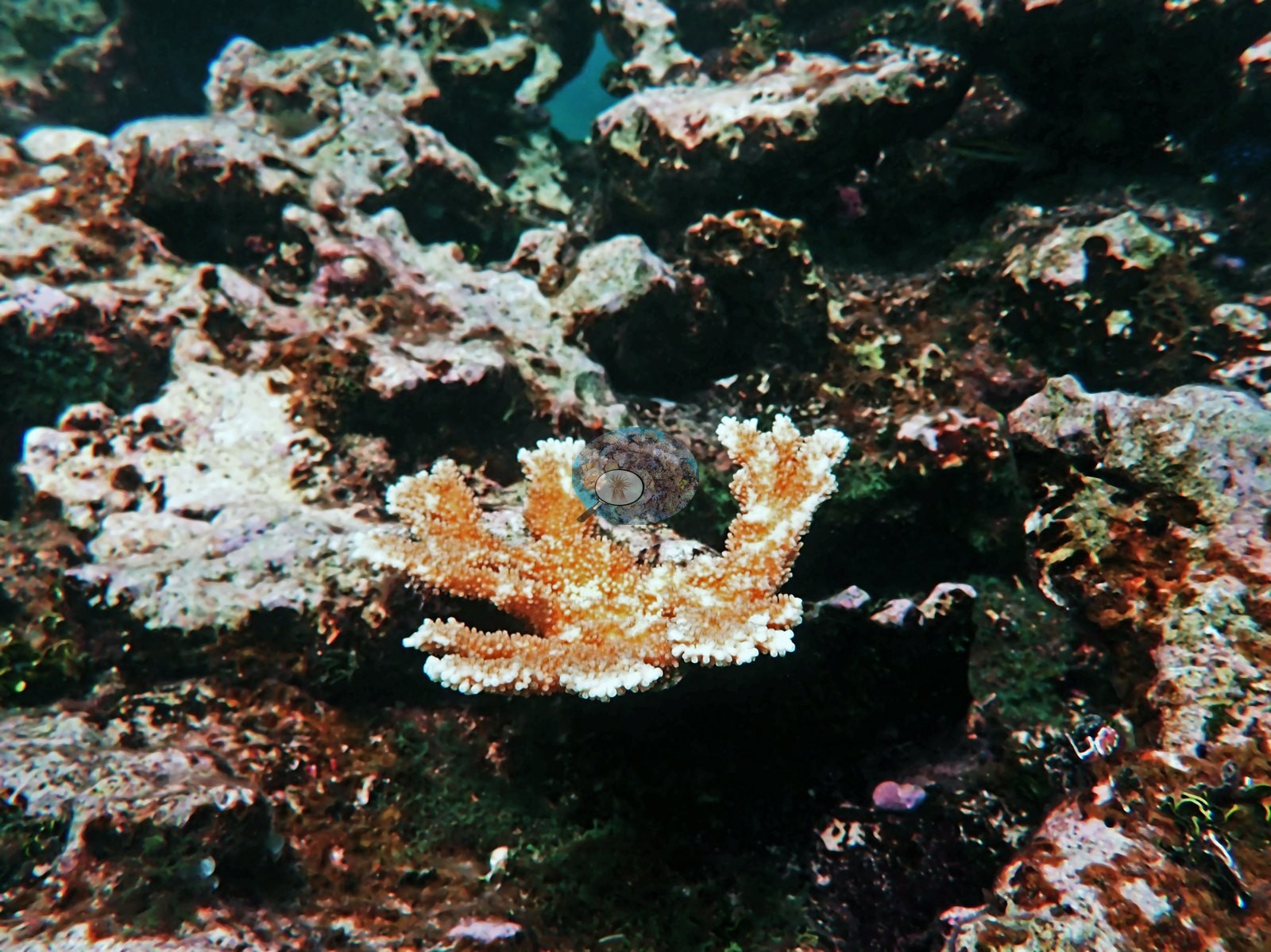
His own labeled frames give the range in one sequence: a whole thicket (e.g., Image 66), the very large colony he calls "monster A. palmata" (e.g., Image 63), a small recruit growing on live rock (e.g., Image 37, "baby Apalm"), a sparse, thin-branched colony he calls "struggling" (e.g., Image 51), and a diver alongside a standing skeleton for scale (e.g., Image 52, "assessing Apalm").
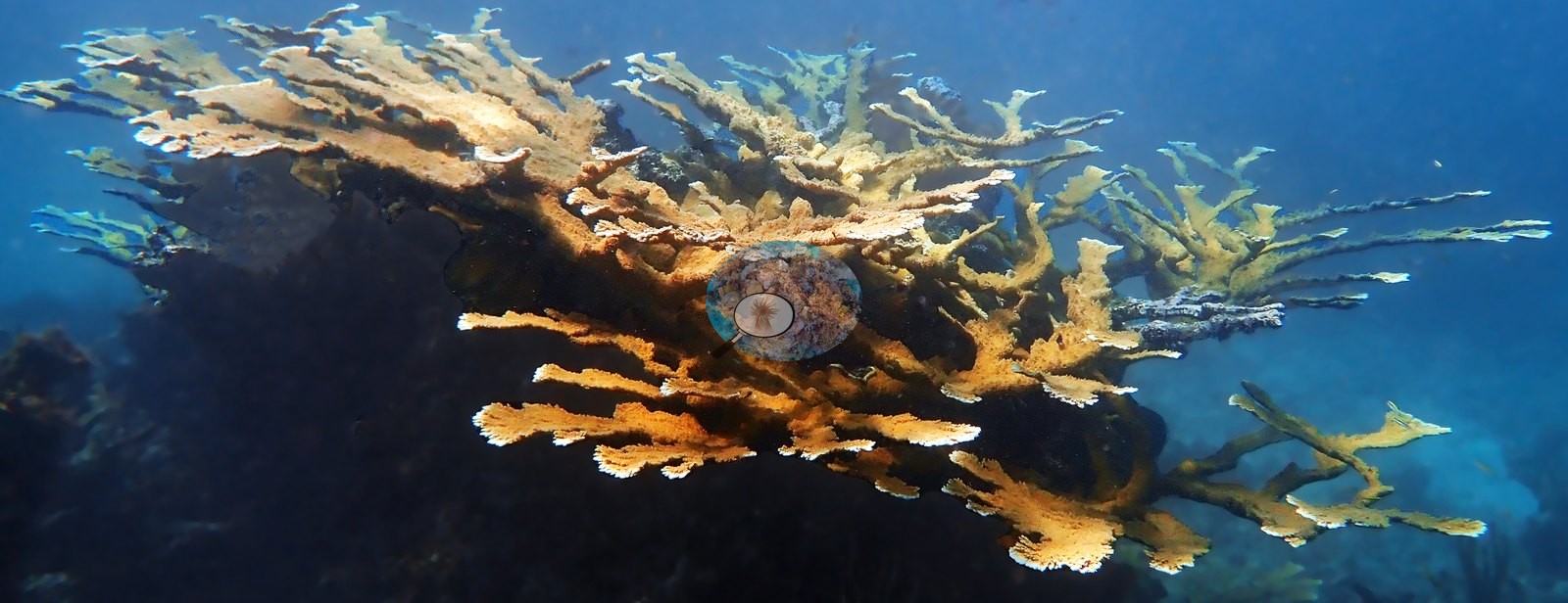
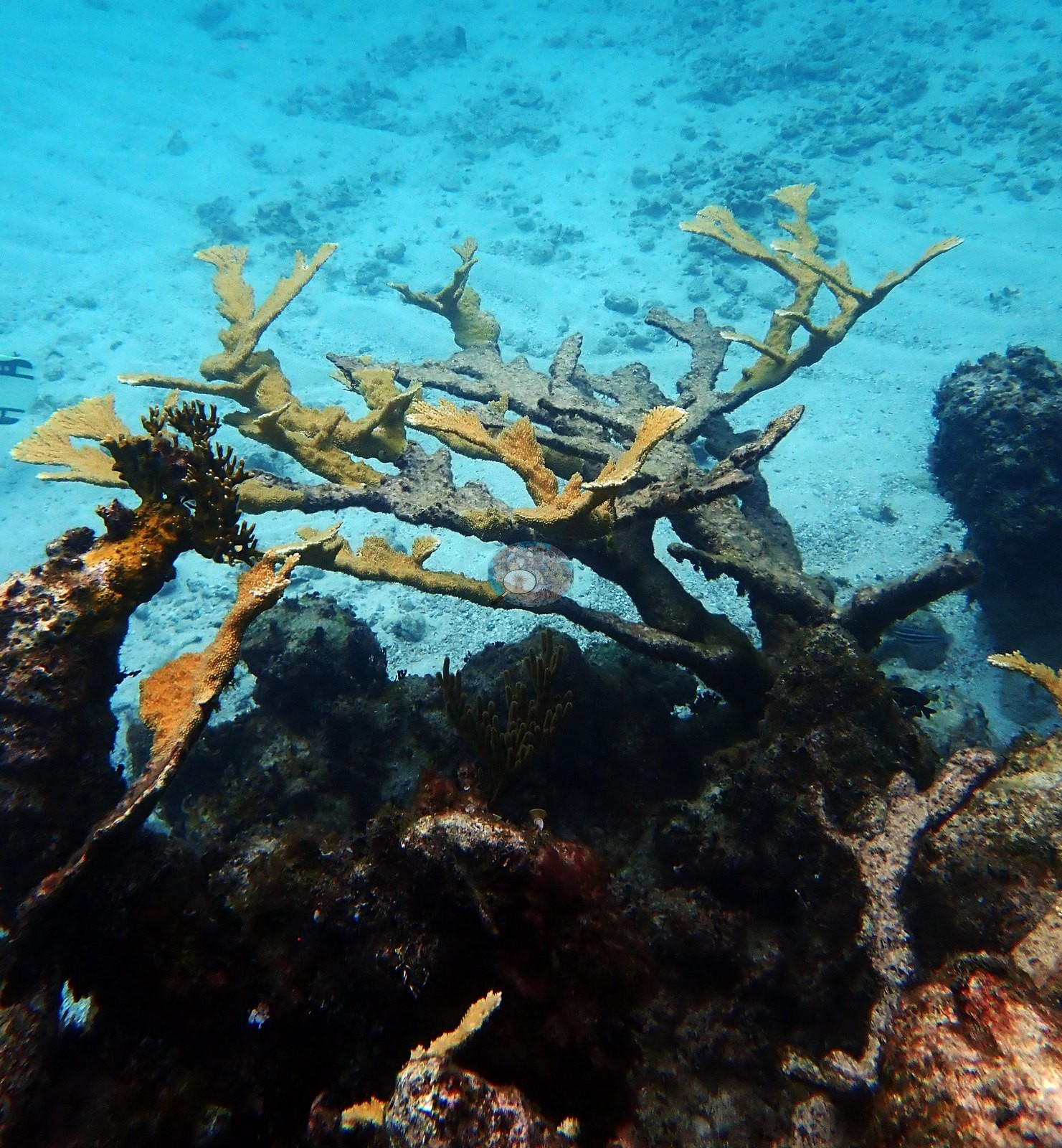
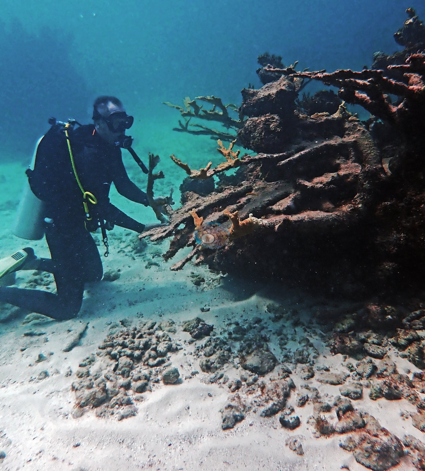
The counterpart to all that live tissue is standing dead framework: bare, turf- and silt-crusted elkhorn branches still in growth position in Image 36, Image 42, Image 43, and Image 39 (his "dead Acropora framework"), and eroded branch stubs mixed into the pavement in Image 38, Image 40, and Image 50.
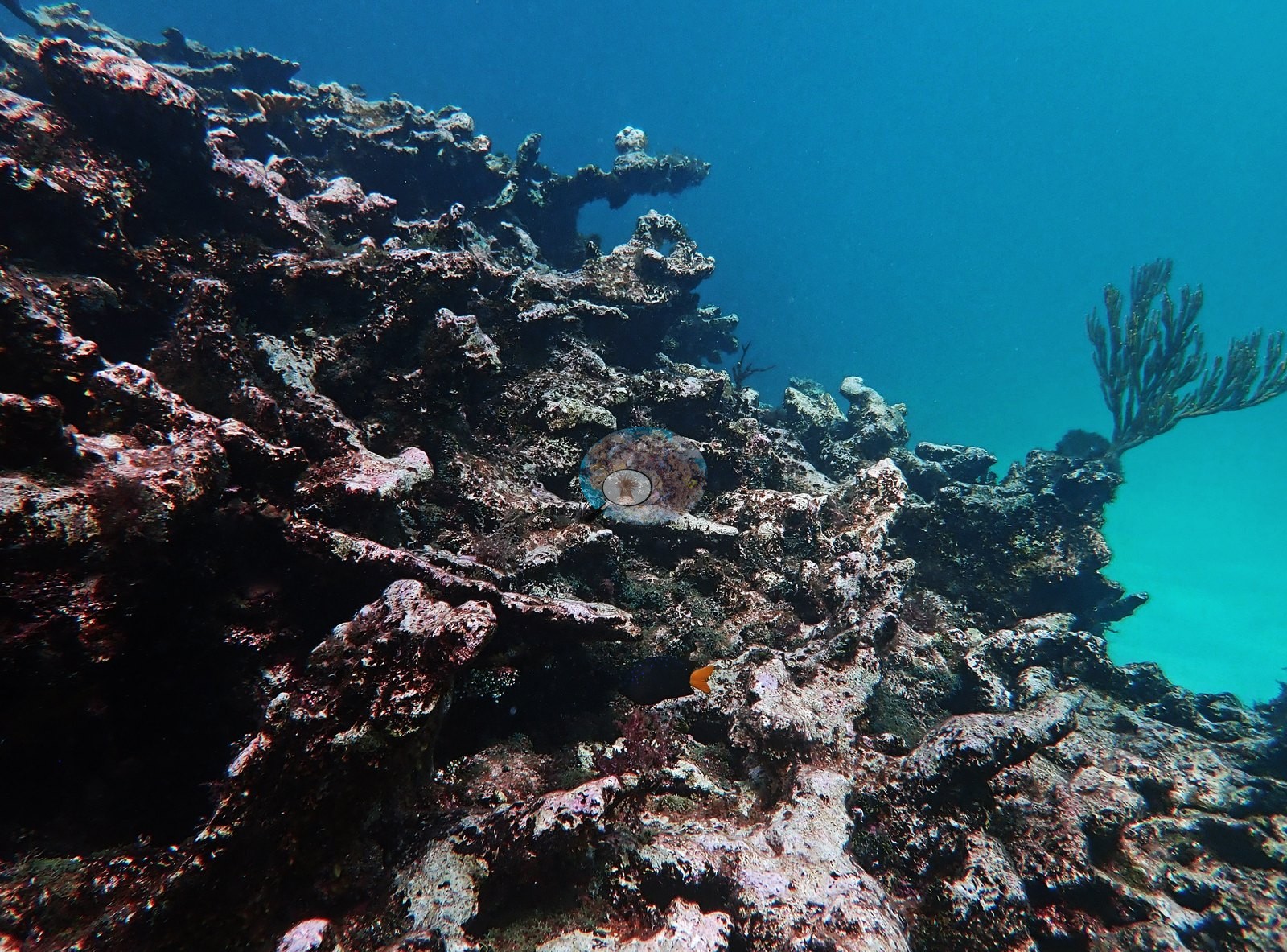
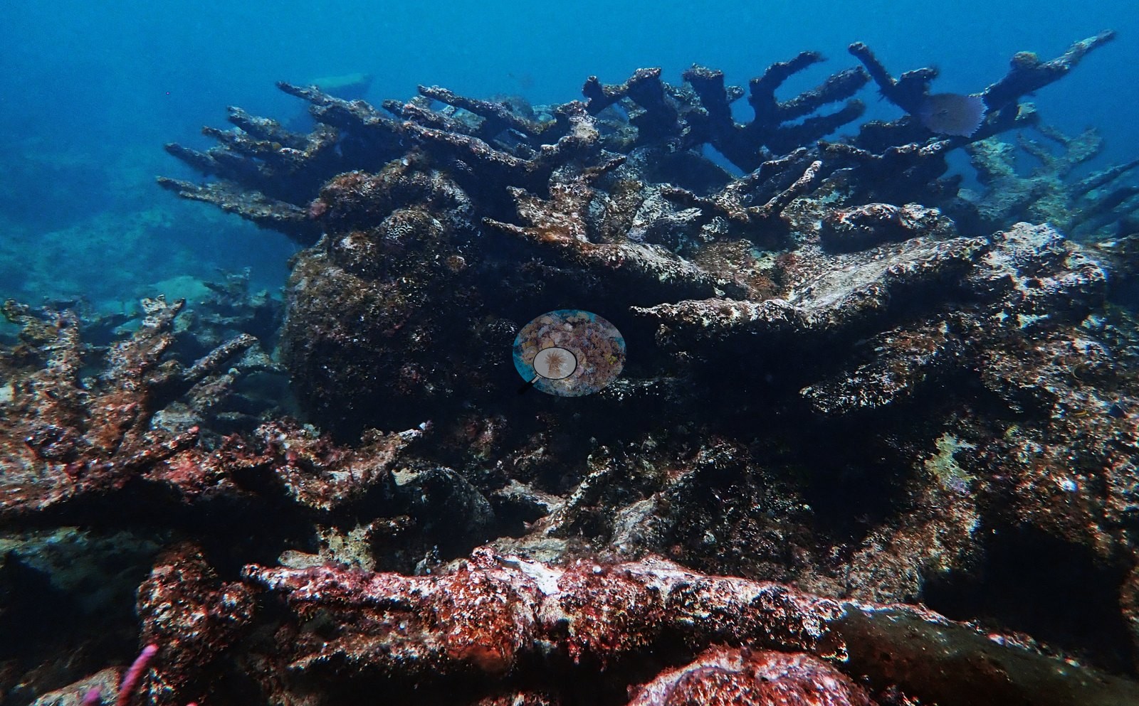
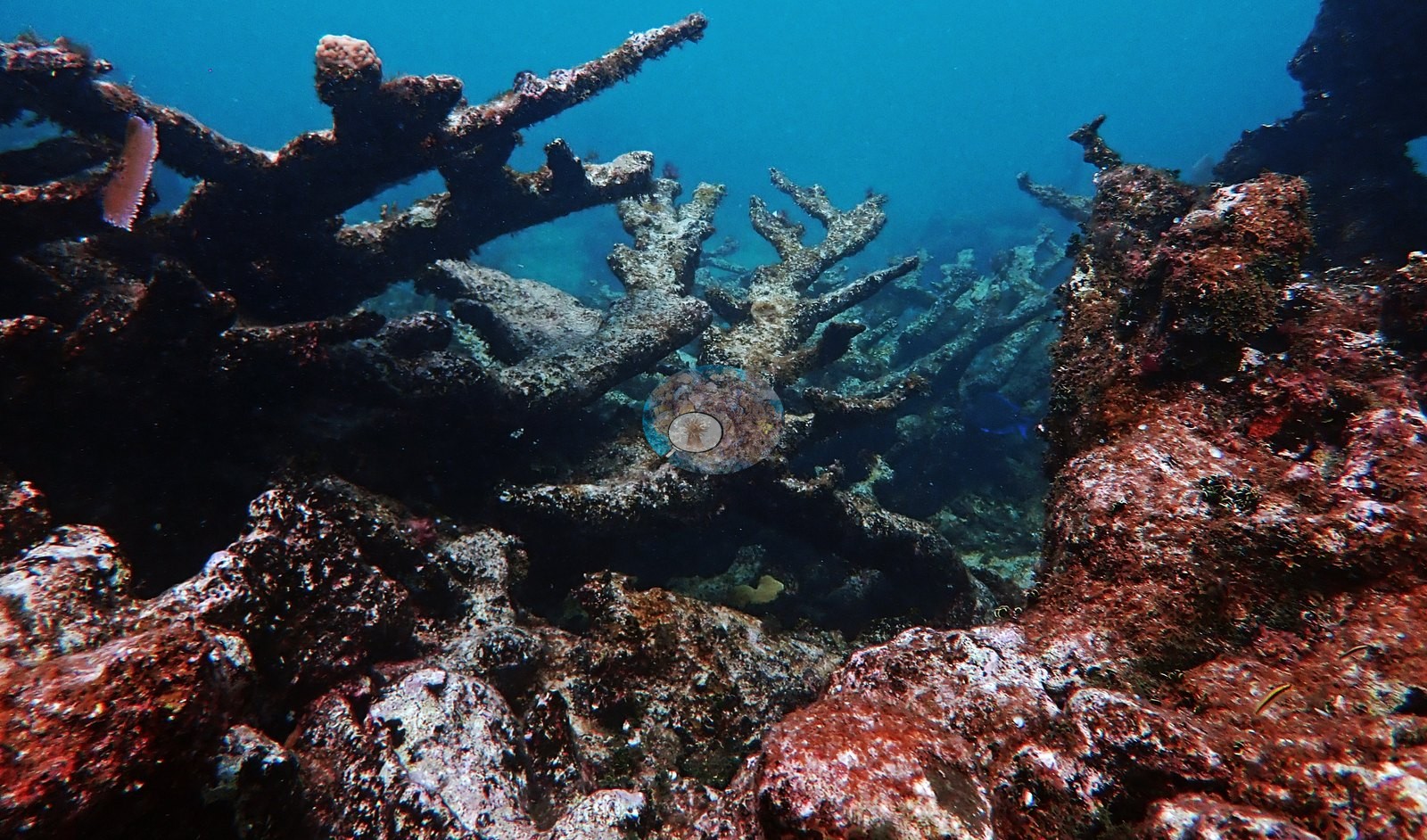
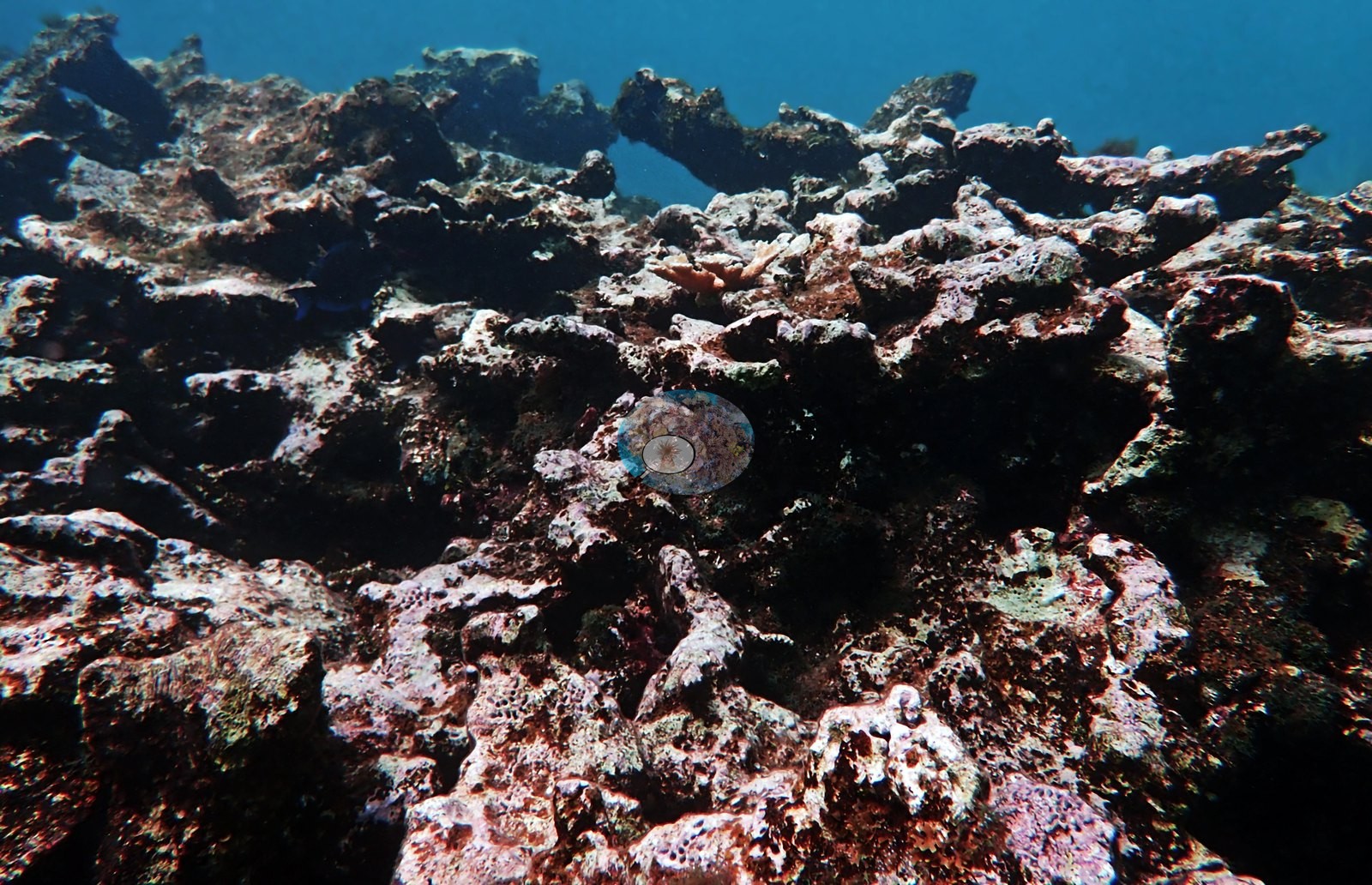
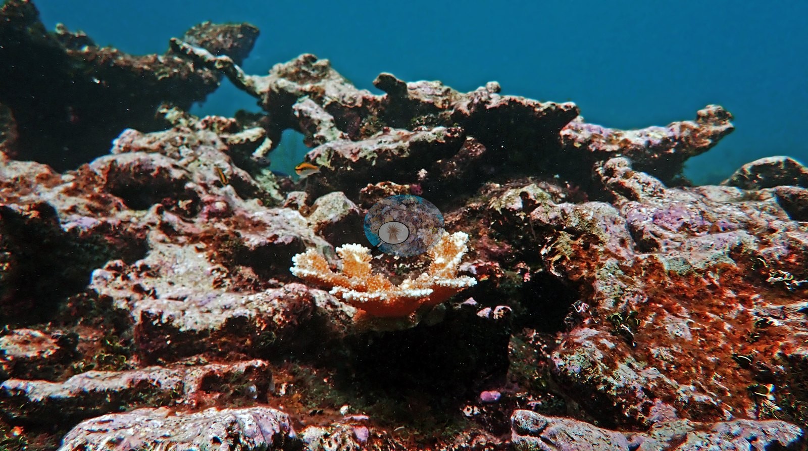
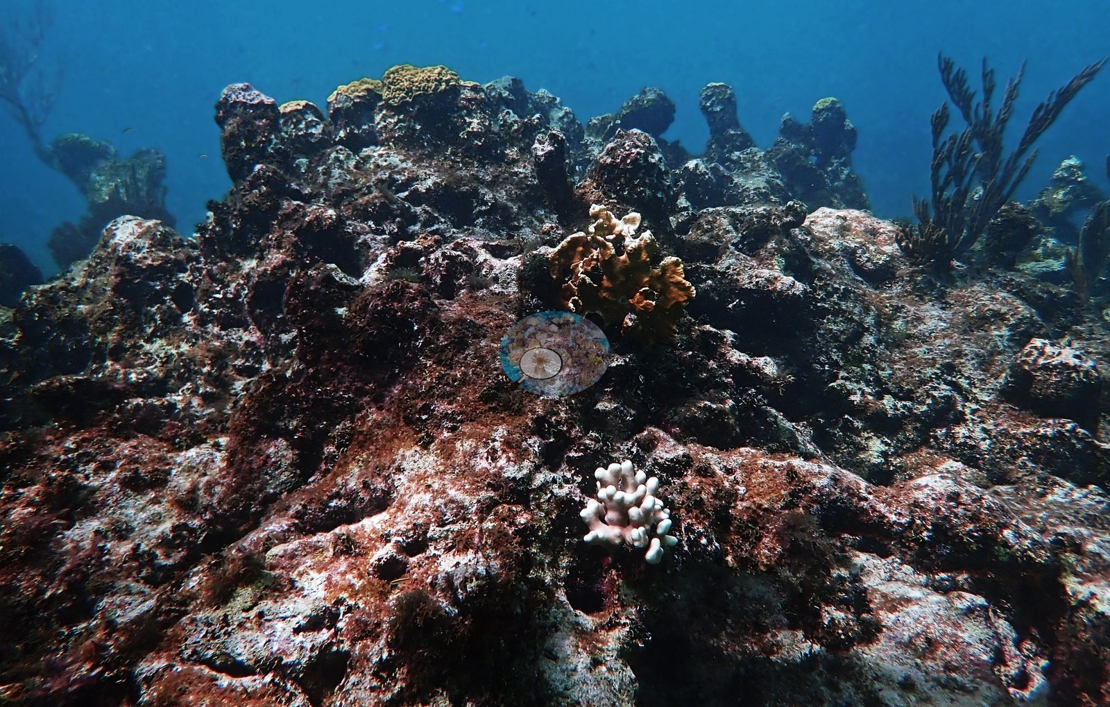
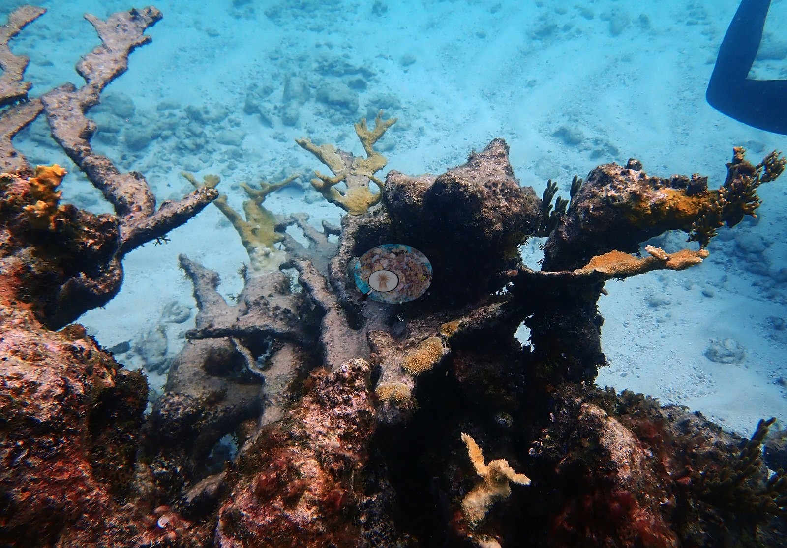
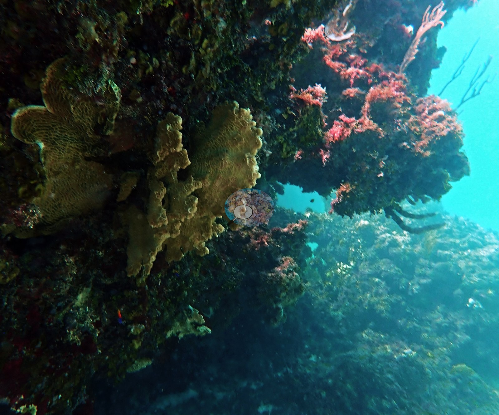
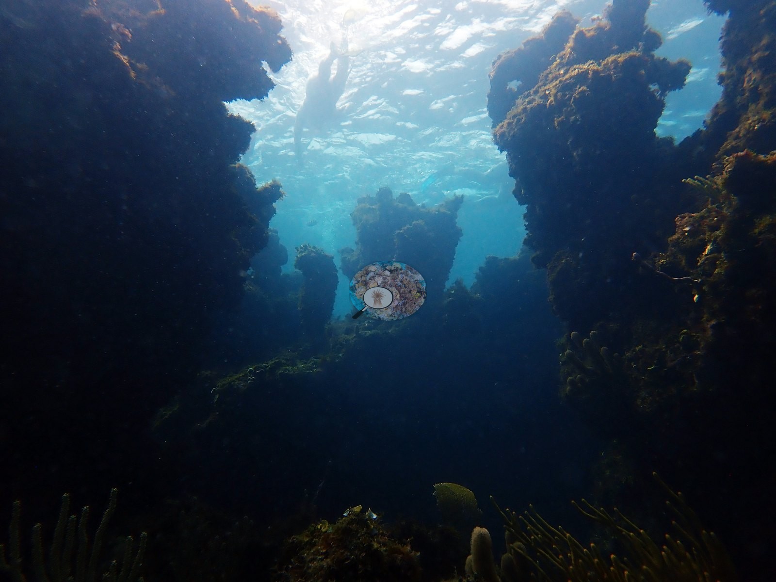
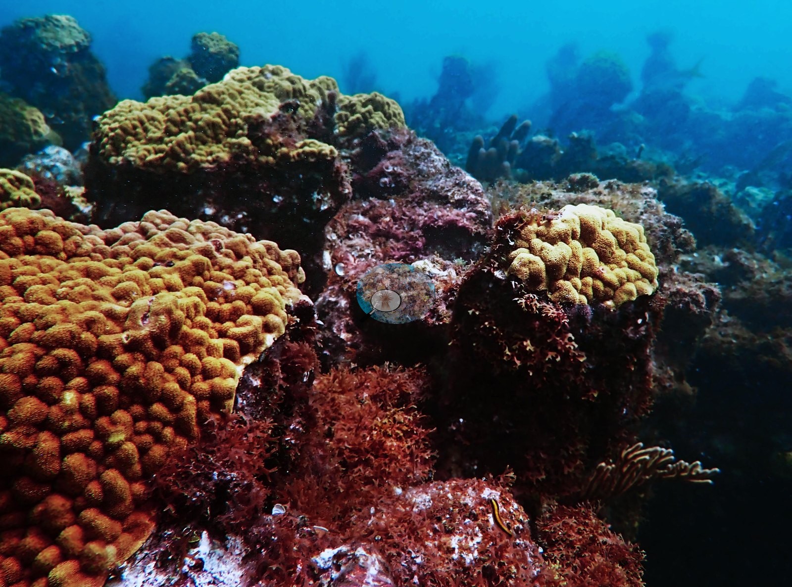
Live and dead elkhorn sit side by side across the site. The reef matrix between the elkhorn is low-relief hardbottom and spur-and-groove pavement carrying a dense red, pink, and mauve crust of coralline algae, turf, and encrusting sponge (e.g., Image 7, Image 8, Image 20, Image 21, Image 31, Image 35, Image 55, Image 56), broken by broad expanses of rippled white sand and rubble (e.g., Image 2, Image 17, Image 18, Image 25, Image 69) and by a couple of narrow shaded swim-throughs and ledges (e.g., Image 22, Image 23, Image 25).
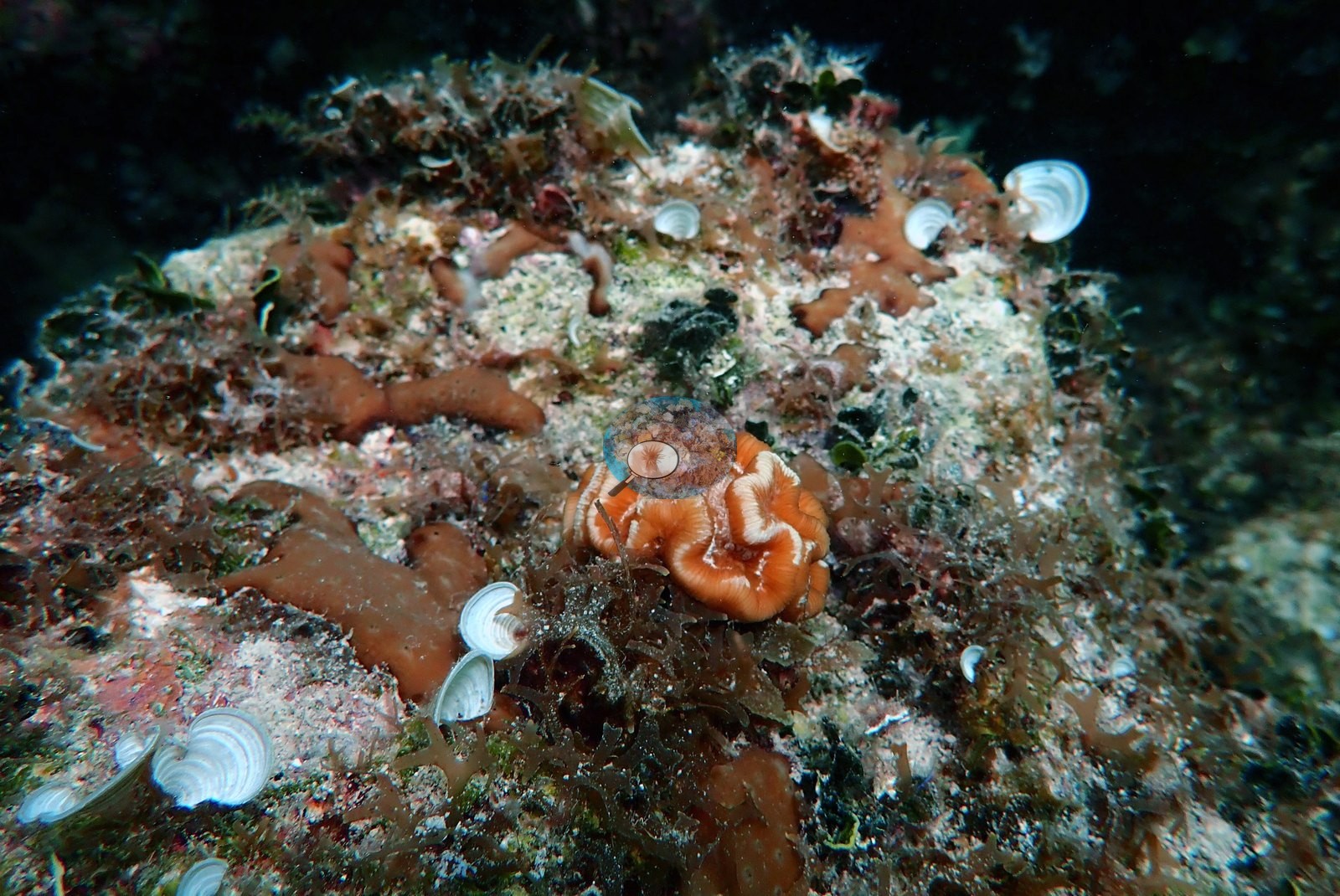
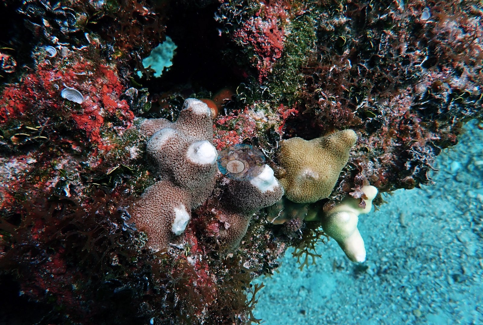
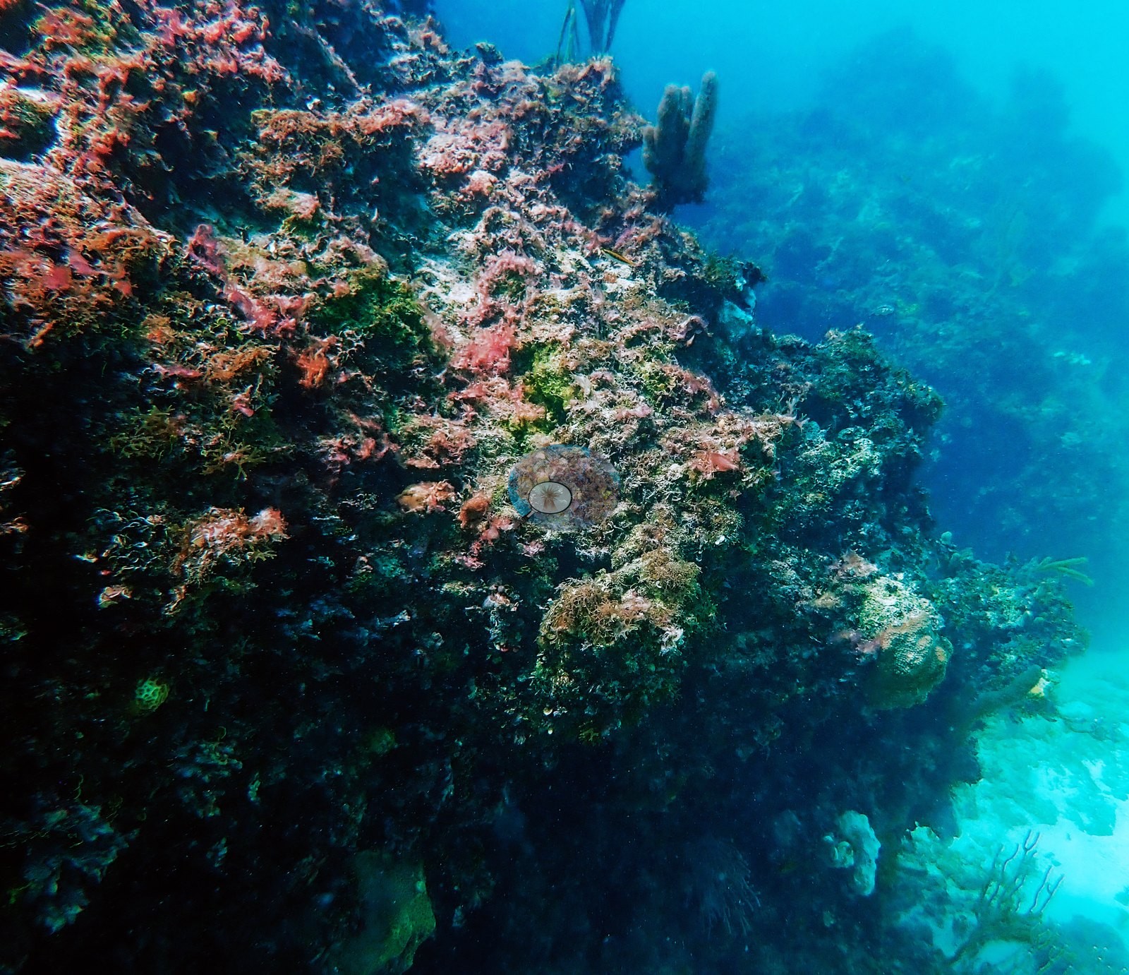
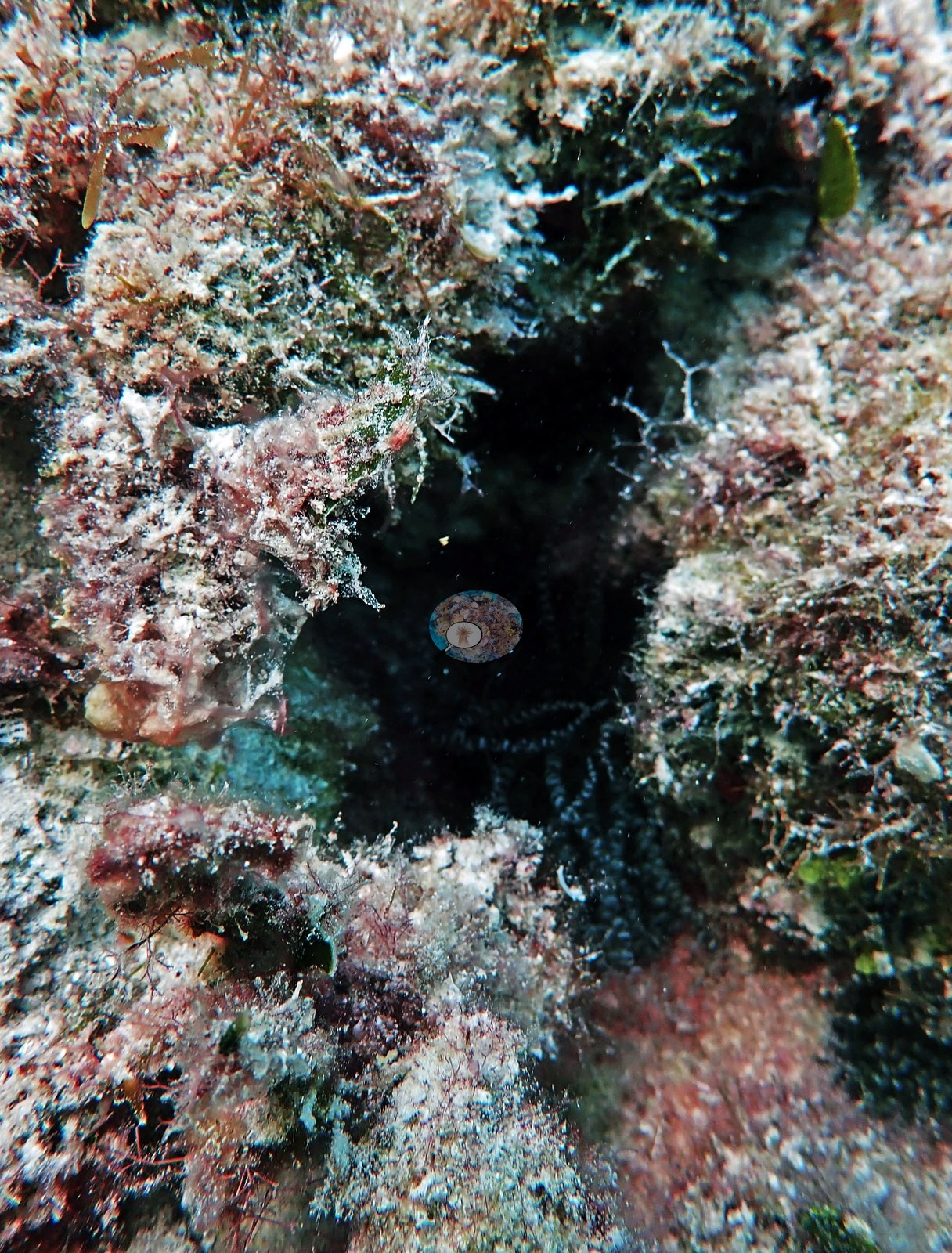
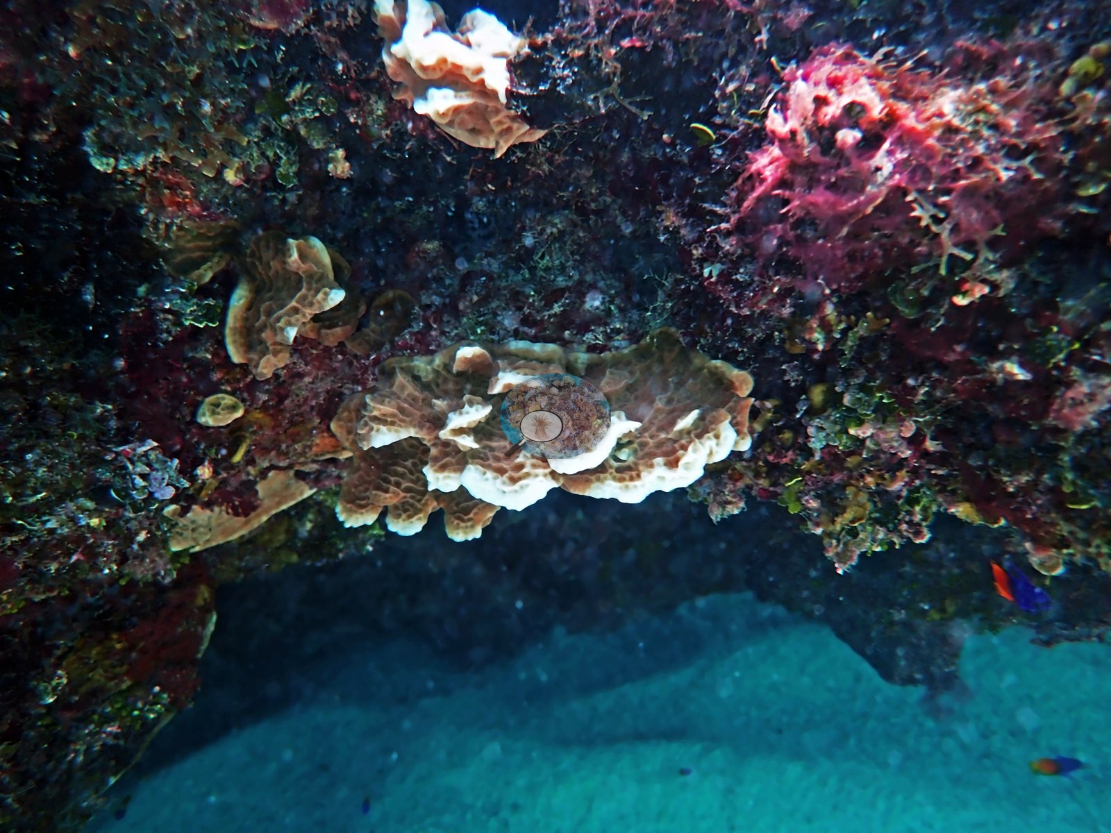
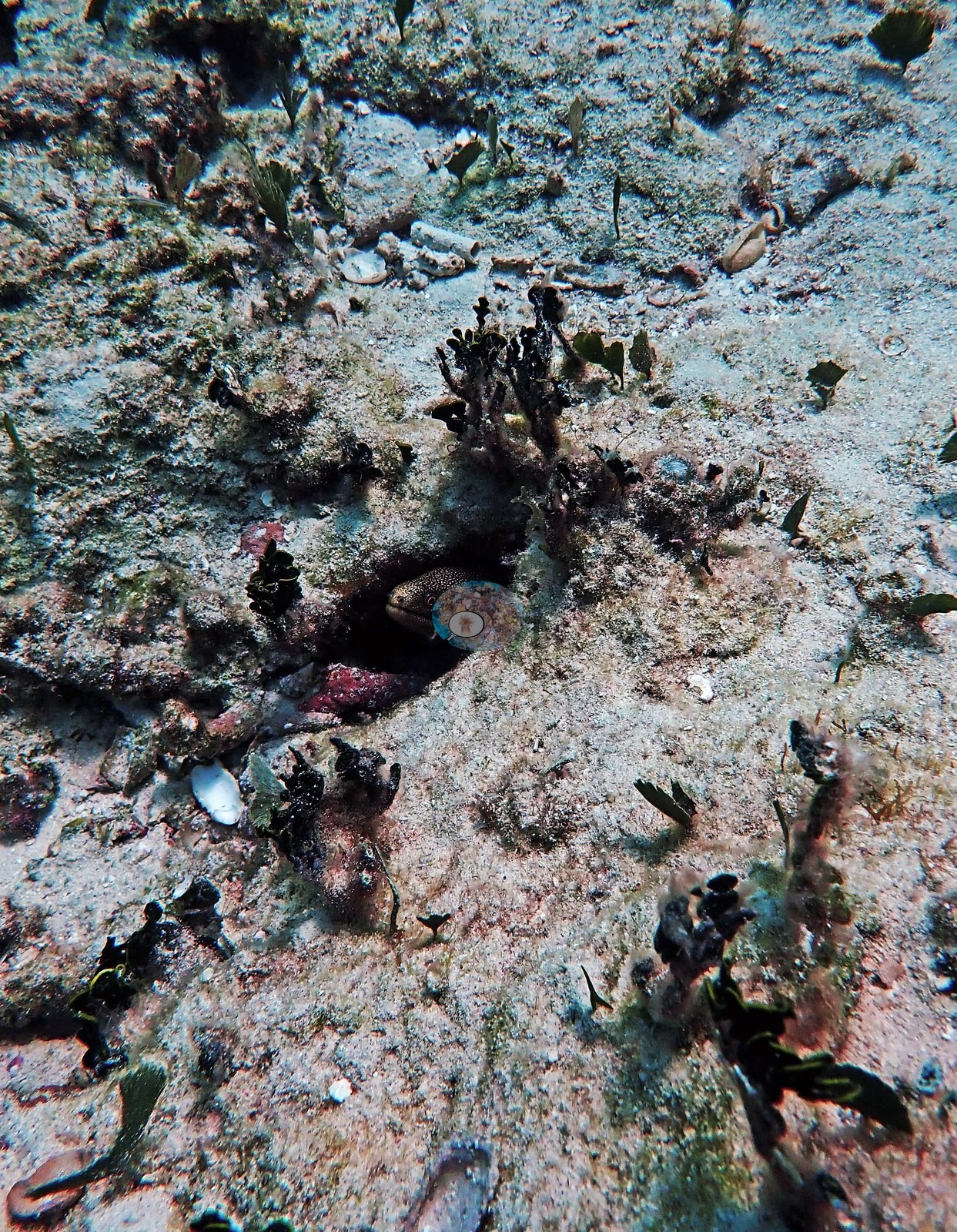
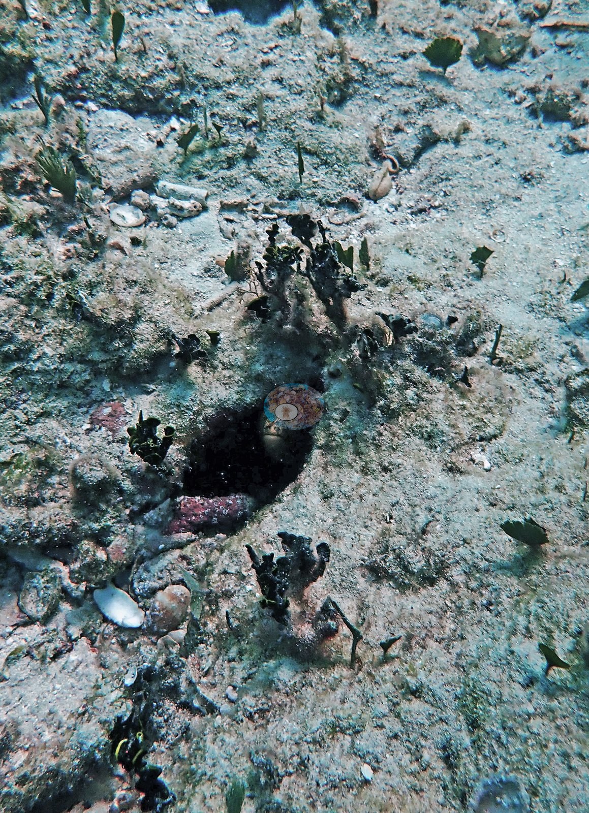
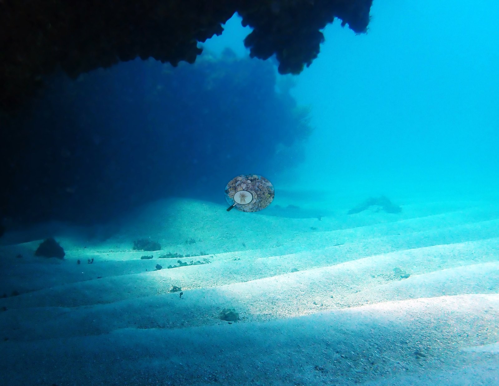
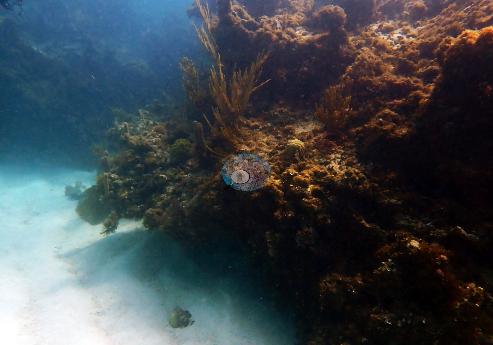
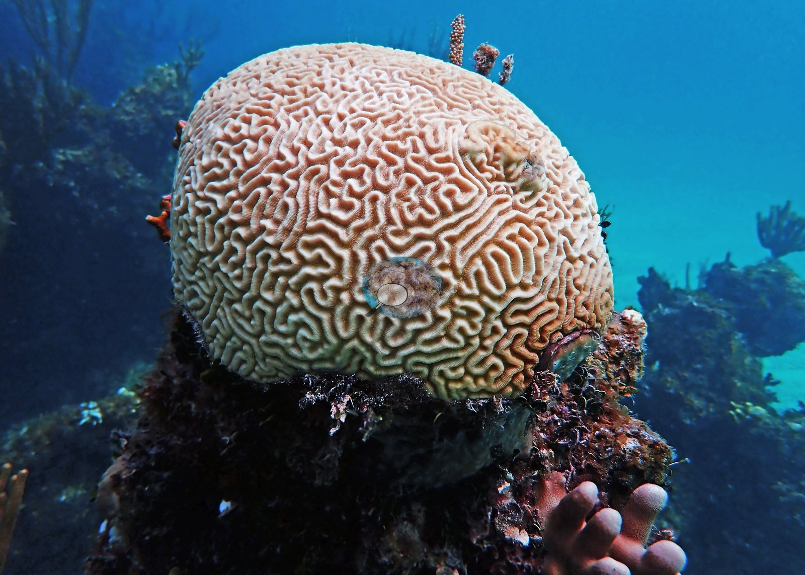
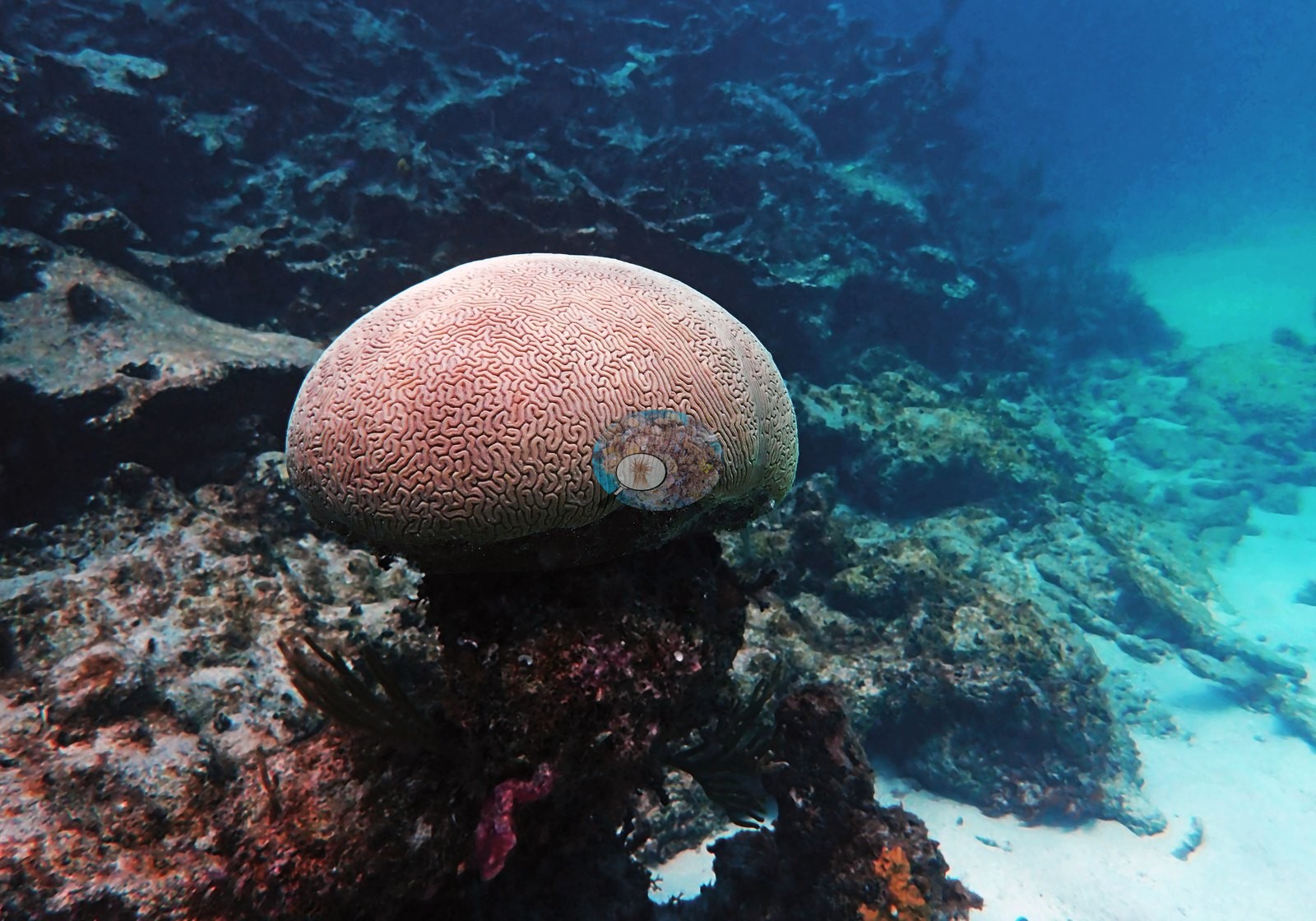
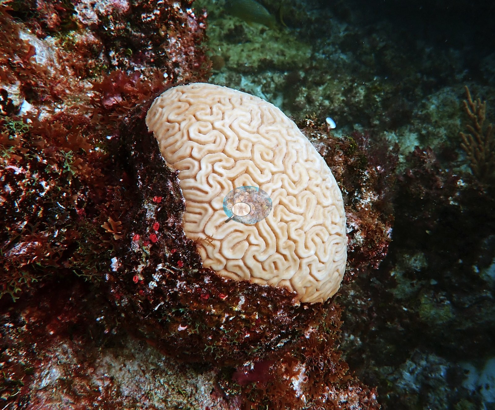
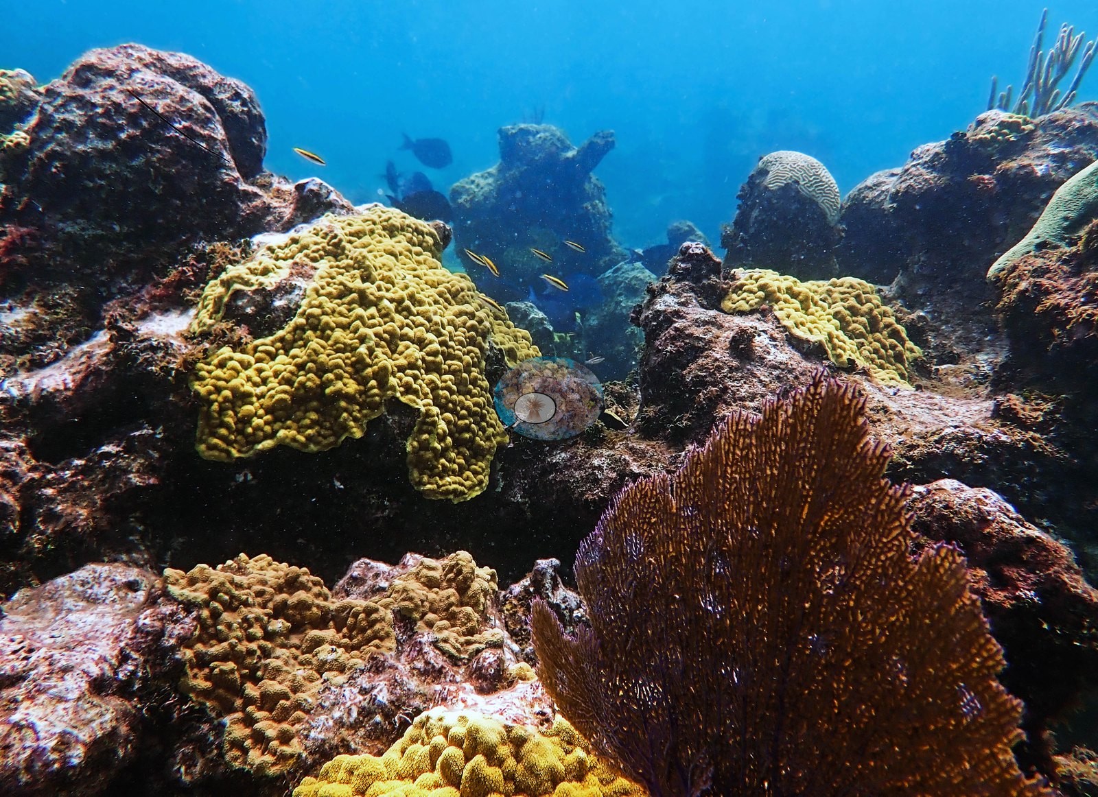
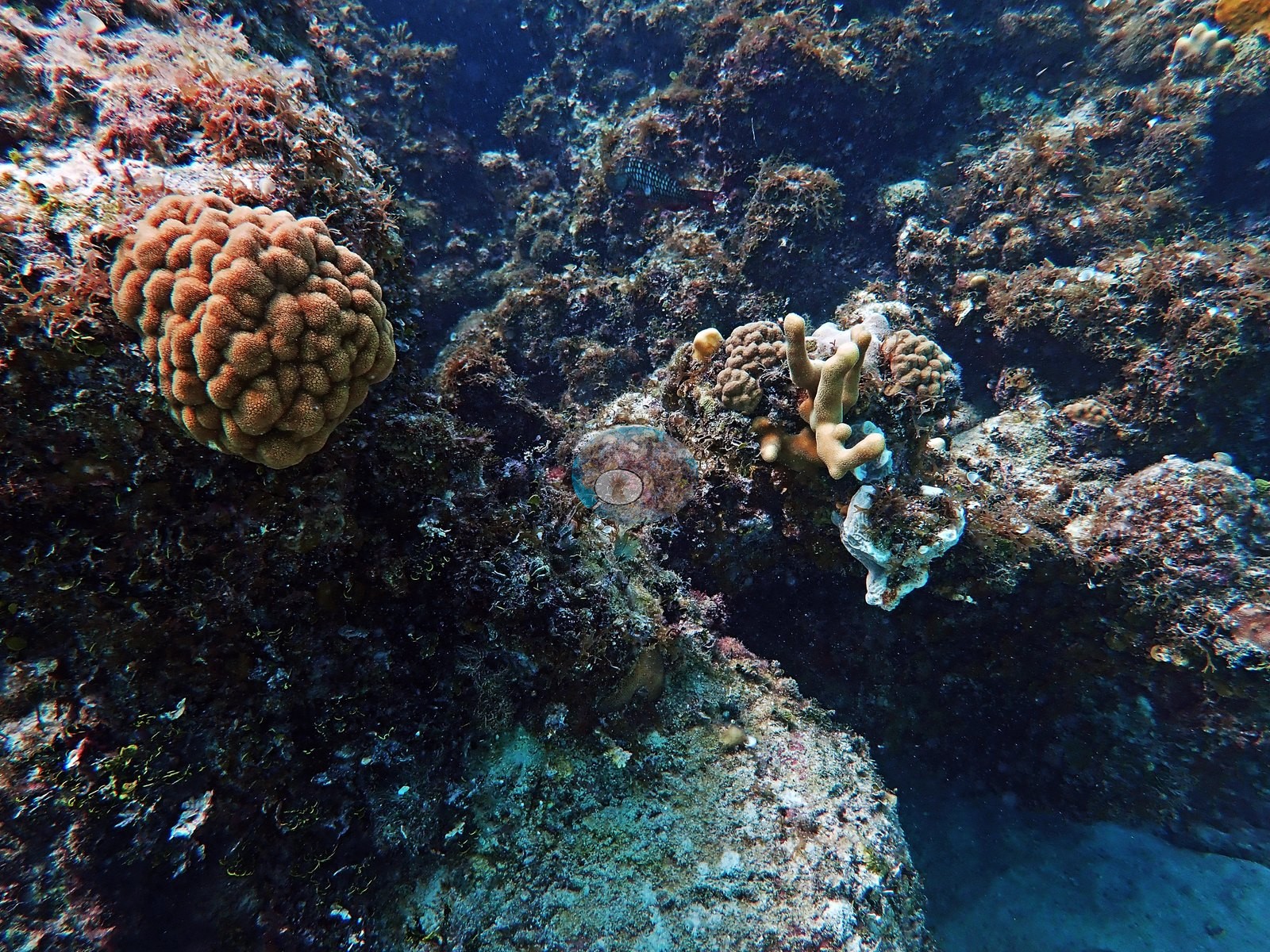
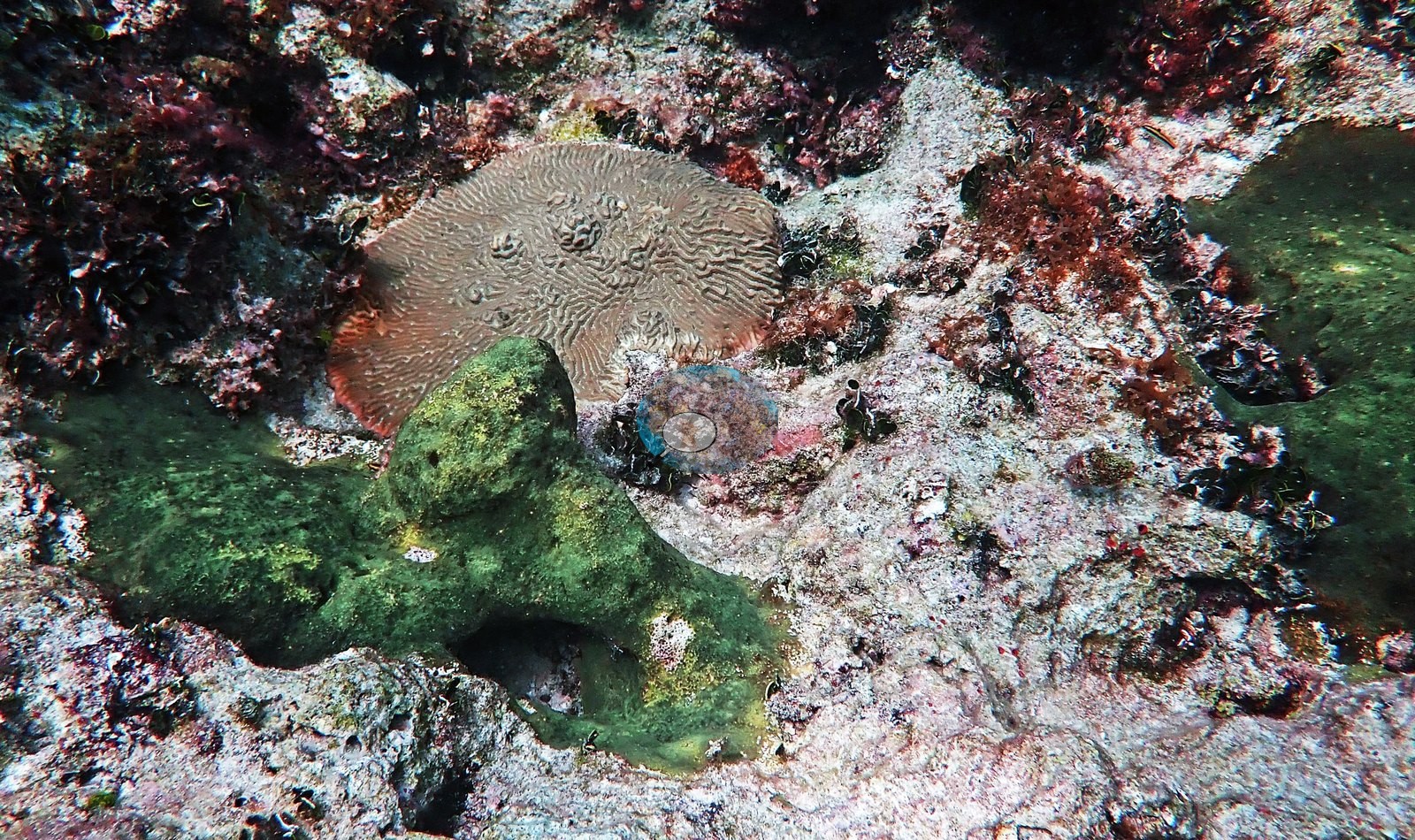
Massive corals are present as scattered discrete heads rather than as a continuous framework: large brain corals with strongly meandering ridges, several of them intact and fully pigmented (e.g., Image 9 "brain coral Bahamas", Image 34 "healthy brain coral", Image 44 "non-diseased brain coral"), against one large gray, algal-covered dead dome (e.g., Image 33, his "dead brain coral").
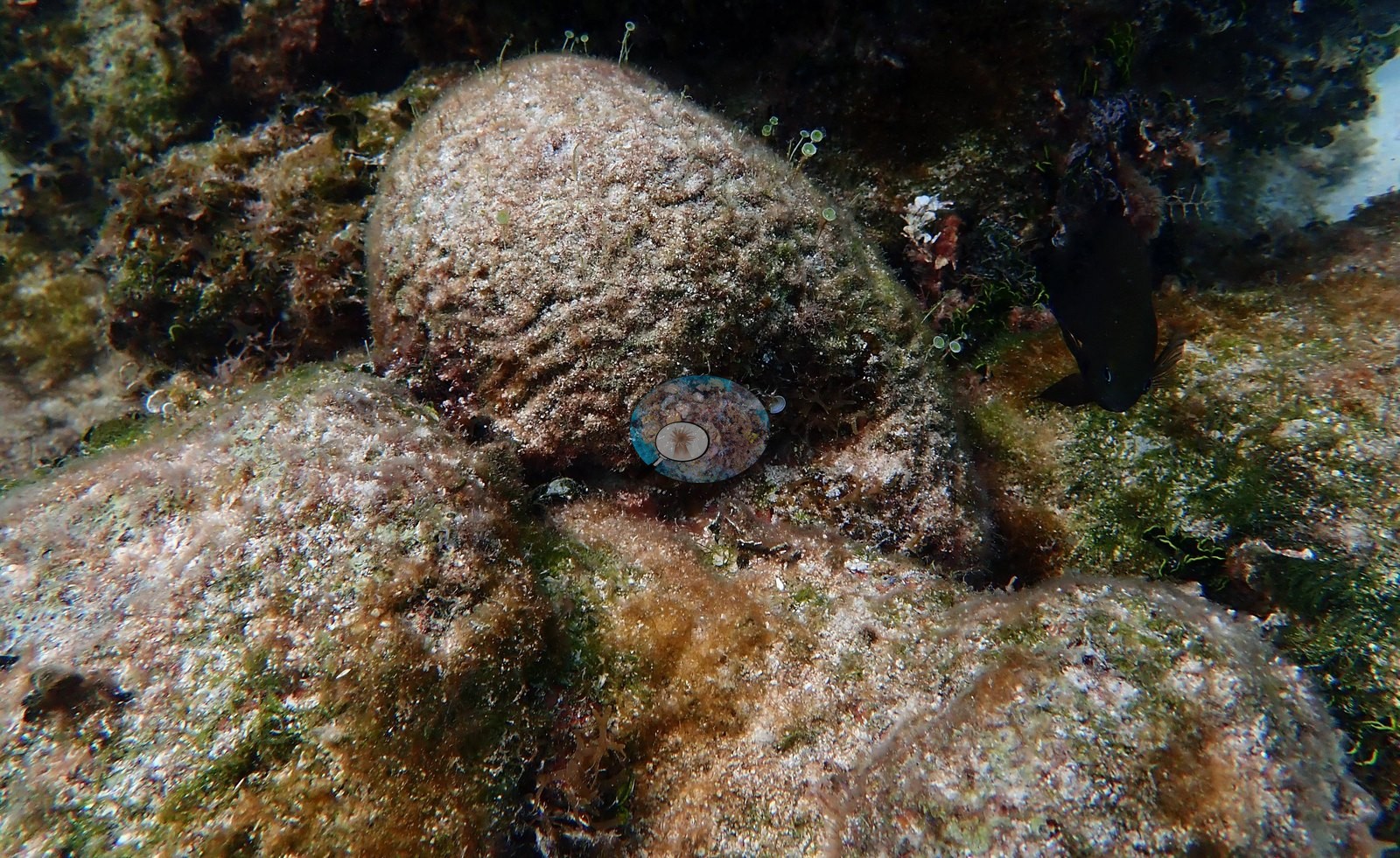
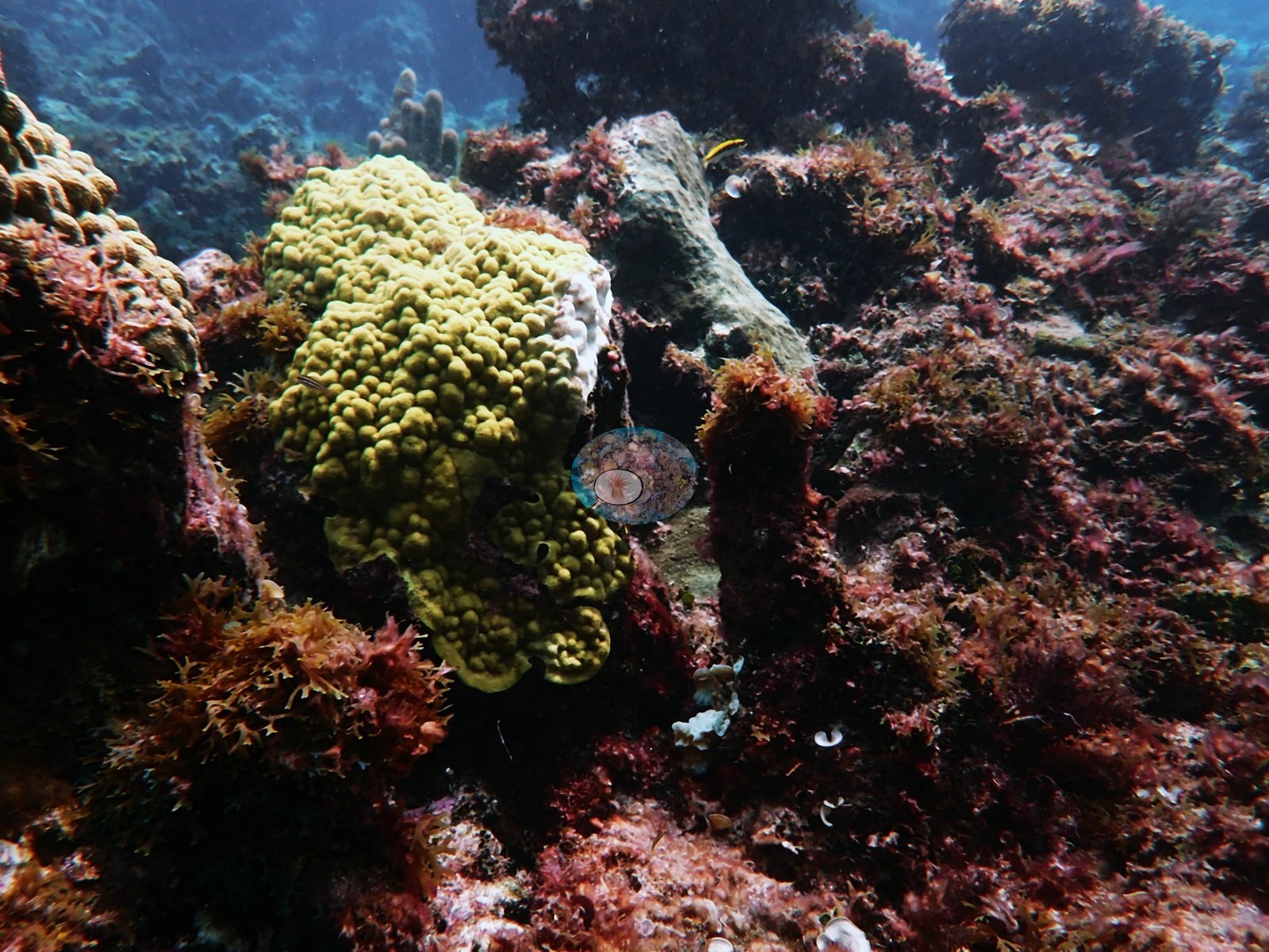
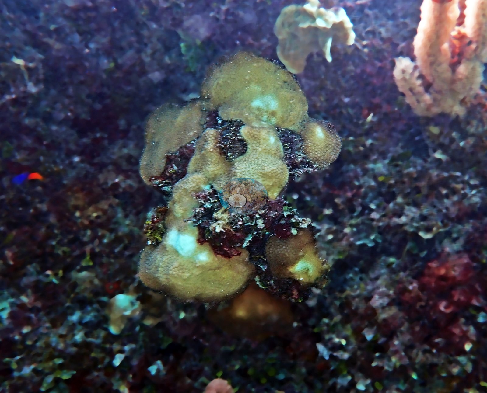
Mustard-yellow to olive mounding colonies recur (e.g., Image 19, Image 24, Image 72, Image 73, Image 70), one of which he labels a "diseased poritid" (e.g., Image 70).
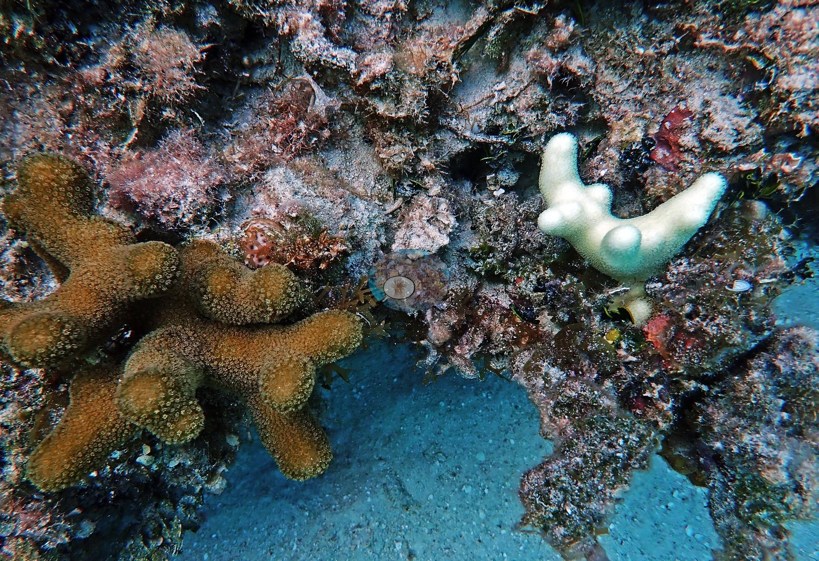
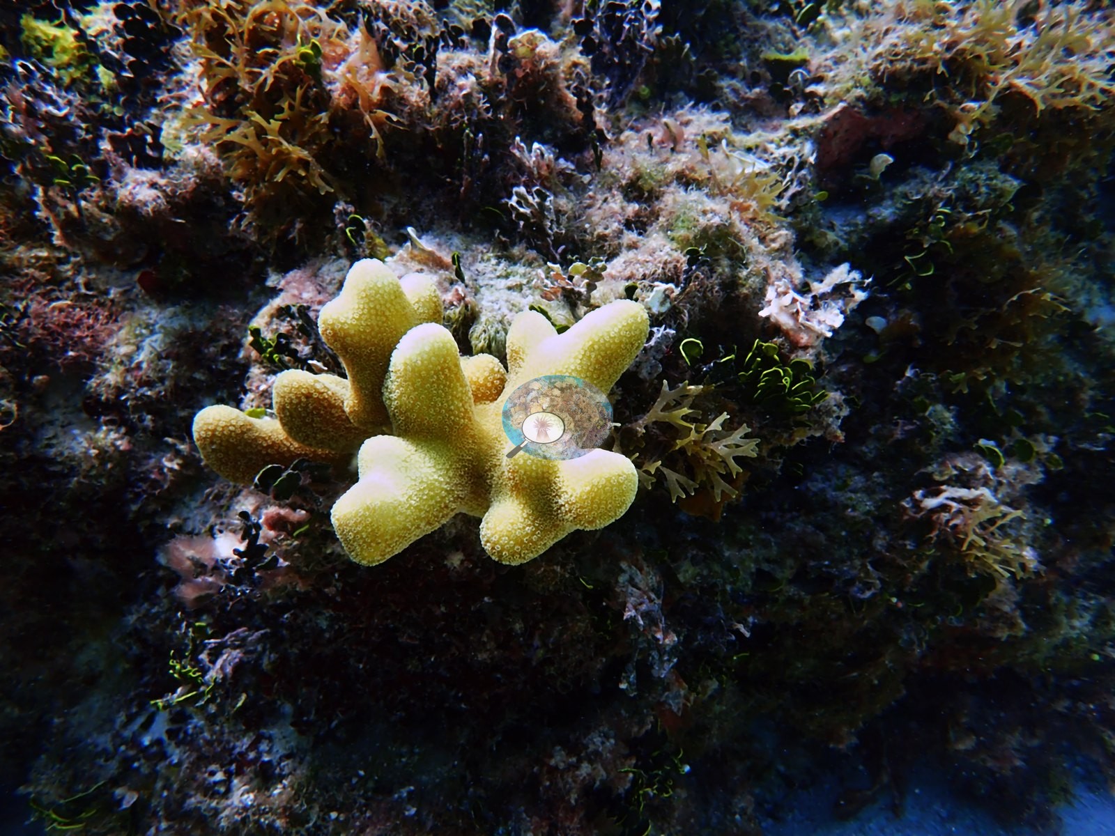
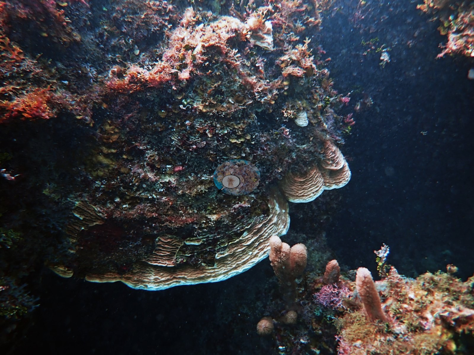
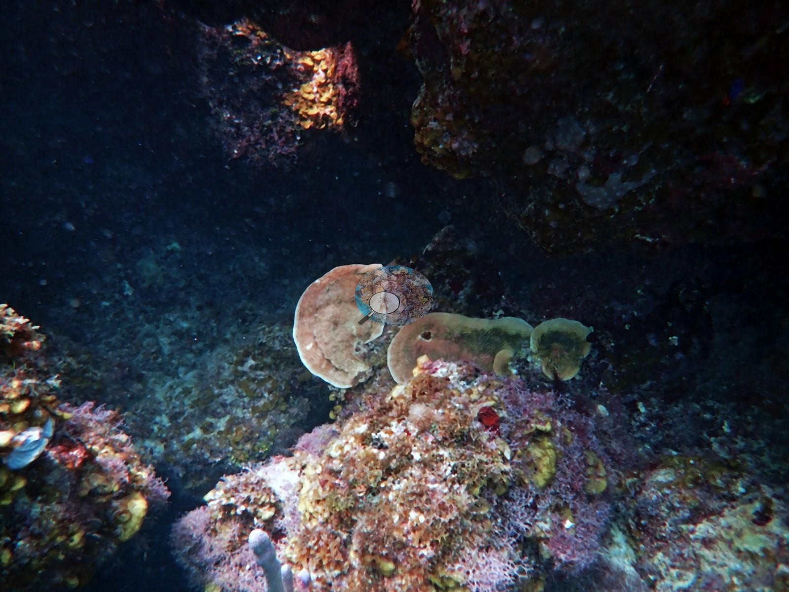
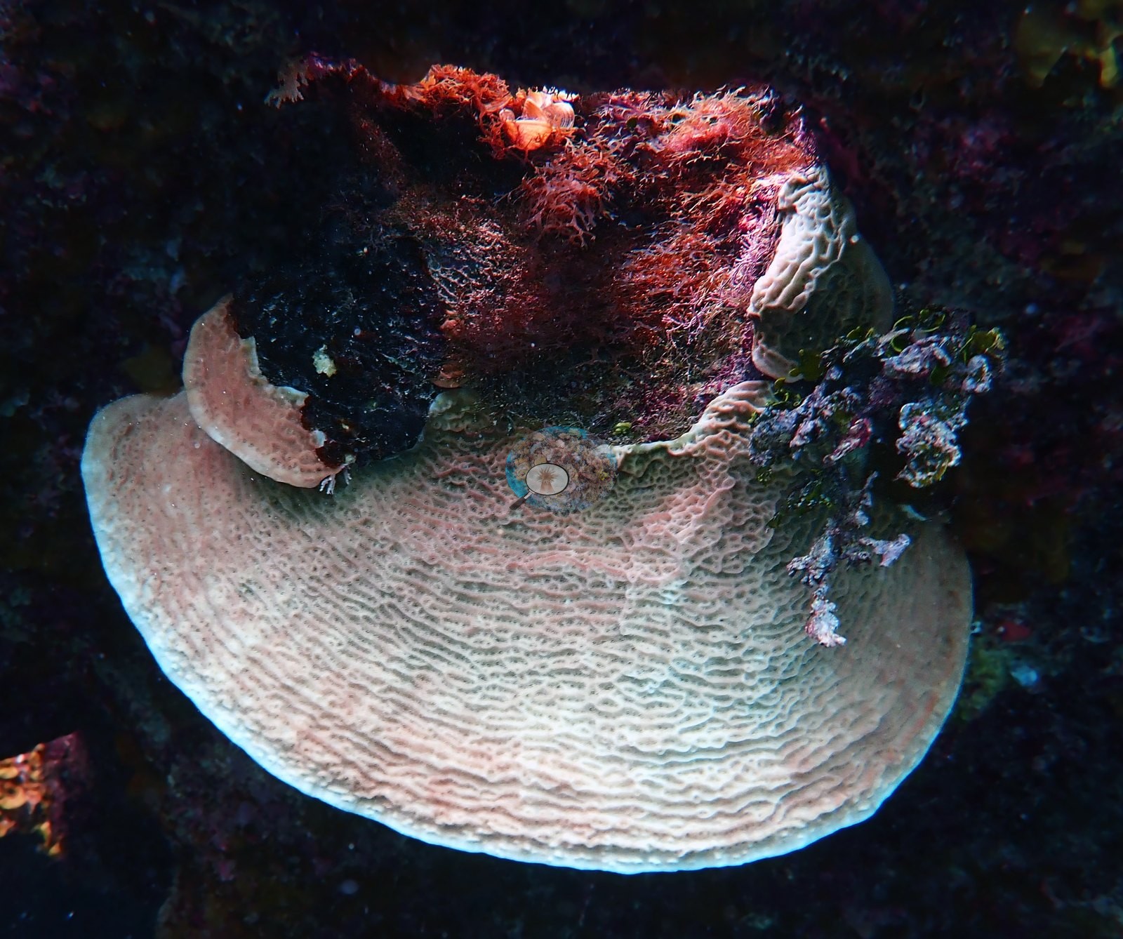
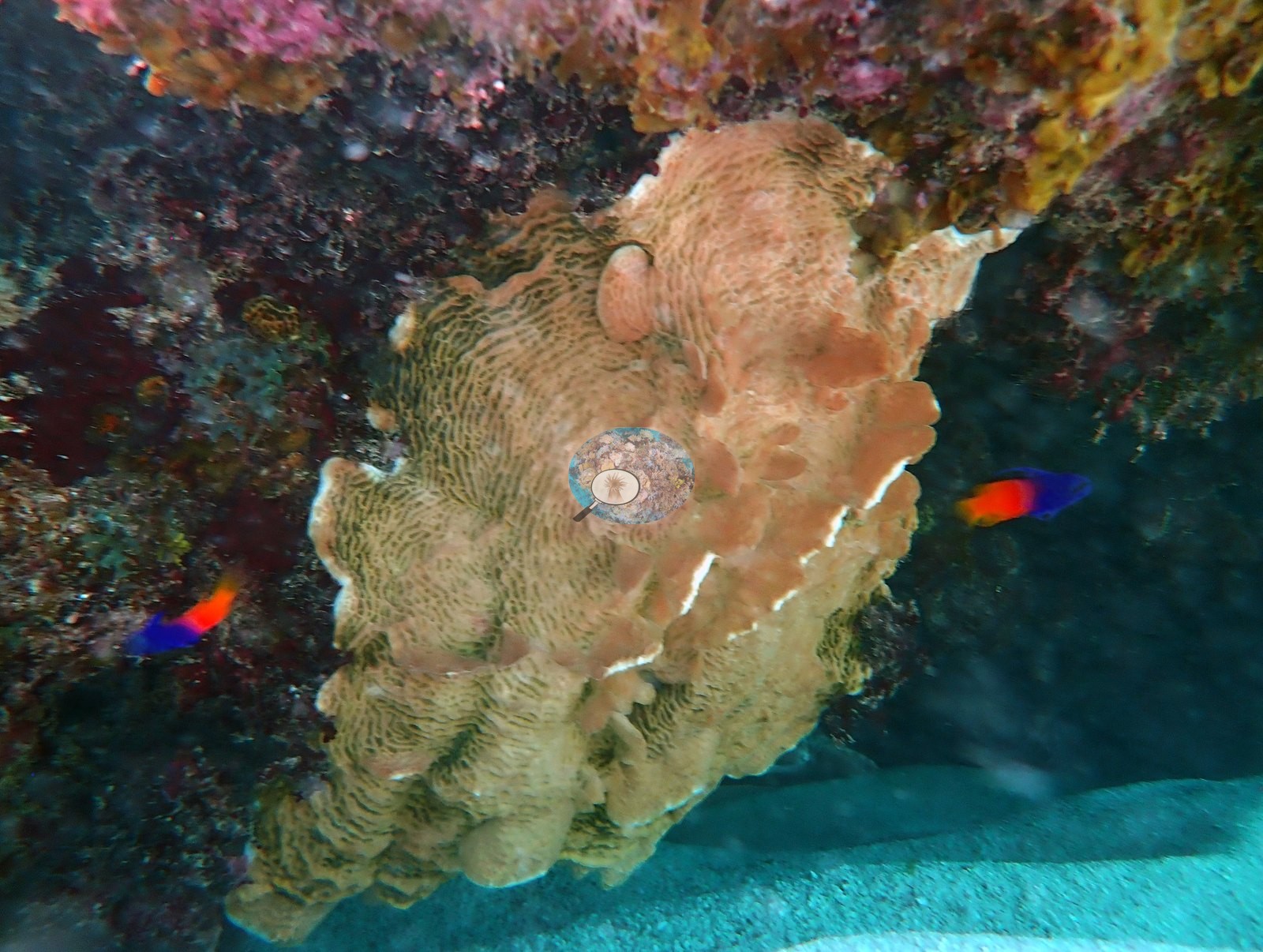
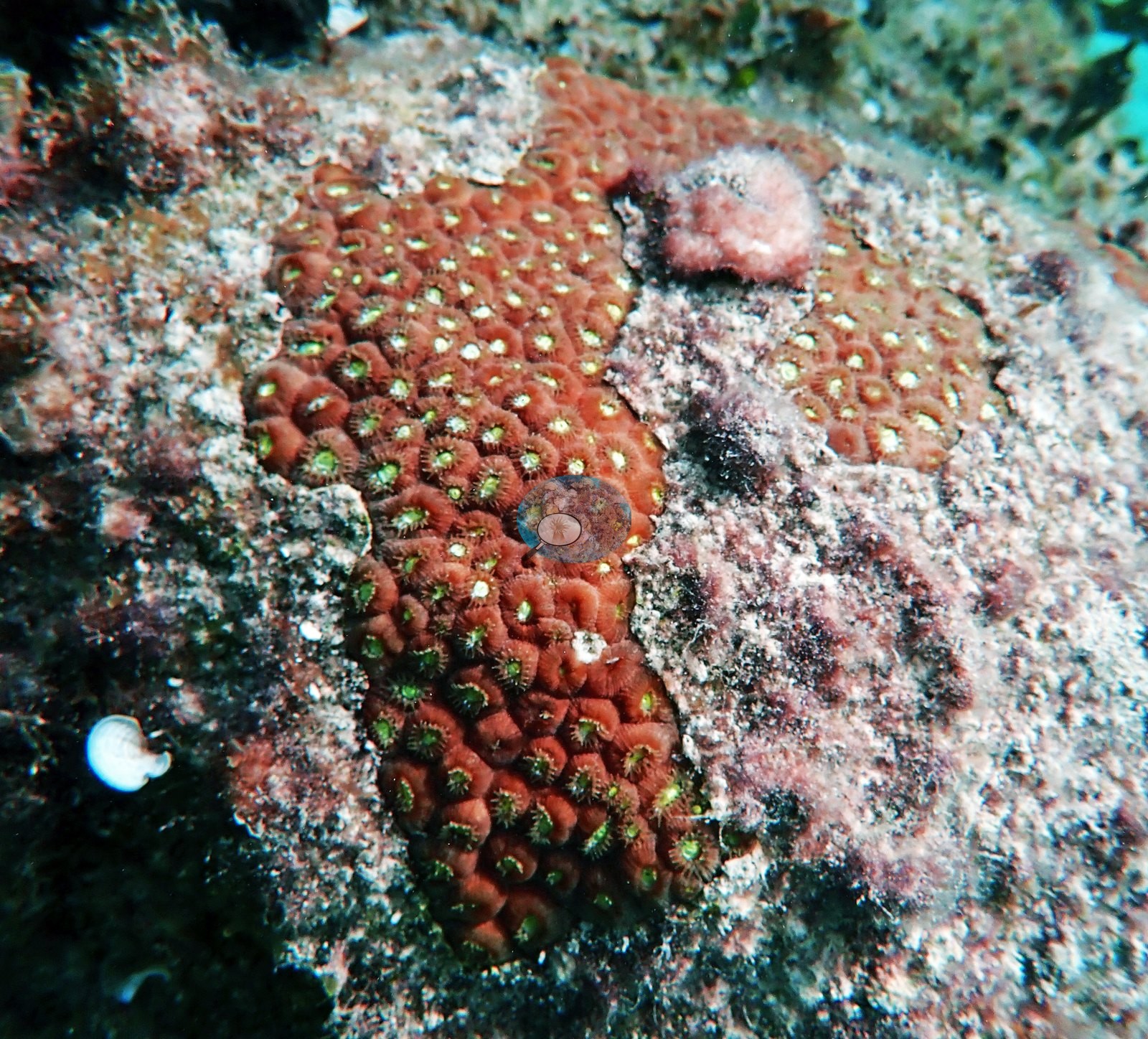
Plating and encrusting agariciid-type sheets stack under ledges and on shaded faces (e.g., Image 11, Image 12, Image 13, Image 31, Image 32), (Image 6) is a close-up of an orange-brown large-calice colony on crusted rock, and small mounding and encrusting recruits are dotted across the pavement (e.g., Image 15, Image 16, Image 27, Image 41).
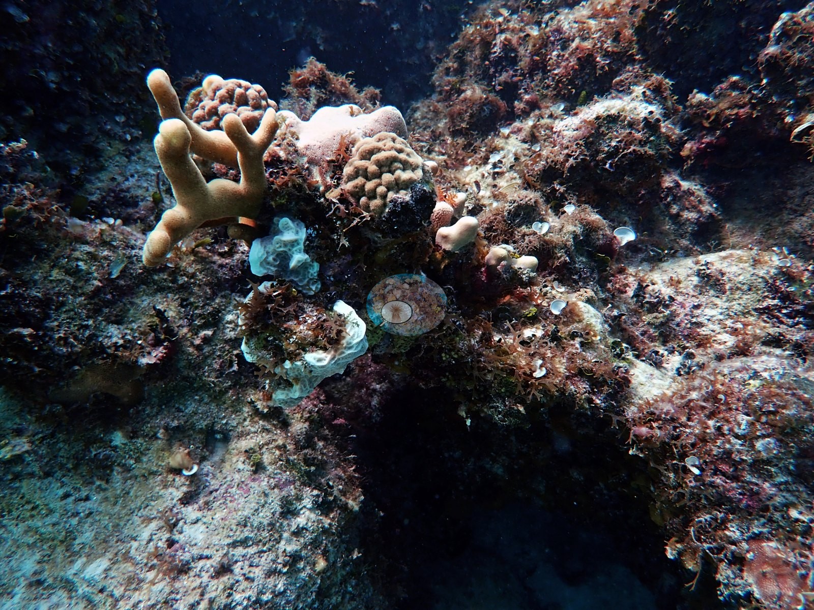
Gorgonians - sea fans, sea rods, and sea plumes - stand along the higher spurs (e.g., Image 58, Image 61, Image 68, Image 72).
Coral cover
- Reference: Claude CV point count (this site's photos)
- Coral cover: 9.1% — a blind Claude computer-vision point count of 60 of Anderson's edited photographs (20 points per photo; water, fish, and obstructions excluded). Other computer-vision methods are listed in the method table.
Stressors
Health signals are reported qualitatively only. Two frames show conspicuously pale, near-cream colonies against otherwise pigmented surroundings - the Orbicella faveolata he labels in Image 45, and a similar pale lobed colony in Image 10 - and pale tissue in a photograph cannot be separated from bleaching, recent recovery, or pigment loss, so nothing is inferred from it. Image 70 he labels as diseased; Image 44 he explicitly labels as NOT diseased, which is a useful pairing. Image 33 and Image 39 record complete mortality on a brain coral and on elkhorn framework respectively.
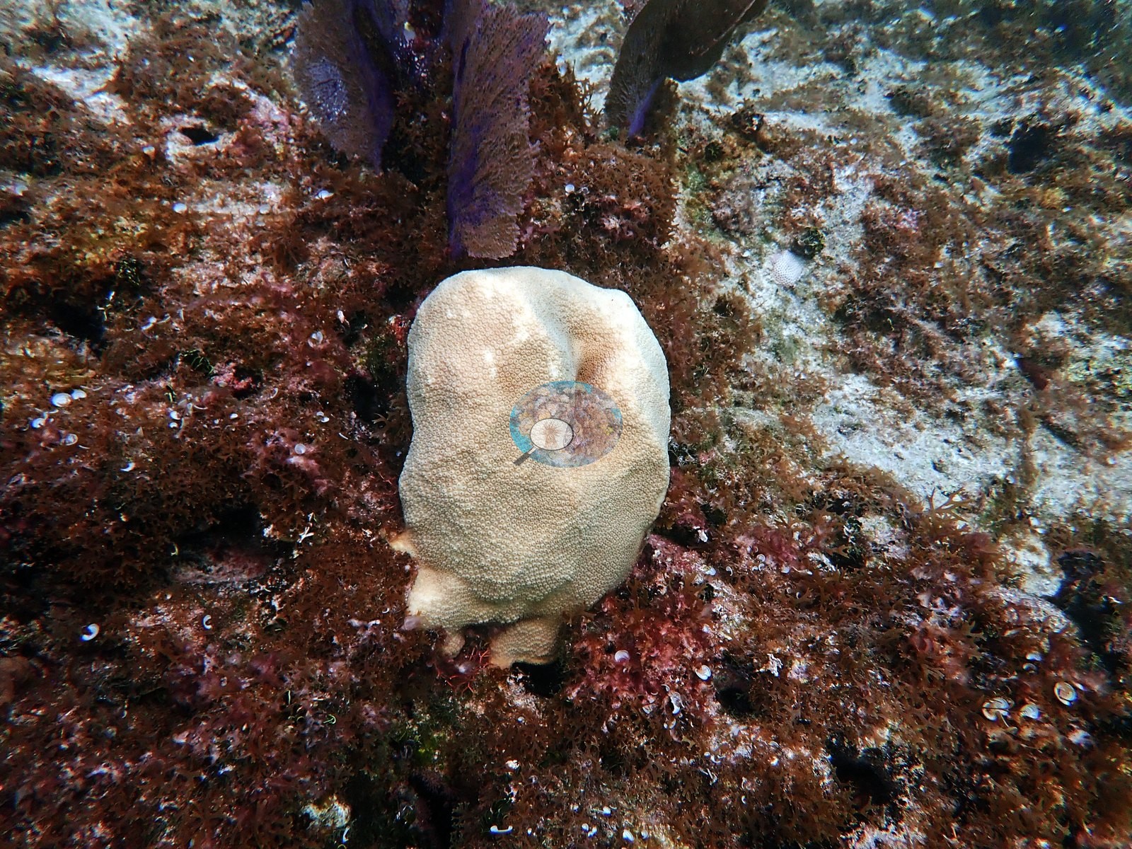
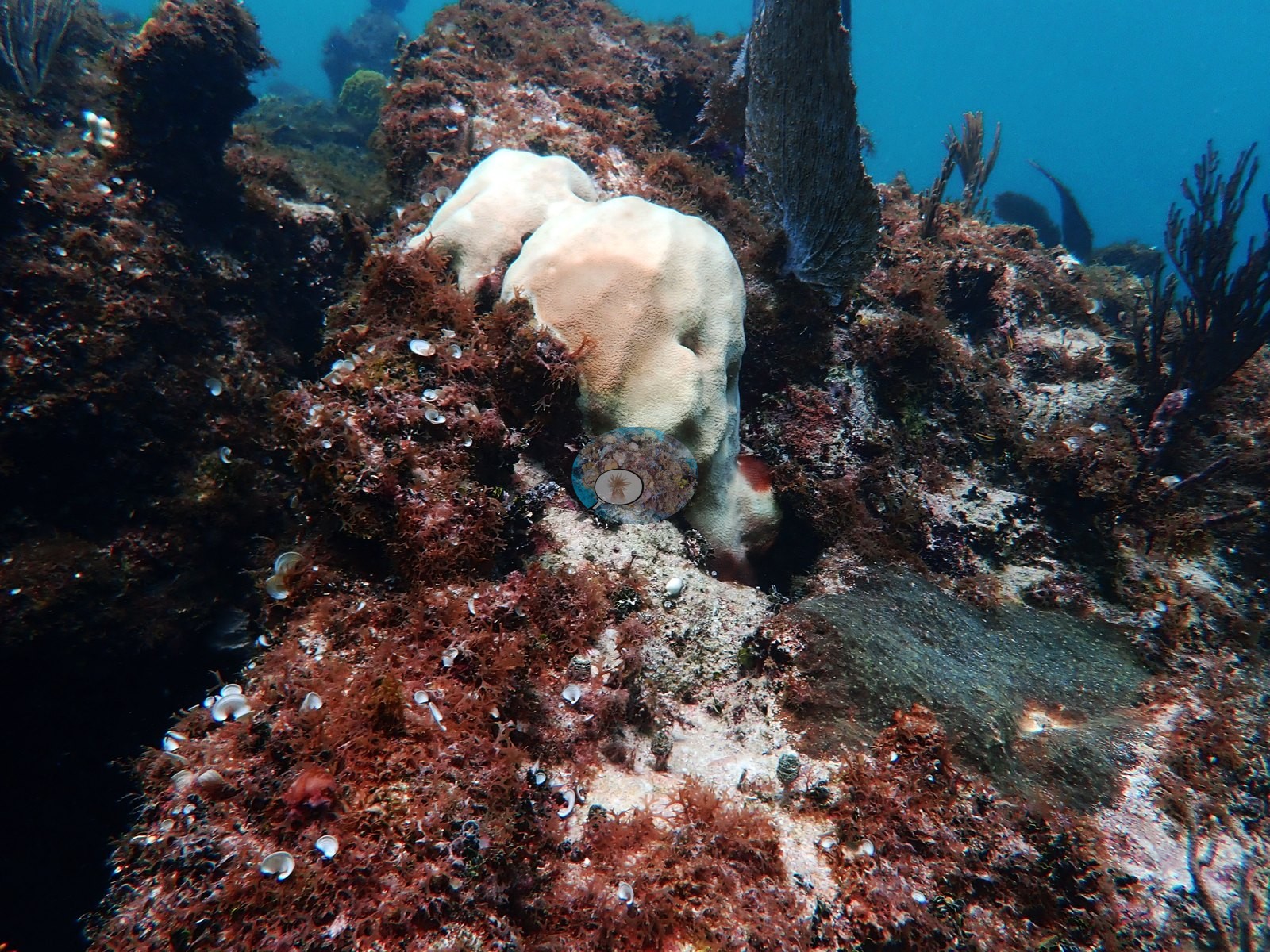
No percentage of bleaching, disease, or mortality is estimated from these frames. No corallivores evident.
Fish and other fauna
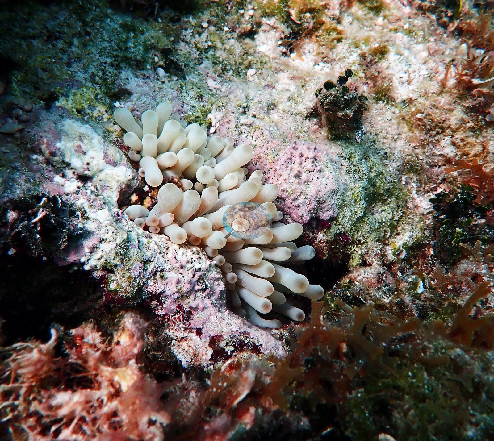
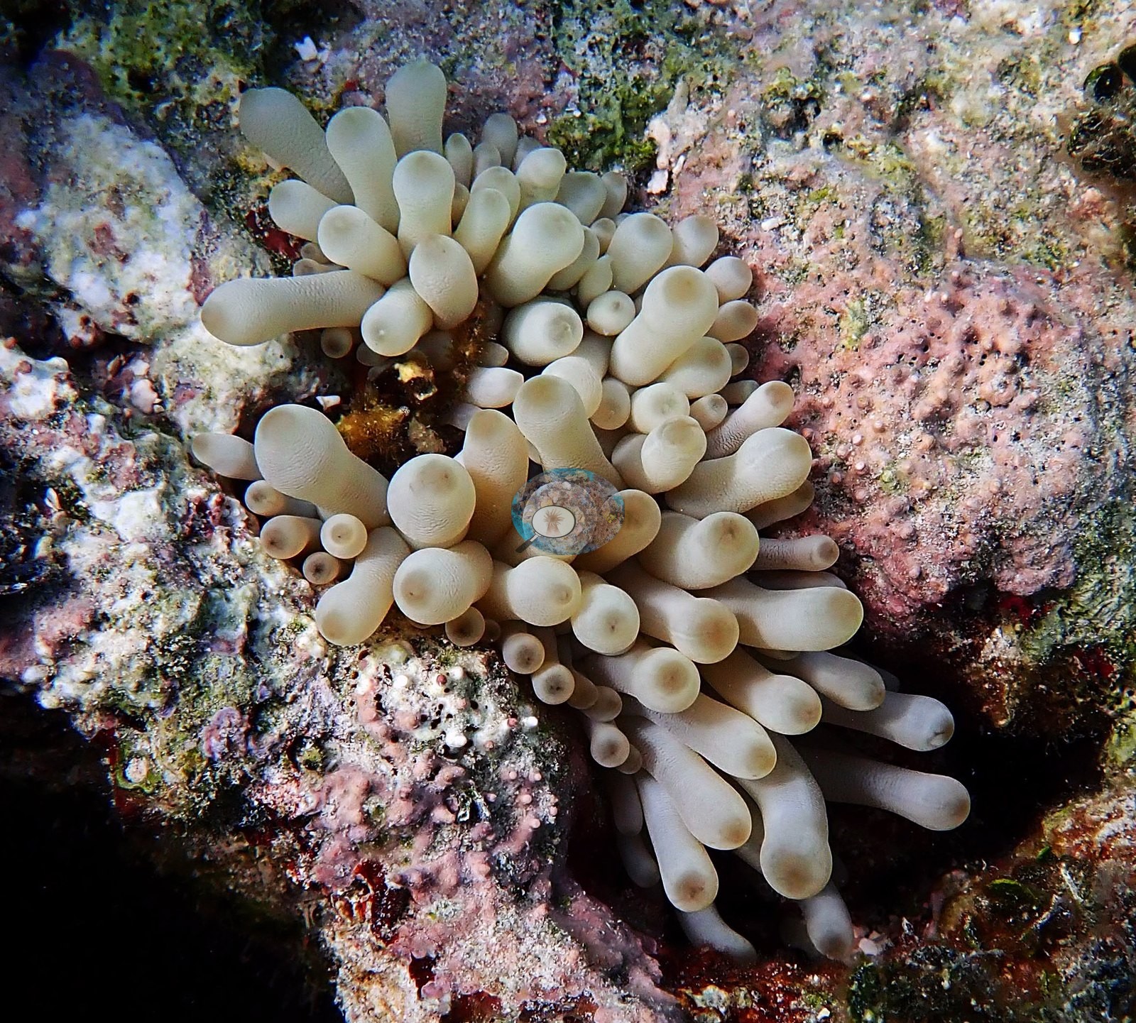
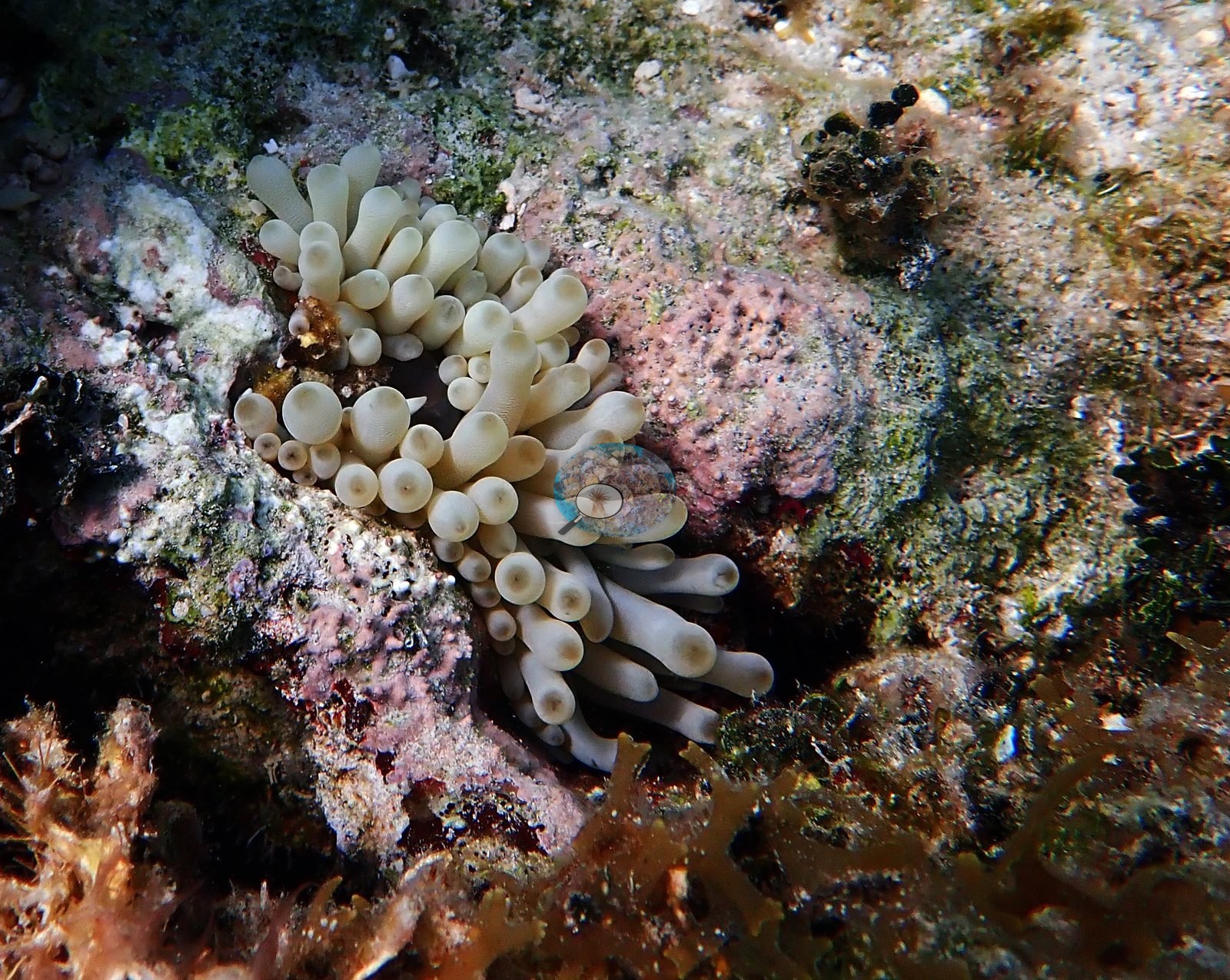
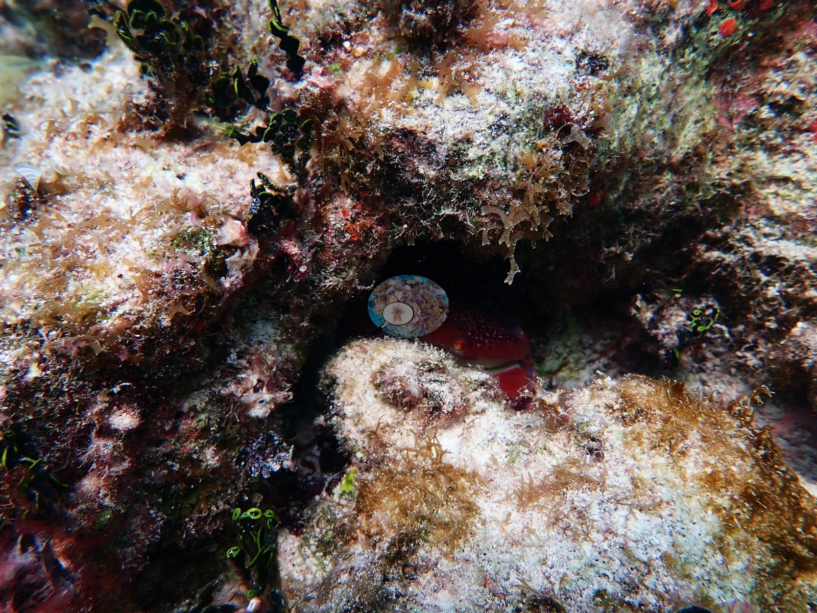
Big Caribbean giant anemones with fat, pale bulb-tipped tentacles are wedged into coralline rock in Image 29, Image 30, and Image 28, and Image 54 is a crab tucked into a crevice.
Comparisons with other data
No other surveys have been conducted at this site to date. Within 5 km, three independent stations (AGRRA and XL Catlin Seaview Survey) recorded 0.5–10% hard coral cover between 2008 and 2019; the nearest, AGRRA at Piece of Cake, 0.41 km away, read 1.0–10% between 2015 and 2019.
Other notes
Frame-level account from Anderson's "Bahamas dive 2" photographs (73 frames); the photographs do not name King Reef, so their match to this site is unconfirmed. The full set corroborates and extends the site's elkhorn story. Broad golden-tan blades fan out over low hardbottom and white sand in F41, F42, F43, and F45; a spreading multi-branched colony stands clear of the bottom in F49, F52, F54, and F56; F51 and F59 show tall candelabra colonies with clean growing margins silhouetted against bright blue water; F55 catches a wide, densely branched colony from below (e.g., Image 49, Image 51, Image 62, Image 63, Image 64).
His own labeled frames give the range in one sequence: a whole thicket (F1), the very large colony he calls "monster A. palmata" (F71), a small recruit growing on live rock (F64, "baby Apalm"), a sparse, thin-branched colony he calls "struggling" (F73), and a diver alongside a standing skeleton for scale (F63, "assessing Apalm"). The counterpart to all that live tissue is standing dead framework: bare, turf- and silt-crusted elkhorn branches still in growth position in F34, F38, F39, and F67 (his "dead Acropora framework"), and eroded branch stubs mixed into the pavement in F35, F36, and F44 (e.g., Image 38, Image 42, Image 43, Image 65). The reef matrix between the elkhorn is low-relief hardbottom and spur-and-groove pavement carrying a dense red, pink, and mauve crust of coralline algae, turf, and encrusting sponge (F9, F10, F21, F22, F31, F33, F46, F47), broken by broad expanses of rippled white sand and rubble (F4, F18, F19, F26, F58) and by a couple of narrow shaded swim-throughs and ledges (F23, F24, F26) (e.g., Image 14, Image 17, Image 21, Image 25, Image 35, Image 39).
Massive corals are present as scattered discrete heads rather than as a continuous framework: large brain corals with strongly meandering ridges, several of them intact and fully pigmented (F65 "brain coral Bahamas", F70 "healthy brain coral", F72 "non-diseased brain coral"), against one large gray, algal-covered dead dome (F68, his "dead brain coral") (e.g., Image 9, Image 34, Image 13). Mustard-yellow to olive mounding colonies recur (F20, F25, F60, F61, F69), one of which he labels a "diseased poritid" (F69) (e.g., Image 24, Image 70). Plating and encrusting agariciid-type sheets stack under ledges and on shaded faces (F12, F13, F14, F31, F32), F8 is a close-up of an orange-brown large-calice colony on crusted rock, and small mounding and encrusting recruits are dotted across the pavement (F16, F17, F28, F37) (e.g., Image 6, Image 11, Image 16, Image 22, Image 31). Gorgonians - sea fans, sea rods, and sea plumes - stand along the higher spurs (F49, F52, F57, F60) (e.g., Image 36, Image 45, Image 72). Big Caribbean giant anemones with fat, pale bulb-tipped tentacles are wedged into coralline rock in F29, F30, and F62, and F66 is a crab tucked into a crevice (e.g., Image 28, Image 29, Image 30). Two frames show conspicuously pale, near-cream colonies against otherwise pigmented surroundings - the Orbicella faveolata he labels in F2, and a similar pale lobed colony in F11 - and pale tissue in a photograph cannot be separated from bleaching, recent recovery, or pigment loss, so nothing is inferred from it (e.g., Image 5, Image 32, Image 44).
F69 he labels as diseased; F72 he explicitly labels as NOT diseased, which is a useful pairing. F68 and F67 record complete mortality on a brain coral and on elkhorn framework respectively (e.g., Image 66).
CV model comparison
More info
A few colony-focused photos raise the mean; the median is the typical photo. No computer-vision method yet estimates coral cover accurately on new images, so treat this as indicative rather than a measurement; it is revised as the methods improve.More info
DINOv3 and CoralSCOP were run on the same photos and are listed for comparison. Coralscapes v2 and v1 were also run but are not shown: the author does not consider Coralscapes reliable for these photos. No diver transect exists for this trip, so agreement between the rows is not accuracy, and no computer-vision method yet estimates coral cover accurately on new images; treat every row as indicative.| Method | Hard-coral cover (%) |
|---|---|
| CoralSCOP edited | 15.5 |
| Claude (edited photos) | 9.1 |
See a more detailed, stand-alone version of this table.
Independent surveys nearby
| Agency / program | Year(s) | Stations / distance | Link |
|---|---|---|---|
| AGRRA | 2008–2019 | 2 stations, 0.41–0.46 km | Records ↓ |
| XL Catlin Seaview Survey | 2013 | 1 station, 1.01 km | Records ↓ |
 Coral Reef Diagnostics
Within 5 km, 3 independent survey stations recorded hard coral cover in 6 records between 2008 and 2019. Of the 1 station surveyed in more than one year, 1 increased between their first and latest surveys. The longest record, Piece of Cake (AGRRA, 0.41 km away), went from 1.0% in 2015 to 10% in 2019. The most recent nearby survey is from 2019, so these values predate recent heat stress. This site's own estimate (9.1%) sits within the range of these surveys (0.5–10%); the methods differ, so the comparison is context, not a like-for-like test.
Collected by other programs using different methods (AGRRA uses benthic point-intercept transects, reported here as live coral cover; the XL Catlin Seaview Survey uses photo-transects with automated image annotation), so these are context for the figures above, not a like-for-like comparison. Where a station was surveyed more than once the range is shown. Station names are not dive-site names.
| Agency | Site / location | Date | Hard coral cover (%) | Distance | Link |
|---|---|---|---|---|---|
| AGRRA | — | 2008 | 9.2% (3 surveys) | 0.46 km | — |
| XL Catlin Seaview Survey | transect 24012 | 2013 | 0.5% | 1.01 km | doi:10.14264/uql.2019.930 |
| AGRRA | Piece of Cake | 2015–2019 | 1.0–10% (2 surveys) | 0.41 km | — |
Data & links
- HeatCRD database ↗
- van Woesik and Kratochwill (2024) ↗
- Photo portfolio ↗
- Coral Reef Diagnostics page for this area ↗
All images from Coral Reef Diagnostics. Data are from Coral Reef Diagnostics and the organizations named in the data sources above.
© Coral Reef Diagnostics.